
NA • 322534
| Product name | B-hPD-L1 plus/hCDH6 MC38 |
|---|---|
| Catalog number | 322534 |
| Strain name | NA |
| Strain background | C57BL/6 |
| NCBI gene ID | 60533,12561 (Human) |
| Chromosome | 19, 15 |
| Aliases | B7h1; Pdl1; Pdcd1l1; Pdcd1lg1; A530045L16Rik; Rcad; R-CAD; R-Cadh |
| Tissue | Colon |
| Disease | Colon carcinoma |
Inoculated cell lines can be suspended with DMEM stock solution.
Before implementing the project, it is recommended to perform tumor growth experiments. The recommended cell inoculation amount is between 5E5~1E6.
In the experiment, it is necessary to ensure that the number of animals inoculated subcutaneously is at least 1.6 times the actual grouping number.
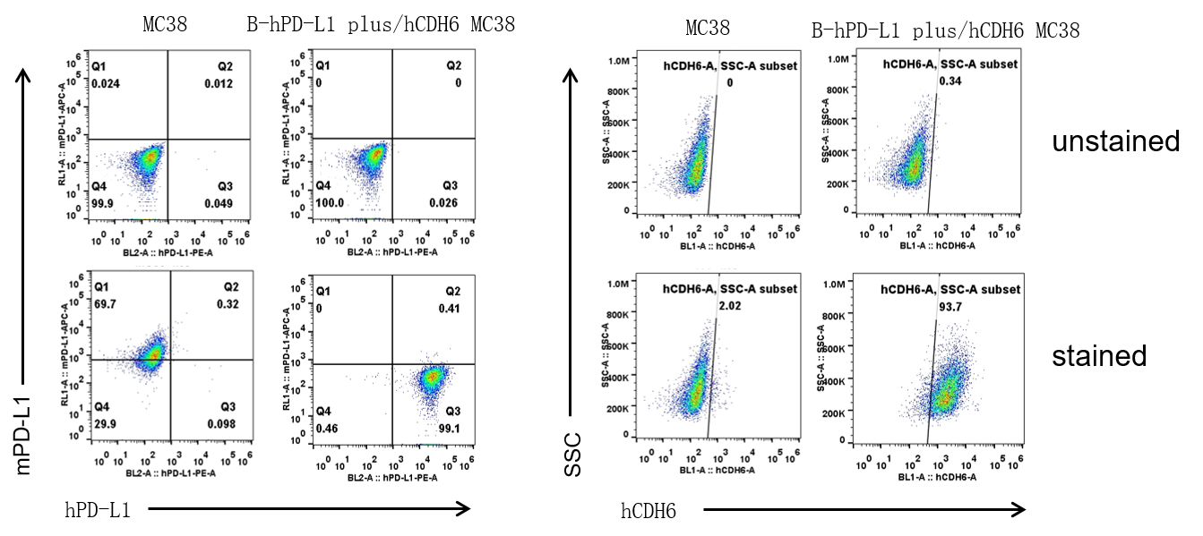
PD-L1 and CDH6 expression analysis in B-hPD-L1 plus/hCDH6 MC38 by flow cytometry. Single cell suspensions from wild-type MC38 and B-hPD-L1 plus/hCDH6 MC38 clone 1-D07 cultures were stained with anti-mouse PD-L1 antibody (Biolegend, 124312), anti-human PD-L1 antibody (Biolegend, 329706), and anti-human CDH6 antibody (R&D, FAB2715G) .

Subcutaneous tumor growth of B-hPD-L1 plus/hCDH6 MC38 cells. B-hPD-L1 plus/hCDH6 MC38 (5×10⁵) and wild-type MC38 cells (5×10⁵) were subcutaneously implanted into C57BL/6 (female, 14-week-old, n=6). Tumor volume and body weight were measured twice a week. (A) Average tumor volume. (B) Body weight. Volume was expressed in mm3 using the formula: V=0.5 × long diameter × short diameter2. Results indicate that B-hPD-L1 plus/hCDH6 MC38 cells were able to establish tumors in vivo and can be used for efficacy studies. Values are expressed as mean ± SEM.

Subcutaneous tumor growth of B-hPD-L1 plus/hCDH6 MC38 cells. B-hPD-L1 plus/hCDH6 MC38 (5×10⁵) and wild-type MC38 cells (5×10⁵) were subcutaneously implanted into C57BL/6 (female, 14-week-old, n=6). Tumor volume and body weight were measured twice a week. (A) Average tumor volume. (B) Body weight. Volume was expressed in mm3 using the formula: V=0.5 × long diameter × short diameter2. Results indicate that B-hPD-L1 plus/hCDH6 MC38 cells were able to establish tumors in vivo and can be used for efficacy studies. Values are expressed as mean ± SEM.
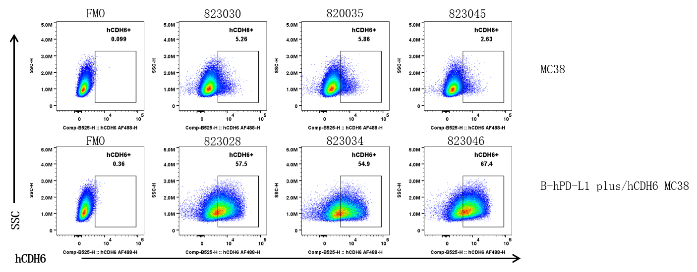
CDH6 expression was evaluated on B-hPD-L1 plus/hCDH6 MC38 by flow cytometry. These cells were subcutaneously transplanted into C57BL/6 mice (n=6). At the end of the experiment, tumor cells were harvested and analyzed for both mouse and human CDH6 expression by flow cytometry.