B-hVEGFA/hCDH17 MC38

• 322600

B-hVEGFA/hCDH17 MC38

Catalog Number: 322600
Strain Background: C57BL/6
NCBI gene ID: 7422,1015 (Human)
Aliases: VPF; VEGF; MVCD1; L-VEGF; HPT1; CDH16; HPT-1
---
ライセンスオプション提供可能

B-hVEGFA/hCDH17 MC38

このページで

  • Description
  • Phenotypic analysis

ポスター

すべて表示

    出版物

      Description
      • Origin: The MC38 cell line is derived from C57BL6 murine colon adenocarcinoma cells. The cell line is a commonly used murine model for colorectal carcinoma.
      •  Background Information: VEGFA is the primary mediator of angiogenesis. It stimulates endothelial cell proliferation, migration, and survival, thereby promoting the growth of new blood vessels. Its overexpression is closely associated with tumor progression. CDH17 is a member of the cadherin superfamily, encoding a calcium-dependent membrane-associated glycoprotein. CDH17 was overexpressed in colorectal adenocarcinoma, gastric adenocarcinoma and pancreatic adenocarcinoma. It drives cancer progression including cell adhesion, proliferation, migration, and invasion and so on.
      •  Gene targeting strategy: The human VEGFA coding sequence was inserted to replace part of murine exon 1 and all of exons 2-5. The exogenous promoter and human CDH17 coding sequence was inserted to replace part of murine exon 3. The insertion disrupts the endogenous murine Cdh17 gene, resulting in a non-functional transcript.
      • Tumorigenicity: Confirmed in C57BL/6JNifdc mice.
      •  Application: The B-hVEGFA/hCDH17 MC38 tumor models can be used for preclinical evaluation of monoclonal antibody drugs targeting human VEGFA, as well as for combination therapy with CDH17-targeting ADC.
      •  Notes:

      ➢ Inoculated cell lines can be suspended with DMEM stock solution.
      ➢ Before implementing the project, it is recommended to perform tumor growth experiments. The recommended cell inoculation amount is 2E6.
      ➢ In the experiment, it is necessary to ensure that the number of animals inoculated subcutaneously is at least 1.4 times the actual grouping number.

      CDH17 Protein Expression Analysis
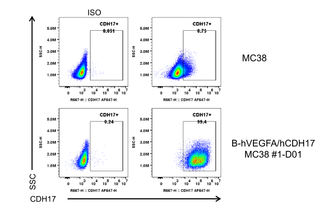
      •  Human CDH17 were detected on the surface of B-hVEGFA/hCDH17 MC38 but not wild-type MC38 cells.

      CDH17 expression analysis in B-hVEGFA/hCDH17 MC38 by flow cytometry. Single cell suspensions from wild-type MC38 and B-hVEGFA/hCDH17MC38 #1-D01 cultures were stained with anti-human CDH17 antibody (in house). This antibody has been validated for cross-reactivity between human and mouse in CHO-CDH17-overexpression cell lines.

      Tumor Growth curve & Body Weight Changes

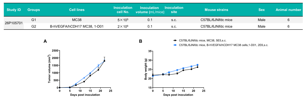
      Subcutaneous tumor growth of B-hVEGFA/hCDH17 MC38 cells. B-hVEGFA/hCDH17 MC38 (2×10⁶) and wild-type MC38 cells (5×10⁵) were subcutaneously implanted into C57BL/6JNifdc mice (male, 7-week-old, n=6). Tumor volume and body weight were measured twice a week. (A) Averagetumor volume. (B) Body weight. Volume was expressed in mm3 using the formula: V=0.5 × long diameter × short diameter2. Results indicate that B-hVEGFA/hCDH17 MC38 cells were able to establish tumors in vivo and can be used for efficacy studies. Values are expressed as mean ± SEM.

      CDH17 Protein Expression Analysis of Tumor Tissue
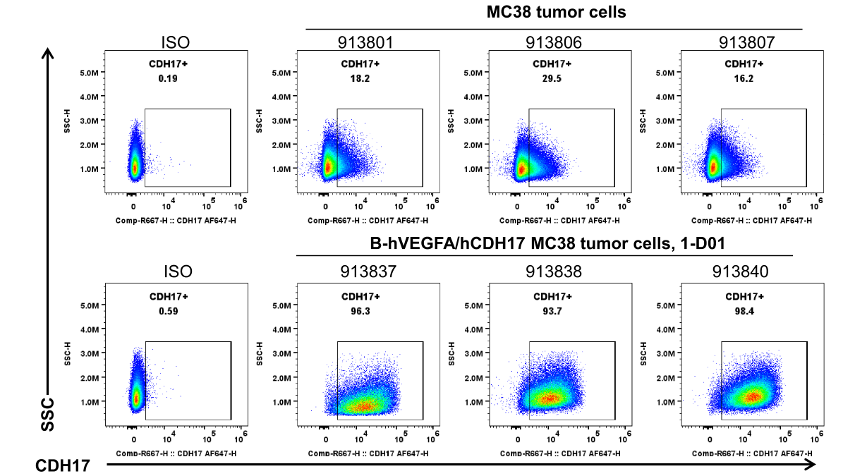
      • Human CDH17 was detected on the surface of B-hVEGFA/hCDH17 MC38 tumors but not wild-type MC38 cells.

      CDH17 expression was evaluated​​ on B-hVEGFA/hCDH17 MC38 by flow cytometry. These cells​​ were subcutaneously transplanted into C57BL/6JNifdc mice (n=6). At the end of the experiment, tumor cells were harvested and ​​analyzed for human CDH17 expression​​ by flow cytometry with anti-human CDH17 antibody (in house). This antibody has been validated for cross-reactivity between human and mouse in CHO-CDH17-overexpression cell lines.

      VEGFA Protein Expression Analysis of Tumor Tissue
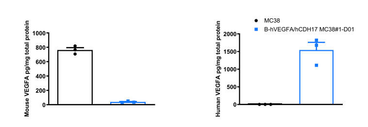
      •  Human VEGFA was consistently highly expressed in the B-hVEGFA/hCDH17 MC38 tumor homogenate, with an expression level of approximately 1500 pg per milligram of total protein. 

      ​​Tumor cells were harvested at the end of ​​the ​​ experiment and ​​assayed​​ for mouse and human VEGFA expression by ELISA. Tumor tissue was homogenized in PBS. The homogenate underwent three freeze-thaw cycles in liquid nitrogen, followed by centrifugation. The supernatant was collected, and the total protein concentration was determined by BCA assay and adjusted to 1 mg/mL. VEGFA levels in the tumor lysates samples were measured using the mouse VEGFA quantikine ELISA Kit (R&D, MMV00) and human VEGFA quantikine ELISA Kit (R&D, DVE00) according to the manufacturer's protocols. Values are expressed as mean±SEM.