

C57BL/6-Il33tm1(IL33)Bcgen Tslptm1(TSLP)Bcgen Crlf2tm2(CRLF2)Bcgen Il7rtm1(IL7R)Bcgen/Bcgen • 112935

IL33: A key cytokine in inflammation and its therapeutic intervention
TSLP: A key cytokine in inflammation and its therapeutic intervention

IL33
TSLP
TSLPR
IL7R

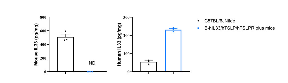
Strain-specific IL33 expression evaluated by ELISA in wild-type C57BL/6 mice and homozygous B-hIL33/hTSLP/hTSLPR plus mice. Lung tissue homogenate were collected from wild-type mice and homozygous B-hIL33/hTSLP/hTSLPR plus mice (female, 6-week-old, n = 3). Mouse and human IL33 levels in lung tissue homogenates were quantified by ELISA (mouse IL33, R&D Systems M3300; human IL33, R&D Systems D3300B).
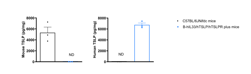
Strain-specific TSLP expression evaluated by ELISA in wild-type C57BL/6 mice and homozygous B-hIL33/hTSLP/hTSLPR plus mice. Calcipotriol (MC903), dissolved in ethanol, was topically applied to the ears of wild-type C57BL/6 mice and homozygous B-hIL33/hTSLP/hTSLPR plus mice for 7 days (female, 6-week-old, n = 3). Mouse and human TSLP levels in ear tissue homogenates were quantified by ELISA (mouse TSLP, BioLegend 434107; human TSLP, BioLegend 434207).
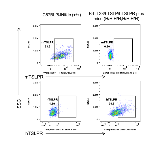
Mouse and human TSLPR expression analysis in bone marrow.Bone marrow cells were collected from wild-type C57BL/6 mice and homozygous B-hIL33/hTSLP/hTSLPR plus mice. TSLPR expressionon BMDCs was analyzed by flow cytometry using anti-mouse TSLPR antibody (Biolegend, 300312) and anti-human TSLP Rantibody (Biolegend, 100312).
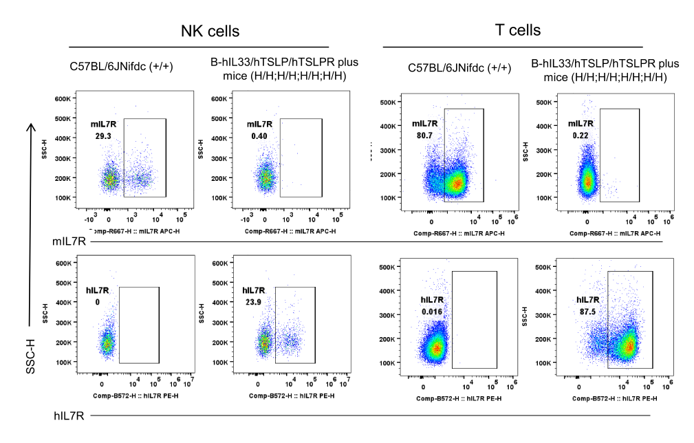
Mouse and human IL7R expression analysis in spleen.Splenocytes were collected from wild-type C57BL/6 mice and homozygous B-hIL33/hTSLP/hTSLPR plus mice. IL7R expression on splenocytes was analyzed by flow cytometry using anti-mouse IL7R antibody (Biolegend, 135011) and anti-human IL7R antibody (Biolegend, 351303).
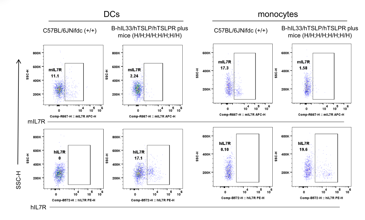
Mouse and human IL7R expression analysis in spleen. Splenocytes were collected from wild-type C57BL/6 mice and homozygous B-hIL33/hTSLP/hTSLPR plus mice. IL7R expression on splenocytes was analyzed by flow cytometry using anti-mouse IL7R antibody (Biolegend, 135011) and anti-human IL7R antibody (Biolegend, 351303).

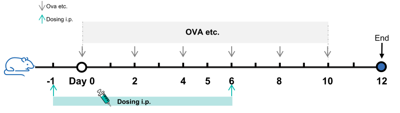
Experimental schedule for the induction of asthma and in vivo efficacy of anti-human IL33 antibody, anti-human TSLP antibody and IL33/TSLP BsAb (the antibodies are provided by a client) in B-hIL33/hTSLP/hTSLPR plus mice. In the OVA etc.-induced model, animals are administered OVA etc. intranasally to induce asthma models. The anti-human IL33 antibody, anti-human TSLP antibody and IL33/TSLP BsAb were administered by intraperitoneal injection (n = 6).
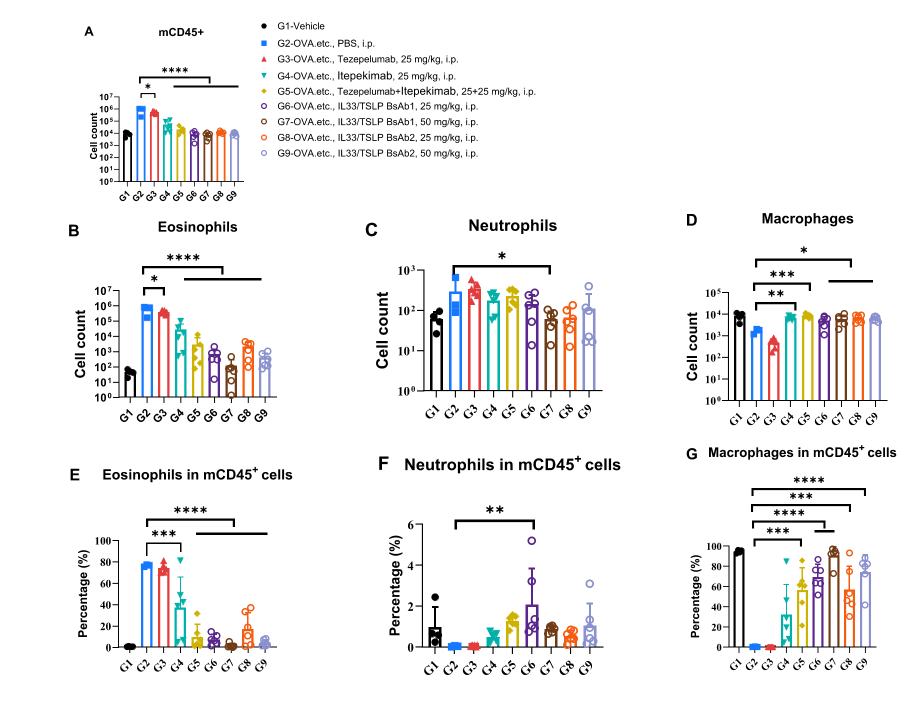
Analysis of immune cells in BALF. B-hIL33/hTSLP/hTSLPR plus mice (female, 11-week-old, n = 6) were immunized with OVA, etc., to induce asthma. Anti-human IL33 antibody, anti-human TSLP antibody, and IL33/TSLP BsAbs (antibodies provided by a client) were intraperitoneally injected into B-hIL33/hTSLP/hTSLPR plus mice. (A, B) The number of CD45⁺ cells and eosinophils in the BALF of the Itepekimab-treated group, the Itepekimab and Tezepelumab combination therapy group, and the IL33/TSLP BsAb-treated group decreased significantly compared with the OVA, etc.-induced PBS-treated group. Values are expressed as mean ± SEM. Significance was determined by two-way ANOVA test. *P < 0.05, P < 0.01, ***P < 0.001.
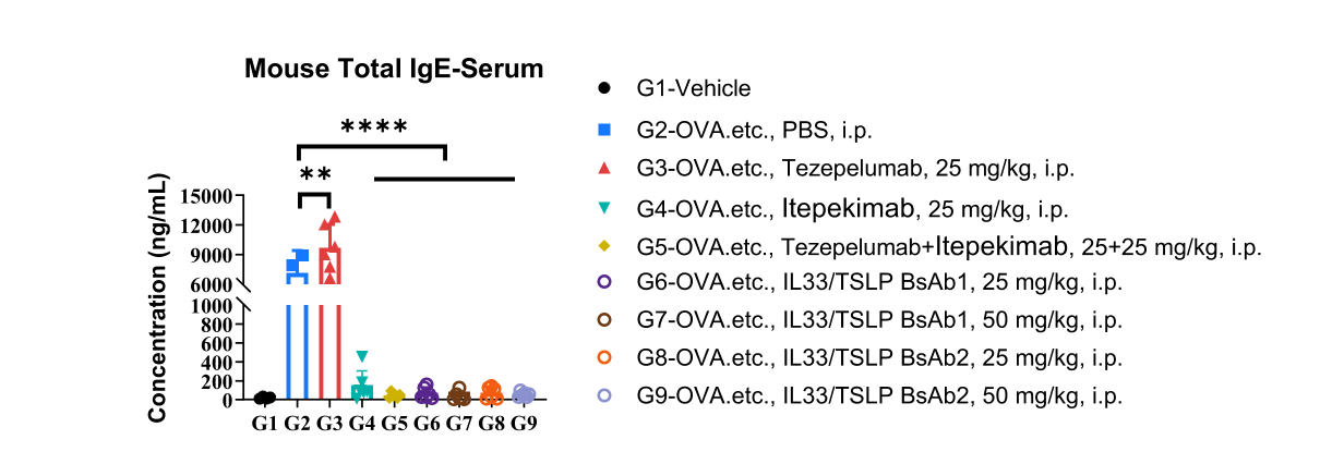
Analysis of mouse total IgE in serum. B-hIL33/hTSLP/hTSLPR plus mice (female, 11-week-old, n = 6) were immunized with OVA, etc., to induce asthma. Anti-human IL33 antibody, anti-human TSLP antibody, and IL33/TSLP BsAbs (antibodies provided by a client) were intraperitoneally injected into B-hIL33/hTSLP/hTSLPR plus mice. Serum was collected at the study endpoint, and IgE levels were analyzed by ELISA. The results showed that the levels of total IgE in the Itepekimab-treated group, the Itepekimab and Tezepelumab combination therapy group, and the IL33/TSLP BsAb-treated group decreased significantly compared with untreated mice. Values are expressed as mean ± SEM. Significance was determined by two-way ANOVA test. *P < 0.05, P < 0.01, ***P < 0.001.
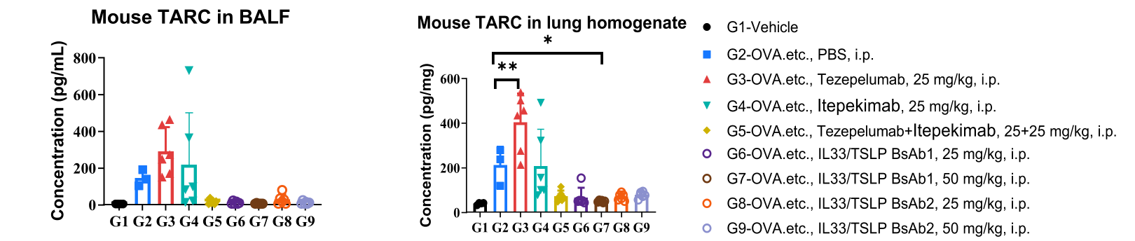
Analysis of mouse TARC in BALF and lung homogenates. B-hIL33/hTSLP/hTSLPR plus mice (female, 11-week-old, n = 6) were immunized with OVA, etc., to induce asthma. Anti-human IL33 antibody, anti-human TSLP antibody, and IL33/TSLP BsAbs (antibodies provided by a client) were intraperitoneally injected into B-hIL33/hTSLP/hTSLPR plus mice. BALF and lung homogenates were collected at the study endpoint, and TARC levels were analyzed by ELISA. The results showed that mouse TARC levels in the Itepekimab-treated group, the Itepekimab and Tezepelumab combination therapy group, and the IL33/TSLP BsAb-treated group were reduced compared with untreated mice. However, no reduction in TARC was observed in the TSLP antibody-treated group. We speculate that this is because even if TSLP signaling is pharmacologically blocked, persistent cellular necrosis and IL-33 release in the tissue can still sustain DC secretion of TARC through the MyD88/NF-κB pathway. This is also why combined or multi-targeted inhibition (e.g., simultaneously targeting TSLP, IL-33, and IL-4Rα) is emerging as a key breakthrough direction in the clinical treatment of some severe type 2 inflammatory conditions. Values are expressed as mean ± SEM. Significance was determined by two-way ANOVA test. *P < 0.05, P < 0.01, ***P < 0.001.
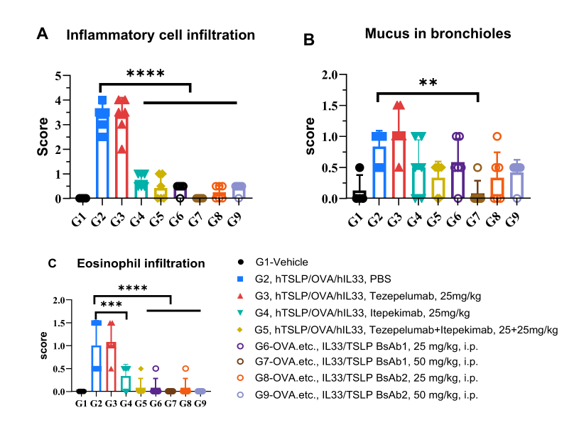
H&E staining of an asthma-like model in B-hIL33/hTSLP/hTSLPR plus mice. Lung tissues were collected at the study endpoint and analyzed by H&E staining. The results showed that inflammatory and eosinophil infiltration in the lung tissues of the Itepekimab-treated group, the Itepekimab and Tezepelumab combination therapy group, and the IL33/TSLP BsAb-treated group were reduced compared with those in untreated mice, indicating that B-hIL33/hTSLP/hTSLPR plus mice provide a powerful preclinical model for the in vivo evaluation of anti-human IL33 and anti-human TSLP antibodies. Values are expressed as mean ± SEM. Significance was determined by unpaired t-test. *P < 0.05, P < 0.01, ***P < 0.001.
