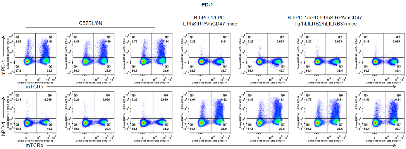
Protein expression analysis-Spleen T cells
Strain specific PD-1 expression analysis in wild-type mice and B-hPD-1/hPD-L1/hSIRPA/hCD47, Tg(hLILRB2/hLILRB3) mice by flow cytometry. Splenocytes were collected from wild-type C57BL/6N mice, B-hPD-1/hPD-L1/hSIRPA/hCD47 mice and B-hPD-1/hPD-L1/hSIRPA/hCD47, Tg(hLILRB2/hLILRB3) mice (female, n=3, 8-week-old), and analyzed by flow cytometry with anti-mouse PD-1 antibody (Biolegend, 109104) and anti-human PD-1 antibody (Biolegend, 329908). Mouse PD-1 was only detectable in wild-type mice. Human PD-1 was only detectable in B-hPD-1/hPD-L1/hSIRPA/hCD47 mice and B-hPD-1/hPD-L1/hSIRPA/hCD47, Tg(hLILRB2/hLILRB3) mice, but not in wild-type mice.
Protein expression analysis-Spleen T cells-Anti-CD3ε-treated
Strain specific PD-1 expression analysis in wild-type mice and B-hPD-1/hPD-L1/hSIRPA/hCD47, Tg(hLILRB2/hLILRB3) mice by flow cytometry. Splenocytes were collected from wild-type C57BL/6N mice, B-hPD-1/hPD-L1/hSIRPA/hCD47 mice and B-hPD-1/hPD-L1/hSIRPA/hCD47, Tg(hLILRB2/hLILRB3) mice stimulated with anti-mouse CD3ε antibody (7.5 μg, i.p.) in vivo for 24 hrs, and analyzed by flow cytometry with anti-mouse PD-1 antibody (Biolegend, 109104) and anti-human PD-1 antibody (Biolegend, 329908). Mouse PD-1 was only detectable in wild-type mice. Human PD-1 was only detectable in B-hPD-1/hPD-L1/hSIRPA/hCD47, Tg(hLILRB2/hLILRB3) mice and B-hPD-1/hPD-L1/hSIRPA/hCD47 mice, but not in wild-type mice.
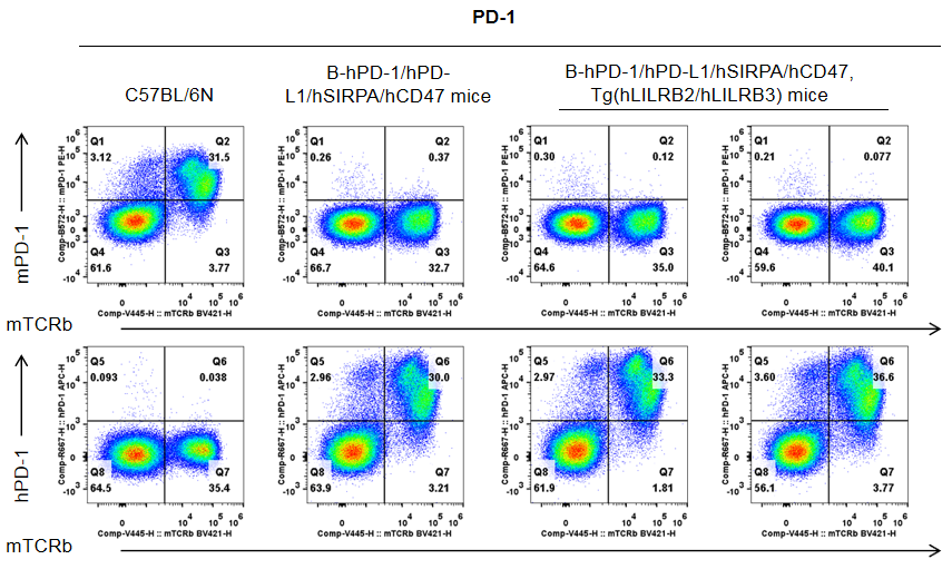
Protein expression analysis-Spleen T cells
Strain specific PD-L1 expression analysis in wild-type mice and B-hPD-1/hPD-L1/hSIRPA/hCD47, Tg(hLILRB2/hLILRB3) mice by flow cytometry. Splenocytes were collected from wild-type C57BL/6N mice, B-hPD-1/hPD-L1/hSIRPA/hCD47 mice and B-hPD-1/hPD-L1/hSIRPA/hCD47, Tg(hLILRB2/hLILRB3) mice (female, n=3, 8-week-old), and analyzed by flow cytometry with anti-mouse PD-L1 antibody (Biolegend, 124312) and anti-human PD-L1 antibody (Biolegend, 329706). Mouse PD-L1 was only detectable in wild-type mice. Human PD-L1 was only detectable in B-hPD-1/hPD-L1/hSIRPA/hCD47 mice and B-hPD-1/hPD-L1/hSIRPA/hCD47, Tg(hLILRB2/hLILRB3) mice, but not in wild-type mice.
Protein expression analysis-Spleen T cells-Anti-CD3
Strain specific PD-L1 expression analysis in wild-type mice and B-hPD-1/hPD-L1/hSIRPA/hCD47, Tg(hLILRB2/hLILRB3) mice by flow cytometry. Splenocytes were collected from wild-type C57BL/6N mice, B-hPD-1/hPD-L1/hSIRPA/hCD47 mice and B-hPD-1/hPD-L1/hSIRPA/hCD47, Tg(hLILRB2/hLILRB3) mice stimulated with anti-mouse CD3ε antibody (7.5 μg, i.p.) in vivo for 24 hrs, and analyzed by flow cytometry with anti-mouse PD-L1 antibody (Biolegend, 124312) and anti-human PD-L1 antibody (Biolegend, 329706). Mouse PD-L1 was only detectable in wild-type mice. Human PD-L1 was only detectable in B-hPD-1/hPD-L1/hSIRPA/hCD47, Tg(hLILRB2/hLILRB3) mice and B-hPD-1/hPD-L1/hSIRPA/hCD47 mice, but not in wild-type mice.
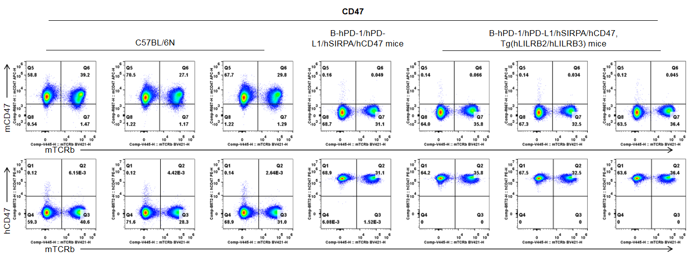
Protein expression analysis-Spleen T cells
Strain specific CD47 expression analysis in wild-type mice and B-hPD-1/hPD-L1/hSIRPA/hCD47, Tg(hLILRB2/hLILRB3) mice by flow cytometry. Splenocytes were collected from wild-type C57BL/6N mice, B-hPD-1/hPD-L1/hSIRPA/hCD47 mice and B-hPD-1/hPD-L1/hSIRPA/hCD47, Tg(hLILRB2/hLILRB3) mice (female, n=3, 8-week-old), and analyzed by flow cytometry with anti-mouse CD47 antibody (Biolegend, 127514) and anti-human CD47 antibody (Biolegend, 323108). Mouse CD47 was only detectable in wild-type mice. Human CD47 was only detectable in B-hPD-1/hPD-L1/hSIRPA/hCD47 mice and B-hPD-1/hPD-L1/hSIRPA/hCD47, Tg(hLILRB2/hLILRB3) mice, but not in wild-type mice.
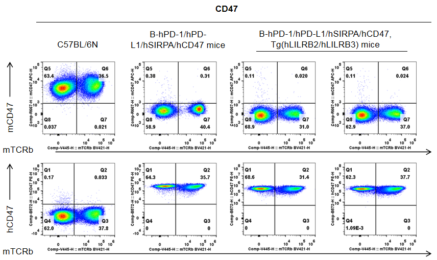
Protein expression analysis-Spleen T cells-Anti-CD3
Strain specific CD47 expression analysis in wild-type mice and B-hPD-1/hPD-L1/hSIRPA/hCD47, Tg(hLILRB2/hLILRB3) mice by flow cytometry. Splenocytes were collected from wild-type C57BL/6N mice, B-hPD-1/hPD-L1/hSIRPA/hCD47 mice and B-hPD-1/hPD-L1/hSIRPA/hCD47, Tg(hLILRB2/hLILRB3) mice stimulated with anti-mouse CD3ε antibody (7.5 μg, i.p.) in vivo for 24 hrs, and analyzed by flow cytometry with anti-mouse CD47 antibody (Biolegend, 127514) and anti-human CD47 antibody (Biolegend, 323108). Mouse CD47 was only detectable in wild-type mice. Human CD47 was only detectable in B-hPD-1/hPD-L1/hSIRPA/hCD47, Tg(hLILRB2/hLILRB3) mice and B-hPD-1/hPD-L1/hSIRPA/hCD47 mice, but not in wild-type mice.
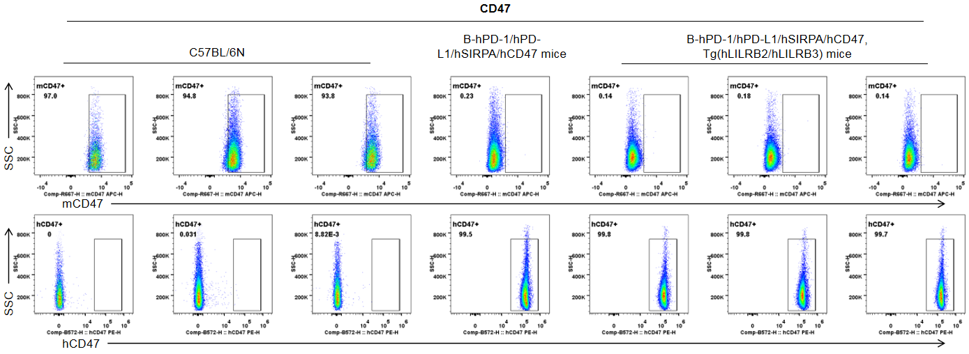
Protein expression analysis-RBC
Strain specific CD47 expression analysis in wild-type mice and B-hPD-1/hPD-L1/hSIRPA/hCD47, Tg(hLILRB2/hLILRB3) mice by flow cytometry. Red blood cells were collected from wild-type C57BL/6N mice, B-hPD-1/hPD-L1/hSIRPA/hCD47 mice and B-hPD-1/hPD-L1/hSIRPA/hCD47, Tg(hLILRB2/hLILRB3) mice (female, n=3, 8-week-old), and analyzed by flow cytometry with anti-mouse CD47 antibody (Biolegend, 127514) and anti-human CD47 antibody (Biolegend, 323108). Mouse CD47 was only detectable in wild-type mice. Human CD47 was only detectable in B-hPD-1/hPD-L1/hSIRPA/hCD47, Tg(hLILRB2/hLILRB3) mice and B-hPD-1/hPD-L1/hSIRPA/hCD47 mice, but not in wild-type mice.
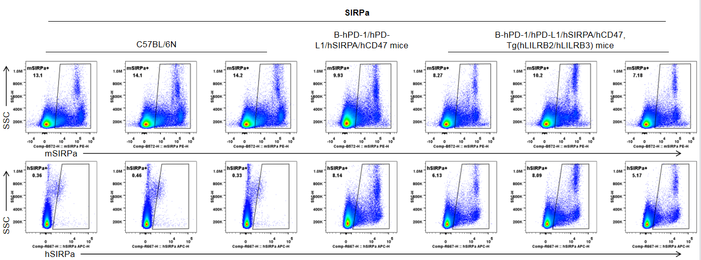
Protein expression analysis-Spleen leukocytes
Strain specific SIRPA expression analysis in wild-type mice and B-hPD-1/hPD-L1/hSIRPA/hCD47, Tg(hLILRB2/hLILRB3) mice by flow cytometry. Splenocytes were collected from wild-type C57BL/6N mice, B-hPD-1/hPD-L1/hSIRPA/hCD47 mice and B-hPD-1/hPD-L1/hSIRPA/hCD47, Tg(hLILRB2/hLILRB3) mice (female, n=3, 8-week-old), and analyzed by flow cytometry with cross-recognized both human and mouse SIRPa antibody (Biolegend, 144011) and anti-human SIRPa antibody (Biolegend, 323810). Human SIRPa was only detectable in B-hPD-1/hPD-L1/hSIRPA/hCD47, Tg(hLILRB2/hLILRB3) mice and B-hPD-1/hPD-L1/hSIRPA/hCD47 mice, but not in wild-type mice.
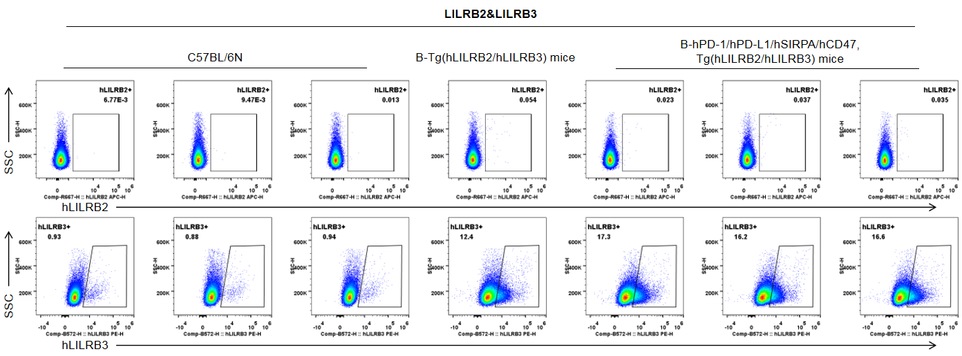
Protein expression analysis-Spleen-T cells
Strain specific LILRB2 and LILRB3 expression analysis in wild-type mice and B-hPD-1/hPD-L1/hSIRPA/hCD47, Tg(hLILRB2/hLILRB3) mice by flow cytometry. Splenocytes were collected from wild-type C57BL/6N mice, B-Tg(hLILRB2/hLILRB3) mice and B-hPD-1/hPD-L1/hSIRPA/hCD47, Tg(hLILRB2/hLILRB3) mice (female, n=3, 8-week-old), and analyzed by flow cytometry with anti-human LILRB2 antibody (Biolegend, 338708) and anti-human LILRB3 antibody (R&D, FAB1806P-100). Human LILRB2 was not detectable in T cells of B-Tg(hLILRB2/hLILRB3) mice and B-hPD-1/hPD-L1/hSIRPA/hCD47, Tg(hLILRB2/hLILRB3) mice. Human LILRB3 was only detectable in T cells of B-Tg(hLILRB2/hLILRB3) mice and B-hPD-1/hPD-L1/hSIRPA/hCD47, Tg(hLILRB2/hLILRB3) mice, but not in wild-type mice.
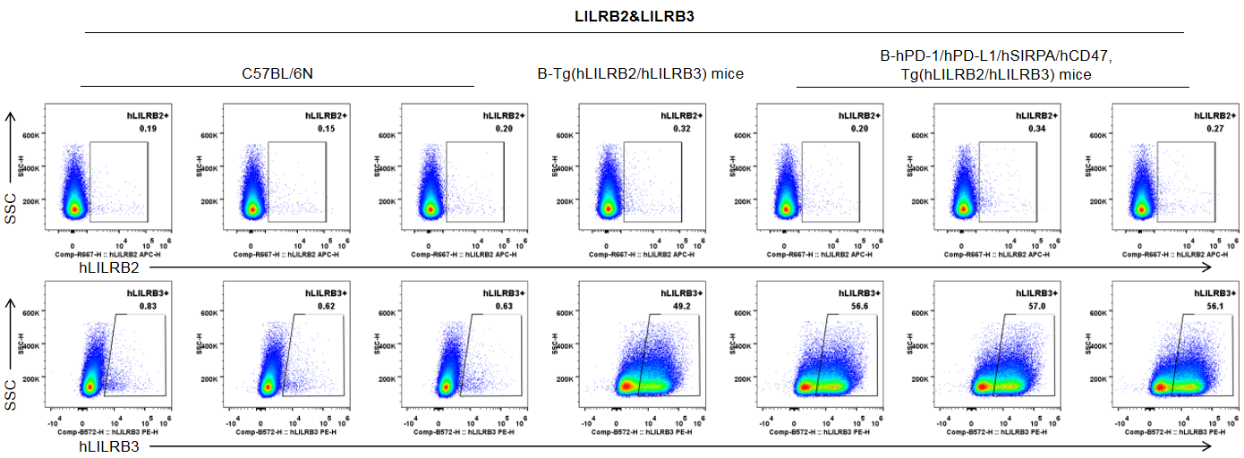
Protein expression analysis-Spleen-B cells
Strain specific LILRB2 and LILRB3 expression analysis in wild-type mice and B-hPD-1/hPD-L1/hSIRPA/hCD47, Tg(hLILRB2/hLILRB3) mice by flow cytometry. Splenocytes were collected from wild-type C57BL/6N mice, B-Tg(hLILRB2/hLILRB3) mice and B-hPD-1/hPD-L1/hSIRPA/hCD47, Tg(hLILRB2/hLILRB3) mice (female, n=3, 8-week-old), and analyzed by flow cytometry with anti-human LILRB2 antibody (Biolegend, 338708) and anti-human LILRB3 antibody (R&D, FAB1806P-100). Human LILRB2 was not detectable in B-Tg(hLILRB2/hLILRB3) mice and B-hPD-1/hPD-L1/hSIRPA/hCD47, Tg(hLILRB2/hLILRB3) mice. Human LILRB3 was only detectable in B cells of B-Tg(hLILRB2/hLILRB3) mice and B-hPD-1/hPD-L1/hSIRPA/hCD47, Tg(hLILRB2/hLILRB3) mice, but not in wild-type mice.
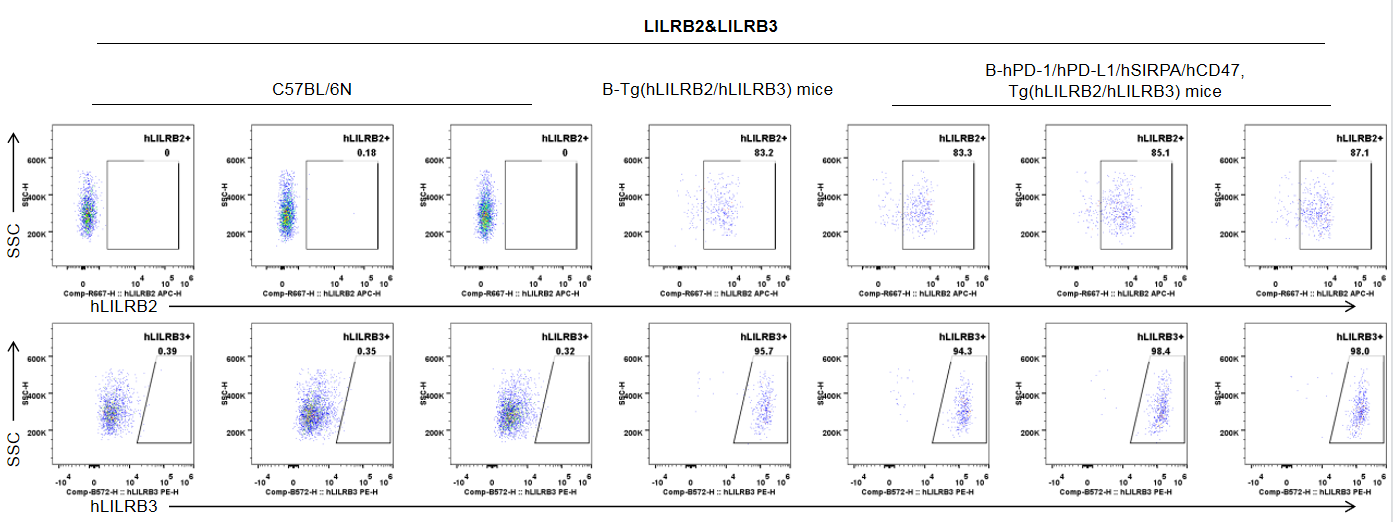
Protein expression analysis-Spleen-Macrophages
Strain specific LILRB2 and LILRB3 expression analysis in wild-type mice and B-hPD-1/hPD-L1/hSIRPA/hCD47, Tg(hLILRB2/hLILRB3) mice by flow cytometry. Splenocytes were collected from wild-type C57BL/6N mice, B-Tg(hLILRB2/hLILRB3) mice and B-hPD-1/hPD-L1/hSIRPA/hCD47, Tg(hLILRB2/hLILRB3) mice (female, n=3, 8-week-old), and analyzed by flow cytometry with anti-human LILRB2 antibody (Biolegend, 338708) and anti-human LILRB3 antibody (R&D, FAB1806P-100). Human LILRB2 was only detectable in macrophages of B-Tg(hLILRB2/hLILRB3) mice and B-hPD-1/hPD-L1/hSIRPA/hCD47, Tg(hLILRB2/hLILRB3) mice. Human LILRB3 was only detectable in macrophages of B-Tg(hLILRB2/hLILRB3) mice and B-hPD-1/hPD-L1/hSIRPA/hCD47, Tg(hLILRB2/hLILRB3) mice, but not in wild-type mice.
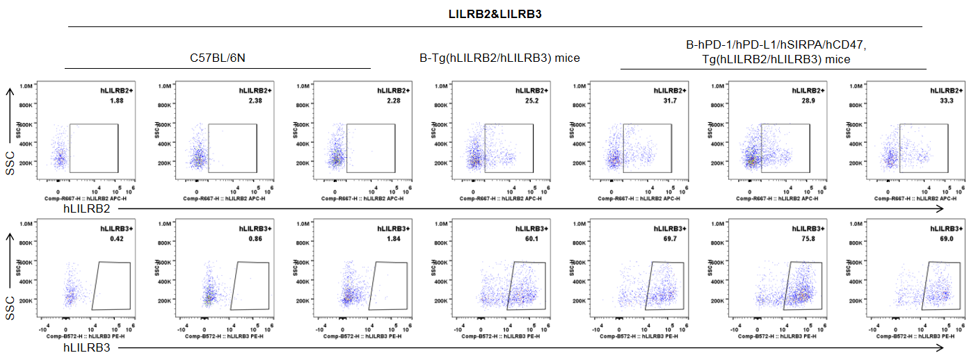
Protein expression analysis-Blood-Monocytes
Strain specific LILRB2 and LILRB3 expression analysis in wild-type mice and B-hPD-1/hPD-L1/hSIRPA/hCD47, Tg(hLILRB2/hLILRB3) mice by flow cytometry. Blood cells were collected from wild-type C57BL/6N mice, B-Tg(hLILRB2/hLILRB3) mice and B-hPD-1/hPD-L1/hSIRPA/hCD47, Tg(hLILRB2/hLILRB3) mice (female, n=3, 8-week-old), and analyzed by flow cytometry with anti-human LILRB2 antibody (Biolegend, 338708) and anti-human LILRB3 antibody (R&D, FAB1806P-100). Human LILRB2 was only detectable in monocytes of B-Tg(hLILRB2/hLILRB3) mice and B-hPD-1/hPD-L1/hSIRPA/hCD47, Tg(hLILRB2/hLILRB3) mice. Human LILRB3 was only detectable in monocytes of B-Tg(hLILRB2/hLILRB3) mice and B-hPD-1/hPD-L1/hSIRPA/hCD47, Tg(hLILRB2/hLILRB3) mice, but not in wild-type mice.
Frequency of leukocyte subpopulations in spleen
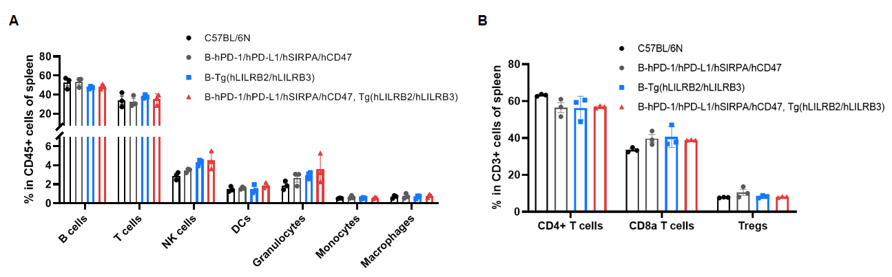
Frequency of leukocyte subpopulations in spleen by flow cytometry. Splenocytes were isolated from wild-type C57BL/6N mice, B-hPD-1/hPD-L1/hSIRPA/hCD47 mice, B-Tg(hLILRB2/hLILRB3) mice and B-hPD-1/hPD-L1/hSIRPA/hCD47, Tg(hLILRB2/hLILRB3) mice (female, n=3, 8-week-old). A. Flow cytometry analysis of the splenocytes was performed to assess the frequency of leukocyte subpopulations. B. Frequency of T cell subpopulations. Frequencies of T cells, B cells, NK cells, DCs, granulocytes, monocytes, macrophages, CD4+ T cells, CD8a+ T cells and Tregs in B-hPD-1/hPD-L1/hSIRPA/hCD47, Tg(hLILRB2/hLILRB3) mice were similar to those in C57BL/6N mice, demonstrating that humanization of hPD-1, hPD-L1, hSIRPA, hCD47 and the transgene of LILRB2 and LILRB3 do not change the frequency or distribution of these cell types in spleen. Values are expressed as mean ± SEM. The frequency of leukocyte subpopulations in lymph node of B-hPD-1/hPD-L1/hSIRPA/hCD47, Tg(hLILRB2/hLILRB3) mice were also comparable to wild-type C57BL/6N mice (Data not shown).
* When publishing results obtained using this animal model, please acknowledge the source as follows: The animal model [B-hPD-1/hPD-L1/hSIRPA/hCD47,Tg(hLILRB2/hLILRB3) mice] (Cat# 113129) was purchased from Biocytogen.