Targeting Strategy
Gene targeting strategy for B-hPD-1/hPD-L1/HLA-A11.1 plus mice.
The exon 2 of mouse Pdcd1 gene that encodes the IgV domain was replaced by human PD-1 exon 2 in B-hPD-1/hPD-L1/HLA-A11.1 plus mice.
The exon 3 of mouse pdl1 gene that encodes the IgV domain was replaced by human PD-L1 exon 3 in B-hPD-1/hPD-L1/HLA-A11.1 plus mice.
The B2M gene (Exon1 to Exon3) of mouse were replaced by the sequence encompassing the human B2M CDS and HLA-A*1101 gene that included leader sequence, α1 and α2 domains ligated to a fragment of the murine H-2Db gene containing the α3, transmembrane and cytoplasmic domains in B-hPD-1/hPD-L1/HLA-A11.1 plus mice.
The mouse B2M gene is knocked into exon 2 of the mouse Fcgrt gene and is fused via a linker to the remaining portion of exon 2, a strategy that enables the co-expression of mouse B2M and Fcgrt in B-hPD-1/hPD-L1/HLA-A11.1 plus mice.
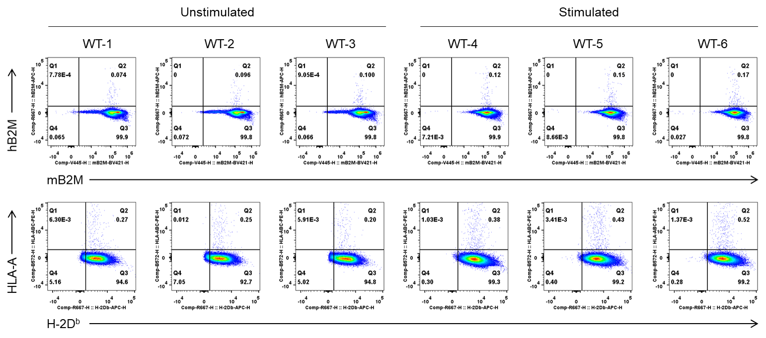
Protein Expression Analysis in Spleen of Wild-type (WT) Mice
Strain specific B2M and HLA-A expression analysis in wild-type mice by flow cytometry. Splenocytes were collected from wild-type C57BL/6JNifdc mice (+/+) after stimulated with anti-mouse CD3ε antibody (7.5 μg, i.p.) in vivo for 24 hrs (male, 8-week-old, n=1) or not. Protein expression was analyzed with anti-mouse B2M antibody (BD Biosciences, 744802), anti-hB2M antibody (Biolegend, 395712), anti-H-2Db antibody (Biolegend, 111513) and anti-HLA-ABC antibody (Biolegend, 311406), by flow cytometry. Mouse B2M and H-2Db were detectable in wild-type C57BL/6JNifdc.
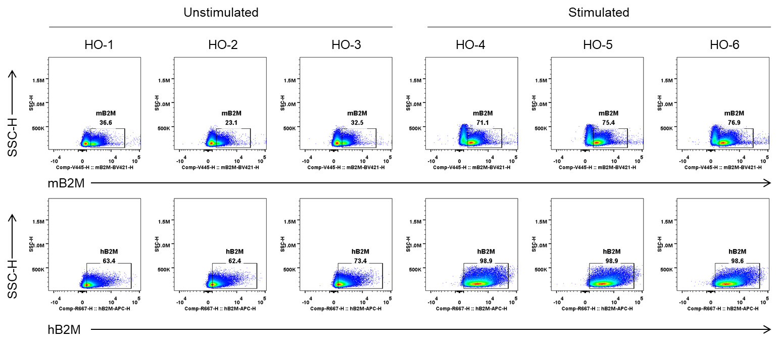
Protein Expression Analysis in Spleen of B-hPD-1/hPD-L1/HLA-A11.1 plus mice
Strain specific B2M expression analysis in B-hPD-1/hPD-L1/HLA-A11.1 plus mice by flow cytometry. Splenocytes were collected from homozygous(HO) B-hPD-1/hPD-L1/HLA-A11.1 plus mice after stimulated with anti-mouse CD3ε antibody (7.5 μg, i.p.) in vivo for 24 hrs (male, 8-week-old, n=1) or not. Protein expression was analyzed with anti-mouse B2M antibody (BD Biosciences, 744802) and anti-hB2M antibody (Biolegend, 395712), by flow cytometry. Mouse B2M and human B2M were detectable in homozygous B-hPD-1/hPD-L1/HLA-A11.1 plus mice because the mouse B2M gene is knocked into exon 2 of the mouse Fcgrt gene and is fused via a linker to the remaining portion of exon 2, a strategy that enables the co-expression of mouse B2M and Fcgrt in B-hPD-1/hPD-L1/HLA-A11.1 plus mice.
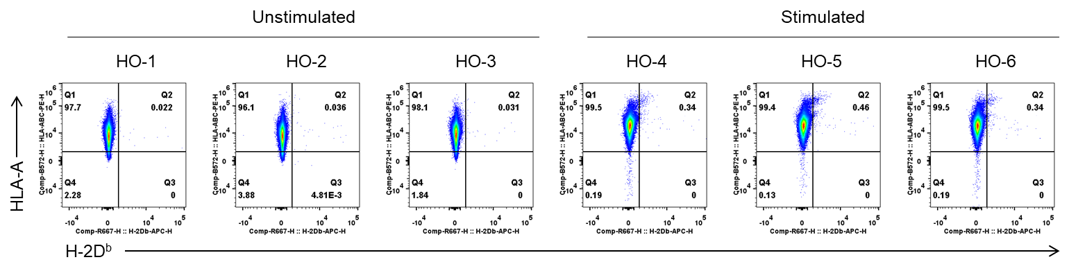
Strain specific HLA-A expression analysis in B-hPD-1/hPD-L1/HLA-A11.1 plus mice by flow cytometry. Splenocytes were collected from homozygous B-hPD-1/hPD-L1/HLA-A11.1 plus mice after stimulated with anti-mouse CD3ε antibody (7.5 μg, i.p.) in vivo for 24 hrs (male, 8-week-old, n=1) or not. Protein expression was analyzed with anti-H-2Db antibody (Biolegend, 111513) and anti-HLA-ABC antibody (Biolegend, 311406), by flow cytometry. Human HLA-A were detectable in B-hPD-1/hPD-L1/HLA-A11.1 plus mice but not in wild-type C57BL/6JNifdc.
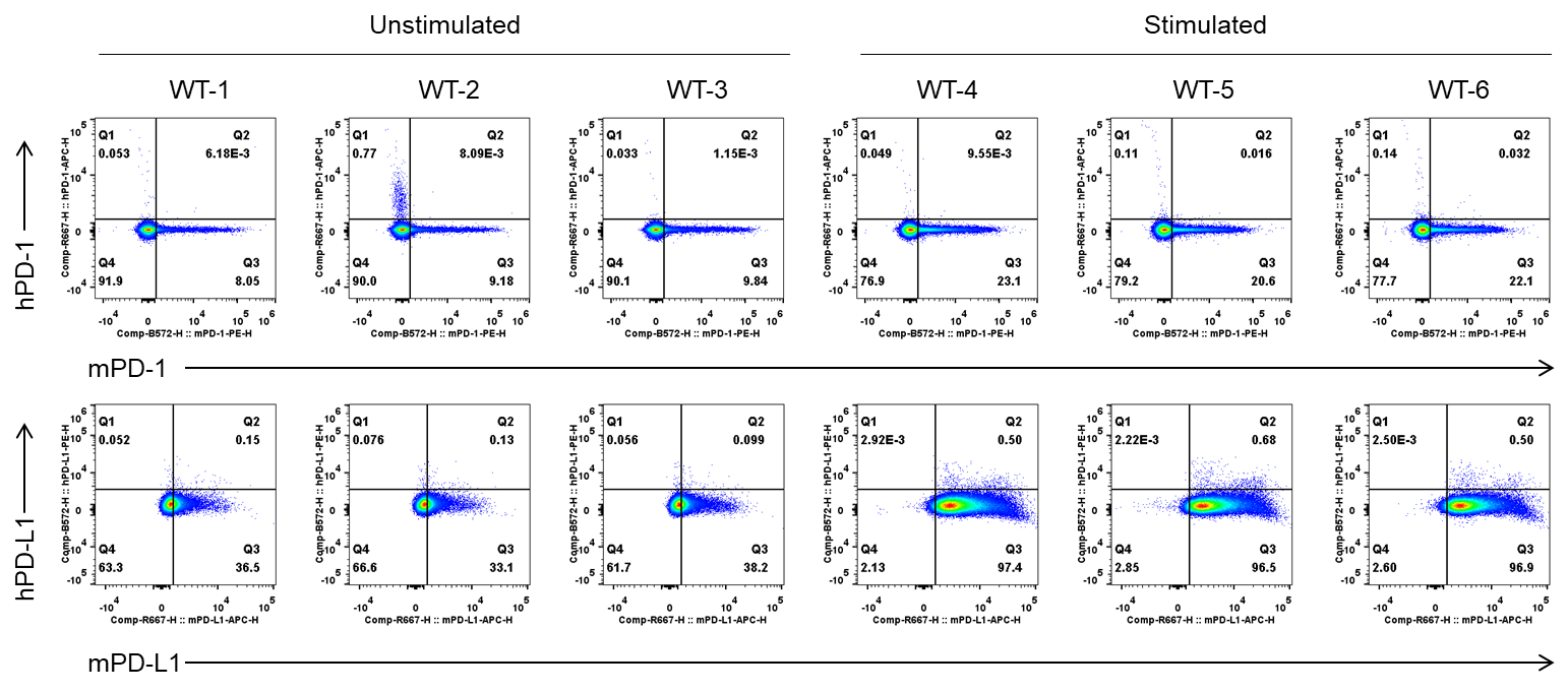
Protein Expression Analysis in Spleen of Wild-type (WT) Mice
Strain specific PD-1 and PD-L1 expression analysis in wild-type mice by flow cytometry. Splenocytes were collected from wild-type C57BL/6JNifdc mice (+/+) after stimulated with anti-mouse CD3ε antibody (7.5 μg, i.p.) in vivo for 24 hrs (male, 8-week-old, n=1) or not. Protein expression was analyzed with anti-mouse PD-1 antibody (Biolegend, 109104), anti-human PD-1 antibody (Biolegend, 329904), anti-mouse PD-L1 antibody (Biolegend, 124312) and anti-human PD-L1 antibody (Biolegend, 329706) by flow cytometry. Mouse PD-1 and PD-L1 were detectable in wild-type C57BL/6JNifdc mice.
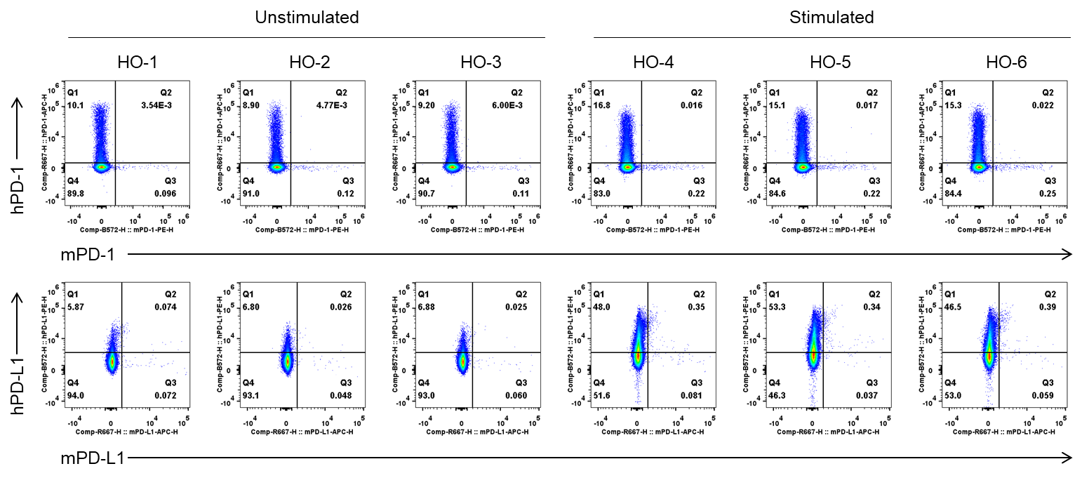
Protein Expression Analysis in Spleen of B-hPD-1/hPD-L1/HLA-A11.1 plus mice
Strain specific PD-1 and PD-L1 expression analysis in B-hPD-1/hPD-L1/HLA-A11.1 plus mice by flow cytometry. Splenocytes were collected from homozygous(HO) B-hPD-1/hPD-L1/HLA-A11.1 plus mice after stimulated with anti-mouse CD3ε antibody (7.5 μg, i.p.) in vivo for 24 hrs (male, 8-week-old, n=1) or not. Protein expression was analyzed with anti-mouse PD-1 antibody (Biolegend, 109104), anti-human PD-1 antibody (Biolegend, 329904), anti-mouse PD-L1 antibody (Biolegend, 124312) and anti-human PD-L1 antibody (Biolegend, 329706) by flow cytometry. Human PD-1 and PD-L1 were exclusively detectable in B-hPD-1/hPD-L1/HLA-A11.1 plus mice.
Frequency of Leukocyte Subpopulations in Spleen
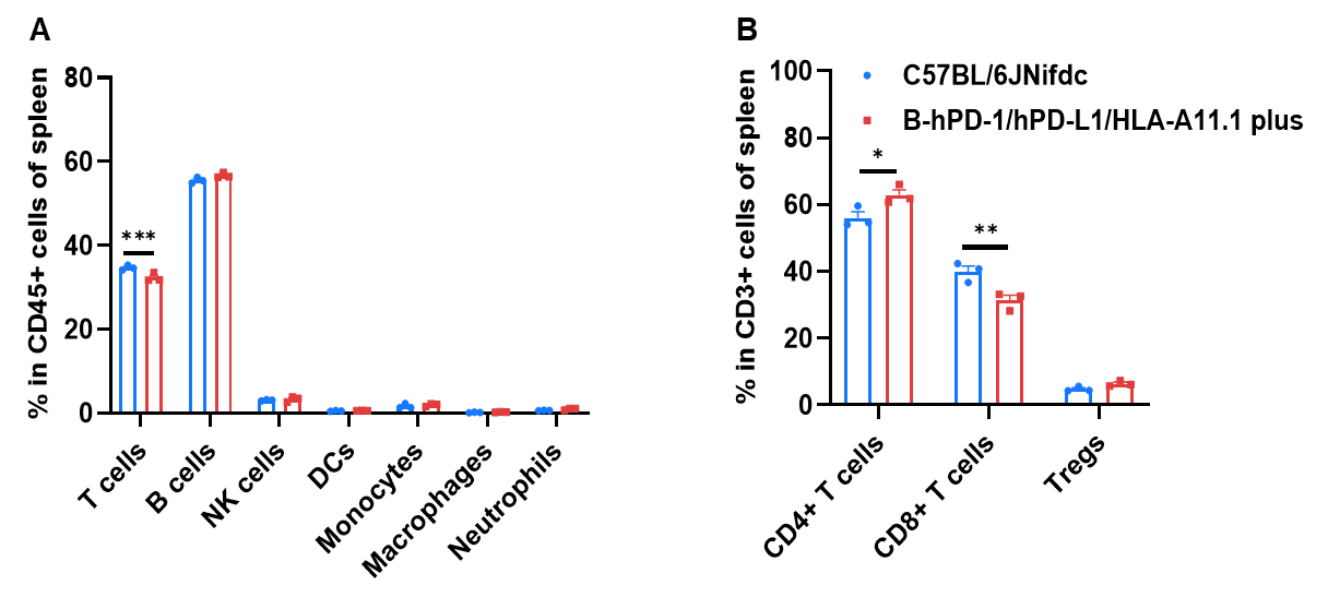
Frequency of leukocyte subpopulations in spleen by flow cytometry. Splenocytes were isolated from wild-type C57BL/6JNifdc mice and homozygous B-hPD-1/hPD-L1/HLA-A11.1 plus mice (male, 8-week-old, n=3). A. Flow cytometry analysis of the splenocytes was performed to assess the frequency of leukocyte subpopulations. B. Frequency of T cell subpopulations. Frequencies of T cells, B cells, NK cells, dendritic cells, monocytes, macrophages, neutrophils, CD4+ T cells, CD8+ T cells, and Tregs in B-hPD-1/hPD-L1/HLA-A11.1 plus mice were similar to those in C57BL/6JNifdc mice. Frequency of T cells in B-hPD-1/hPD-L1/HLA-A11.1 plus mice were lower than in C57BL/6JNifdc mice. Frequency of CD8+ T cells in B-hPD-1/hPD-L1/HLA-A11.1 plus mice were lower than that in C57BL/6JNifdc mice, whereas the frequency of CD4+ T cells B-hPD-1/hPD-L1/HLA-A11.1 plus mice were higher than that in C57BL/6JNifdc mice. Values are expressed as mean ± SEM. Significance was determined by two-way ANOVA test. *P < 0.05, **P < 0.01, ***p < 0.001.
Frequency of Leukocyte Subpopulations in Blood
Frequency of leukocyte subpopulations in blood by flow cytometry. Blood were isolated from wild-type C57BL/6JNifdc mice and homozygous B-hPD-1/hPD-L1/HLA-A11.1 plus mice (male, 8-week-old, n=3). A. Flow cytometry analysis of the blood was performed to assess the frequency of leukocyte subpopulations. B. Frequency of T cell subpopulations. Frequencies of T cells, B cells, NK cells, dendritic cells, monocytes, macrophages, neutrophils, CD4+ T cells, CD8+ T cells, and Tregs in B-hPD-1/hPD-L1/HLA-A11.1 plus mice were similar to those in C57BL/6JNifdc mice. Frequency of CD8+ T cells in B-hPD-1/hPD-L1/HLA-A11.1 plus mice were lower than that in C57BL/6JNifdc mice, whereas the frequency of CD4+ T cells in B-hPD-1/hPD-L1/HLA-A11.1 plus mice were higher than that in C57BL/6JNifdc mice. Values are expressed as mean ± SEM. Significance was determined by two-way ANOVA test. *P < 0.05, **P < 0.01, ***p < 0.001.
Frequency of Leukocyte Subpopulations in Lymph Node
Frequency of leukocyte subpopulations in lymph node by flow cytometry. Lymph node cells were isolated from wild-type C57BL/6JNifdc mice and homozygous B-hPD-1/hPD-L1/HLA-A11.1 plus mice (male, 8-week-old, n=3). A. Flow cytometry analysis of the lymph node was performed to assess the frequency of leukocyte subpopulations. B. Frequency of T cell subpopulations. Frequencies of T cells, B cells, and NK cells in B-hPD-1/hPD-L1/HLA-A11.1 plus mice were similar to those in C57BL/6JNifdc mice. Frequency of CD8+ T cells in B-hPD-1/hPD-L1/HLA-A11.1 plus mice were lower than that in C57BL/6JNifdc mice, whereas the frequency of CD4+ T cells in B-hPD-1/hPD-L1/HLA-A11.1 plus mice were higher than that in C57BL/6JNifdc mice. Values are expressed as mean ± SEM. Significance was determined by two-way ANOVA test. *P < 0.05, **P < 0.01, ***p < 0.001.
* When publishing results obtained using this animal model, please acknowledge the source as follows: The animal model [B-hPD-1/hPD-L1/HLA-A11.1 plus mice] (Cat# 113952) was purchased from Biocytogen.