Targeting Strategy
Gene targeting strategy for B-hPD-1 plus/hPD-L1/hANG2 mice.
Human PD-1 gene encoding the extracellular region and mouse Pd-1 gene encoding the transmembrane and cytoplasmic region were inserted after the initiation codon ATG of mouse Pd-1 gene in B-hPD-1 plus/hPD-L1/hANG2 mice.
The exon 3 of mouse Pdl1 gene that encodes the IgV domain was replaced by human PD-L1 exon 3 in B-hPD-1 plus/hPD-L1/hANG2 mice.
The exons 1-9 of mouse Ang2 gene that encode signal peptide, extracellular domain, transmembrane domain and cytoplasmic region are replaced by human counterparts in B-hPD-1 plus/hPD-L1/hANG2 mice. The promoter, 5’UTR and 3’UTR region of the mouse gene are also retained. The chimeric ANG2 expression is driven by endogenous mouse Ang2 promoter, while mouse Ang2 gene transcription and translation will be disrupted.
Protein Expression Analysis
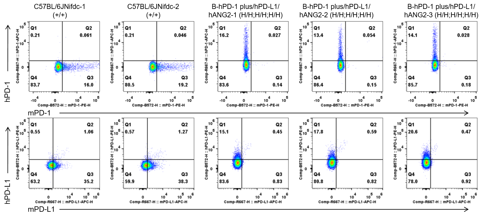
Strain specific PD-1 and PD-L1 expression analysis in homozygous B-hPD-1 plus/hPD-L1/hANG2 mice by flow cytometry. Splenocytes were collected from wild-type C57BL/6 mice (+/+) and homozygous B-hPD-1 plus/hPD-L1/hANG2 mice (H/H;H/H;H/H), and analyzed by flow cytometry with species-specific anti-mouse PD-1 antibody (Biolegend,109104), anti-human PD-1 antibody (Biolegend,329908), anti-mouse PD-L1 antibody (Biolegend,124312) and anti-human PD-L1 antibody (Biolegend,329706). Mouse PD-1 and PD-L1 were detectable in wild-type mice. Human PD-1 and PD-L1 were exclusively detectable in homozygous B-hPD-1 plus/hPD-L1/hANG2 mice but not in wild-type mice.
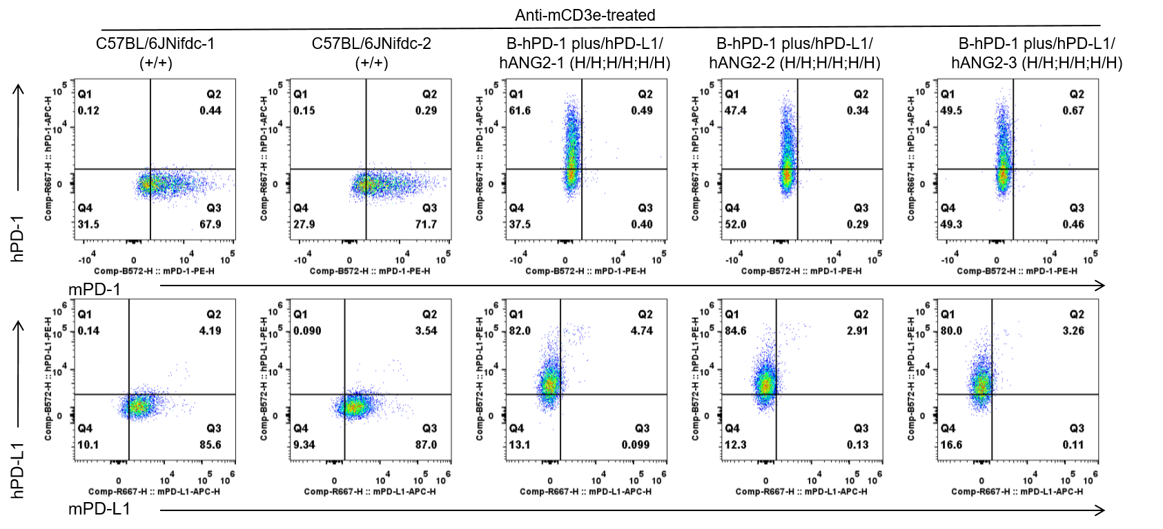
Strain specific PD-1 and PD-L1 expression analysis in homozygous B-hPD-1 plus/hPD-L1/hANG2 mice by flow cytometry. Splenocytes were collected from wild-type C57BL/6 mice (+/+) and homozygous B-hPD-1 plus/hPD-L1/hANG2 mice (H/H;H/H;H/H) stimulated with anti-CD3ε in vivo (7.5 μg/mice, stimulation for 24 hours, i.p.), and analyzed by flow cytometry with species-specific anti-mouse PD-1 antibody (Biolegend,109104), anti-human PD-1 antibody (Biolegend,329908), anti-mouse PD-L1 antibody (Biolegend,124312) and anti-human PD-L1 antibody (Biolegend,329706). Mouse PD-1 and PD-L1 were detectable in wild-type mice. Human PD-1 and PD-L1 were exclusively detectable in homozygous B-hPD-1 plus/hPD-L1/hANG2 mice but not in wild-type mice.
Protein Expression Analysis in Plasma
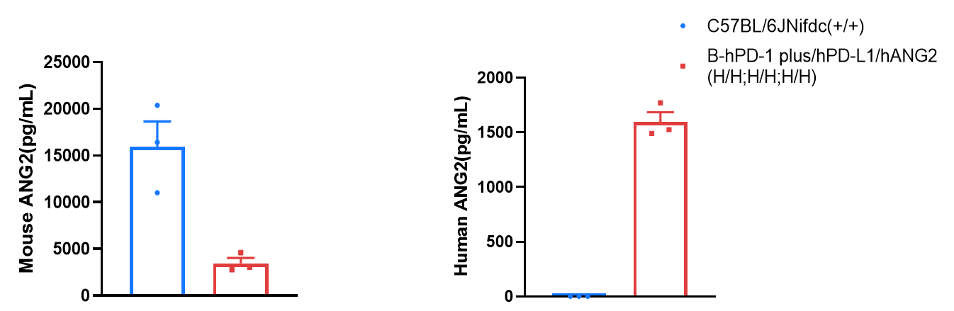
Strain specific ANG2 expression analysis in wild-type C57BL/6JNifdc mice and homozygous B-hPD-1 plus/hPD-L1/hANG2 mice by ELISA. Plasma was collected from wild-type C57BL/6JNifdc mice (+/+) (female, n=3, 9-week-old) and homozygous B-hPD-1 plus/hPD-L1/hANG2 mice (H/H;H/H;H/H) (female, n=3, 9-week-old), and expression level of mouse and human ANG2 were analyzed by ELISA (anti-mouse ANG2 ELISA kit: abcam, ab209883; anti-human ANG2 ELISA kit: R&D, DANG20). Mouse ANG2 was detectable in wild-type C57BL/6 mice and homozygous B-hPD-1 plus/hPD-L1/hANG2 mice as the ELISA kit weakly recognizes human ANG2. Human ANG2 was exclusively detectable in homozygous B-hPD-1 plus/hPD-L1/hANG2 mice. Values are expressed as mean ± SEM.
Frequency of Leukocyte Subpopulations in Spleen
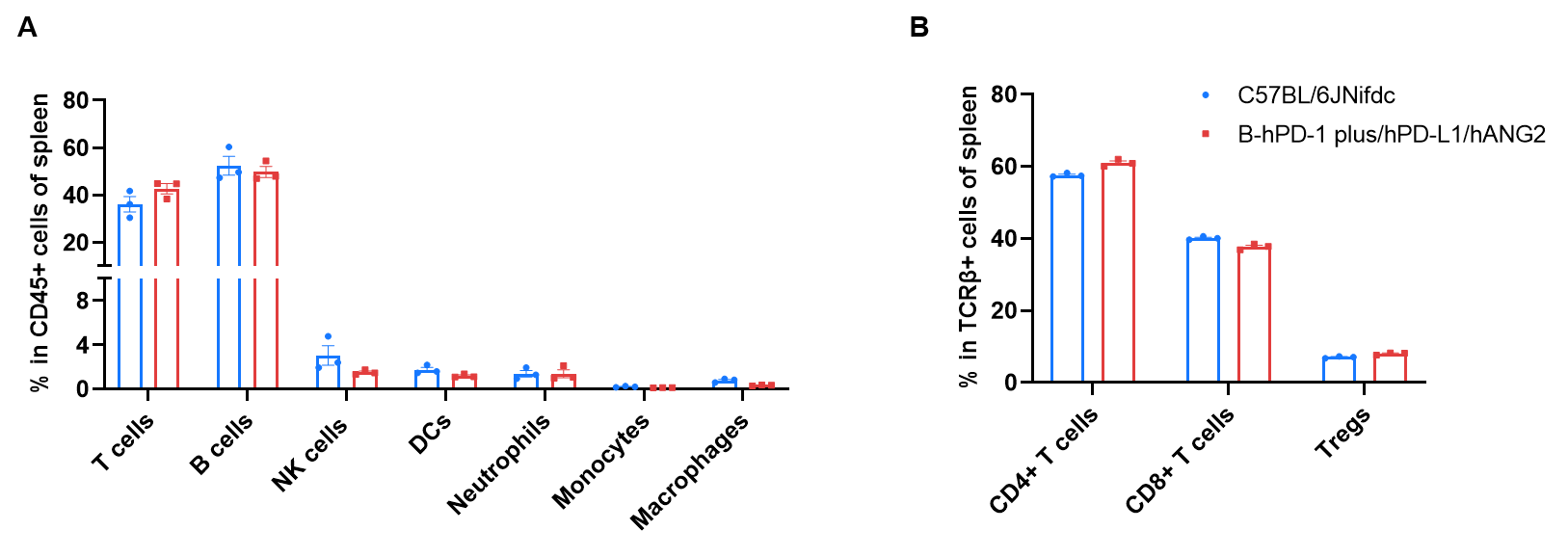
Frequency of leukocyte subpopulations in spleen by flow cytometry. Splenocytes were isolated from wild-type C57BL/6JNifdc mice (female, n=3, 9-week-old), homozygous B-hPD-1 plus/hPD-L1/hANG2 mice (female, n=3, 9-week-old). A. Flow cytometry analysis of the splenocytes was performed to assess the frequency of leukocyte subpopulations. B. Frequency of T cell subpopulations. Frequencies of T cells, B cells, NK cells, dendritic cells, neutrophils, monocytes, macrophages, CD4+ T cells, CD8+ T cells and Tregs in B-hPD-1 plus/hPD-L1/hANG2 mice were similar to those in C57BL/6JNifdc. The frequency of leukocyte subpopulations in blood and lympy nodes of B-hPD-1 plus/hPD-L1/hANG2 mice were also comparable to wild-type C57BL/6JNifdc mice (Data not shown). Values are expressed as mean ± SEM.
* When publishing results obtained using this animal model, please acknowledge the source as follows: The animal model [B-hPD-1 plus/hPD-L1/hANG2 mice] (Cat# 114041) was purchased from Biocytogen.