あなたもお好きかもしれません
The immune system maintains a delicate balance, vigilantly protecting the body from pathogens while avoiding self-inflicted damage. Disruptions in this balance can lead to a spectrum of diseases, such as asthma, atopic dermatitis, inflammatory bowel disease, and rheumatoid arthritis, that have long puzzled researchers. Central to this enigma are cytokines: small but powerful proteins that regulate immune signaling. Among them, interleukin-4 (IL-4) and IL-13 are key orchestrators of type 2 immune responses, while IL-31 contributes to the pathophysiology of certain inflammatory skin conditions (Karo-Atar et al. 2018). As our understanding of cytokine function deepens, so too does the potential for more precisely targeted therapies.
The Pivotal Roles of IL-4, IL-13, and IL-31 in Autoimmune Diseases
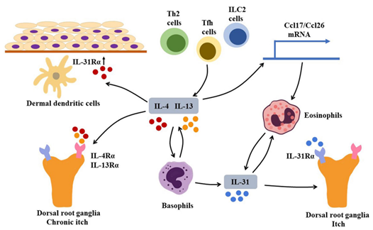
Type 2 immunity plays a vital role in clearing parasitic infections; however, it is also heavily involved in the development of allergic diseases. IL-4 and IL-13 are hallmark Th2 (CD4+ T helper) cytokines that drive type 2 inflammation. They also inhibit Th17 cell development and counteract Th1 responses, thereby exerting “anti-inflammatory” effects in certain autoimmune contexts by dampening pathogenic inflammation (Iwaszko et al. 2021). Despite their functional similarities, IL-4 and IL-13 share only about 25% amino acid sequence homology. Both cytokines signal through receptor complexes that include the shared type II receptor composed of IL-4Rα and IL-13Rα1 subunits (Bernstein et al. 2023; Chen et al. 2023).
IL-31, another Th2-associated cytokine, contributes to skin inflammation and pruritus. Its receptor, a heterodimer consisting of IL-31RA and oncostatin M receptor beta (OSMRβ), is expressed on various cell types, including peripheral sensory neurons, keratinocytes, and immune cells. Upon binding, IL-31 activates JAK/STAT, PI3K/AKT, and MAPK pathways, leading to chronic itch and skin inflammation (Bağci and Ruzicka et al. 2018; Murdaca et al. 2019; Dubin et al. 2021).
IL-4, IL-13 and IL-31 are inter-related in the pathogenesis of autoimmune diseases (Chen et al. 2023)
Advancements in Preclinical Research: Humanized Mouse Models
Given the central role of cytokines in inflammation and autoimmune diseases, there is a growing need for human-relevant in vivo models to study these pathways and evaluate therapeutic interventions. Biocytogen has addressed this need by developing a series of humanized mouse models using advanced gene-editing technologies. These models express human cytokines and their receptors, enabling researchers to more accurately assess the pharmacodynamics and therapeutic potential of candidate drugs targeting the IL-4, IL-13, and IL-31 pathways.
These innovative humanized mouse models serve as powerful platforms to bridge the gap between basic research and clinical application, accelerating the development of next-generation therapies for inflammatory and autoimmune diseases.
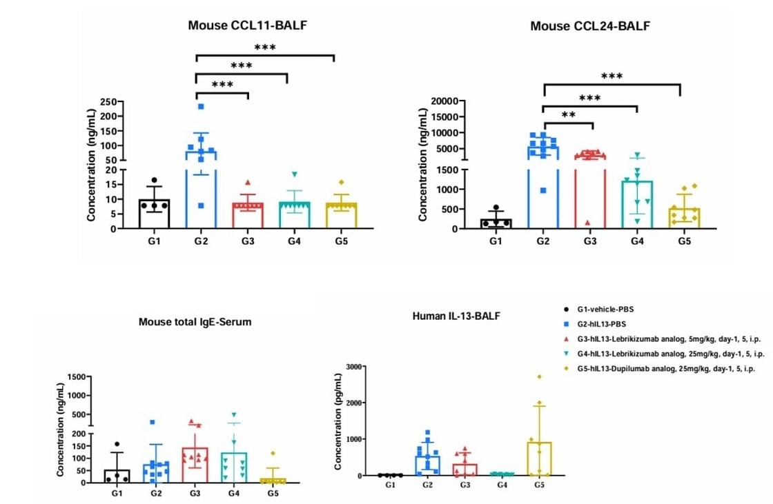
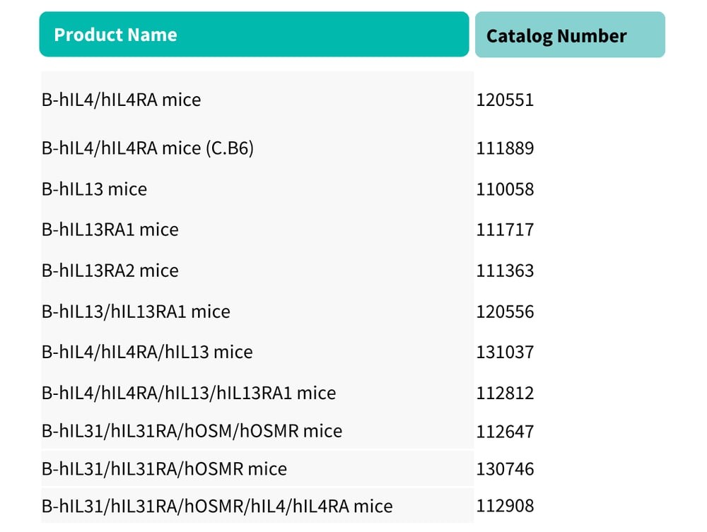
Case Study 1: B-hIL4/hIL4RA/hIL13/hIL13RA1 Mice |
|
Cytokine and IgE analysis in humanized asthma mouse model. B-hIL4/hIL4R/hIL13/hIL13RA1 mice were challenged with human IL-13 to induce asthma, and treated intraperitoneally with in-house analogs of dupilumab (anti-IL-4Rα) or lebrikizumab (anti-IL-13). BALF analysis showed reduced CCL11 and CCL24 levels in both treatment groups vs. PBS controls. Human IL-13 decreased in the high-dose lebrikizumab group but increased in the dupilumab group, likely due to antibody-bound cytokine. Serum IgE levels were significantly reduced in the dupilumab group. Mean ± SEM. |
Case Study 2: B-hIL31/hIL31A/hOSMR/hIL4/hIL4RA Mice |
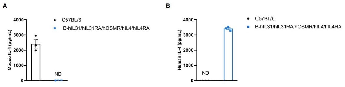
Strain-specific IL-4 expression in humanized mice. Wild-type C57BL/6 and B-hIL31/hIL31RA/hOSMR/hIL4/hIL4RA mice were stimulated with anti-mCD3ε and anti-mCD28 to activate T cells for IL-4 production. Serum collected 3 h post-injection was analyzed by ELISA. (A) Mouse IL-4 was detected only in wild-type mice. (B) Human IL-4 was detected only in humanized mice, confirming strain-specific expression. |
Explore our featured humanized mice targeting the IL-4, IL-13, and IL-31 pathways! |
|
Reference:
Karo-Atar, Danielle, et al. "Therapeutic targeting of the interleukin-4/interleukin-13 signaling pathway: in allergy and beyond." BioDrugs 32.3 (2018): 201-220.
Iwaszko, Milena, Sylwia Biały, and Katarzyna Bogunia-Kubik. "Significance of interleukin (IL)-4 and IL-13 in inflammatory arthritis." Cells 10.11 (2021): 3000.
Bernstein, Zachary J., et al. "Engineering the IL‐4/IL‐13 axis for targeted immune modulation." Immunological reviews 320.1 (2023): 29-57.
Chen, Fangyuan, et al. "Targeting interleukin 4 and interleukin 13: a novel therapeutic approach in bullous pemphigoid." Annals of Medicine 55.1 (2023): 1156-1170.
Bağci, Işın Sinem, and Thomas Ruzicka. "IL-31: A new key player in dermatology and beyond." Journal of Allergy and Clinical Immunology 141.3 (2018): 858-866.
Murdaca, Giuseppe, et al. "IL-33/IL-31 axis in immune-mediated and allergic diseases." International journal of molecular sciences 20.23 (2019): 5856.
Dubin, Celina, Ester Del Duca, and Emma Guttman-Yassky. "The IL-4, IL-13 and IL-31 pathways in atopic dermatitis." Expert review of clinical immunology 17.8 (2021): 835-852.