
B-hPD-L1 plus/Tg(hCDH17) MC38

NA • 322473

B-hPD-L1 plus/Tg(hCDH17) MC38

Catalog Number: 322473
Strain Name: NA
Strain Background: C57BL/6
NCBI gene ID: 60533,12557 (Human)
Aliases: B7h1; Pdl1; Pdcd1l1; Pdcd1lg1; A530045L16Rik; HPT-1; HPT-1/LI
---
ライセンスオプション提供可能
B-hPD-L1 plus/Tg(hCDH17) MC38

このページで

  • Description
  • Phenotypic analysis
  • Tumorigenicity

ポスター

すべて表示

    出版物

      Description
      • Origin: The MC38 cell line is derived from C57BL/6 murine colon adenocarcinoma cells. The cell line is a commonly used murine model for colorectal carcinoma.
      • Background Information: PD-L1 is an immune checkpoint protein expressed on the surface of various cells, including tumor cells and immune cells. It binds to PD-1 on T cells, leading to T cell exhaustion, reduced cytokine production, and impaired anti-tumor immunity. CDH17 (Cadherin-17) is a calcium-dependent cell adhesion protein primarily expressed in intestinal epithelial cells and highly upregulated in several gastrointestinal cancers. It promotes tumor cell adhesion, proliferation, and survival, making it a potential therapeutic target and biomarker in digestive system malignancies.
      • Gene targeting strategy: The exogenous promoter, human PD-L1 and luciferase coding sequences were inserted to replace part of murine exon 3. The insertion disrupts the endogenous murine Pdl1 gene, resulting in a non-functional transcript. The human CDH17 coding sequence was randomly inserted into the genome MC38.
      • Tumorigenicity: Confirmed in B-hCDH17 mice.
      • Application: The B-hPD-L1 plus/Tg(hCDH17) MC38 tumor models can be used for preclinical evaluation of monoclonal antibody drugs and bispecific antibody drugs targeting human CDH17 and PD-1/PD-L1 pathway.
      • Notes:

      In the experiment, it is necessary to ensure that the number of animals inoculated subcutaneously is at least 1.6 times the actual grouping number.

      PD-L1 and CDH17 Protein Expression Analysis
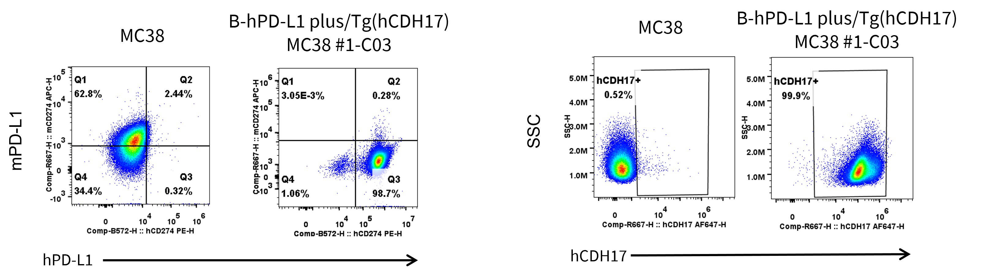
      • Human PD-L1 and CDH17 were detected on the surface of B-hPD-L1 plus/Tg(hCDH17) MC38 but not wild-type MC38 cells.
      • Mouse PD-L1 was not detected on the surface of B-hPD-L1 plus/Tg(hCDH17) MC38.

      PD-L1 and CDH17 expression analysis in B-hPD-L1 plus/Tg(hCDH17) MC38 by flow cytometry. Single cell suspensions from wild-type MC38 and B-hPD-L1 plus/Tg(hCDH17) MC38 #1-C03 cultures were stained with anti-mouse PD-L1 antibody (Biolegend, 124312), anti-human PD-L1 antibody (Biolegend, 329706), and anti-human CDH17 antibody (in house) .


      Tumor Growth Curve & Body Weight Changes

      Subcutaneous tumor growth of B-hPD-L1 plus/Tg(hCDH17) MC38 cells. B-hPD-L1 plus/Tg(hCDH17) MC38 (1×10⁶) and wild-type MC38 cells (5×10⁵) were subcutaneously implanted into B-hCDH17 mice (female, 8-week-old, n=6). Tumor volume and body weight were measured twice a week. (A) Average tumor volume. (B) Body weight. Volume was expressed in mm3 using the formula: V=0.5 × long diameter × short diameter2. Results indicate that B-hPD-L1 plus/Tg(hCDH17) MC38 cells were able to establish tumors in vivo and can be used for efficacy studies. Values are expressed as mean ± SEM.
      PD-L1 Protein Expression Analysis of Tumor Tissue
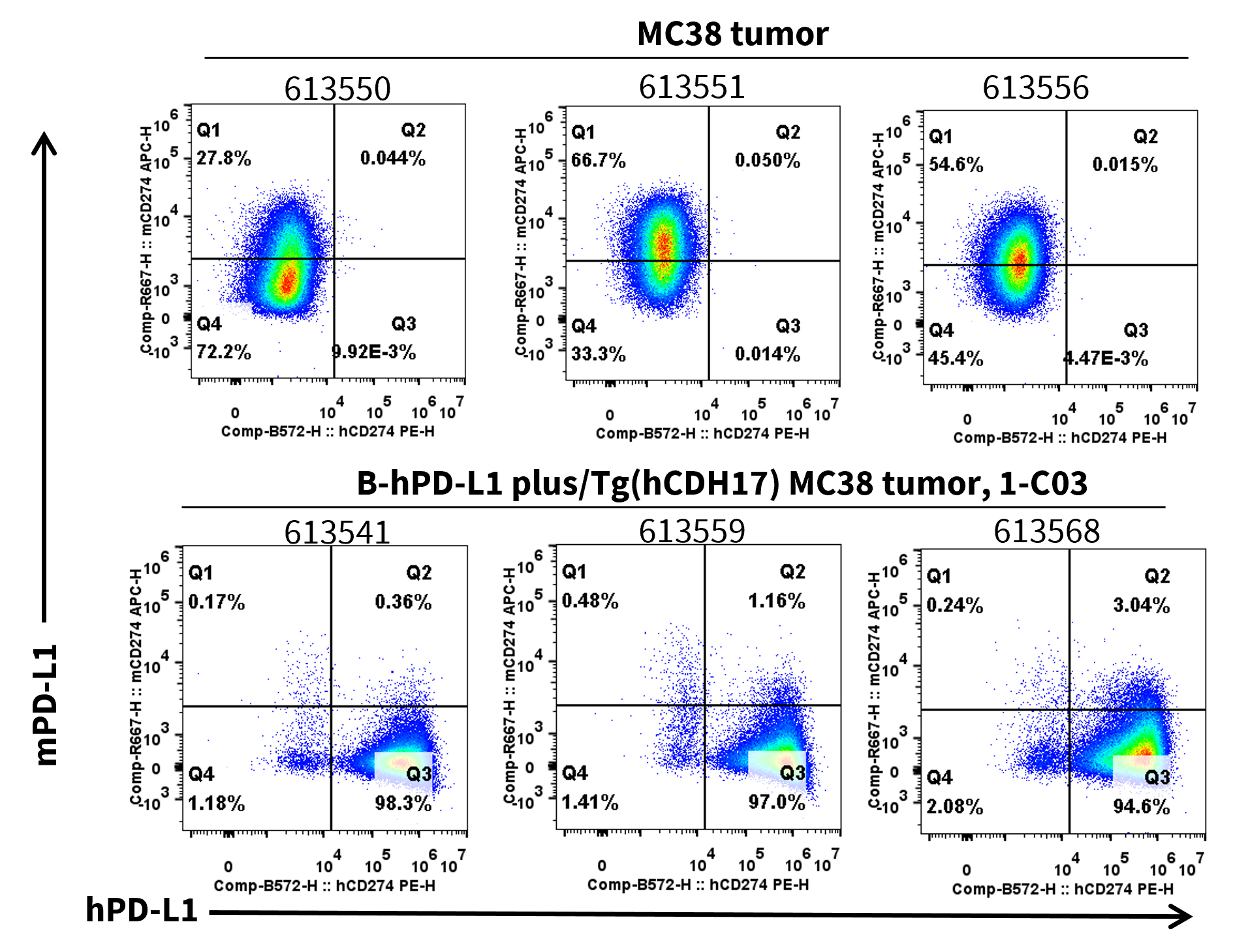
      • Human PD-L1 was detected on the surface of B-hPD-L1 plus/Tg(hCDH17) MC38 tumors but not wild-type MC38 tumors.
      • Mouse PD-L1 was not detected on the surface of B-hPD-L1 plus/Tg(hCDH17) MC38 tumors.

      PD-L1 expression was evaluated​​ on B-hPD-L1 plus/Tg(hCDH17) MC38​​ by flow cytometry. ​​These cells​​ were subcutaneously transplanted into B-hCDH17 mice (n=6). At the end of the experiment, tumor cells were harvested and ​​analyzed for both mouse and human PD-L1 expression​​ by flow cytometry. ​​

      CDH17 Protein Expression Analysis of Tumor Tissue
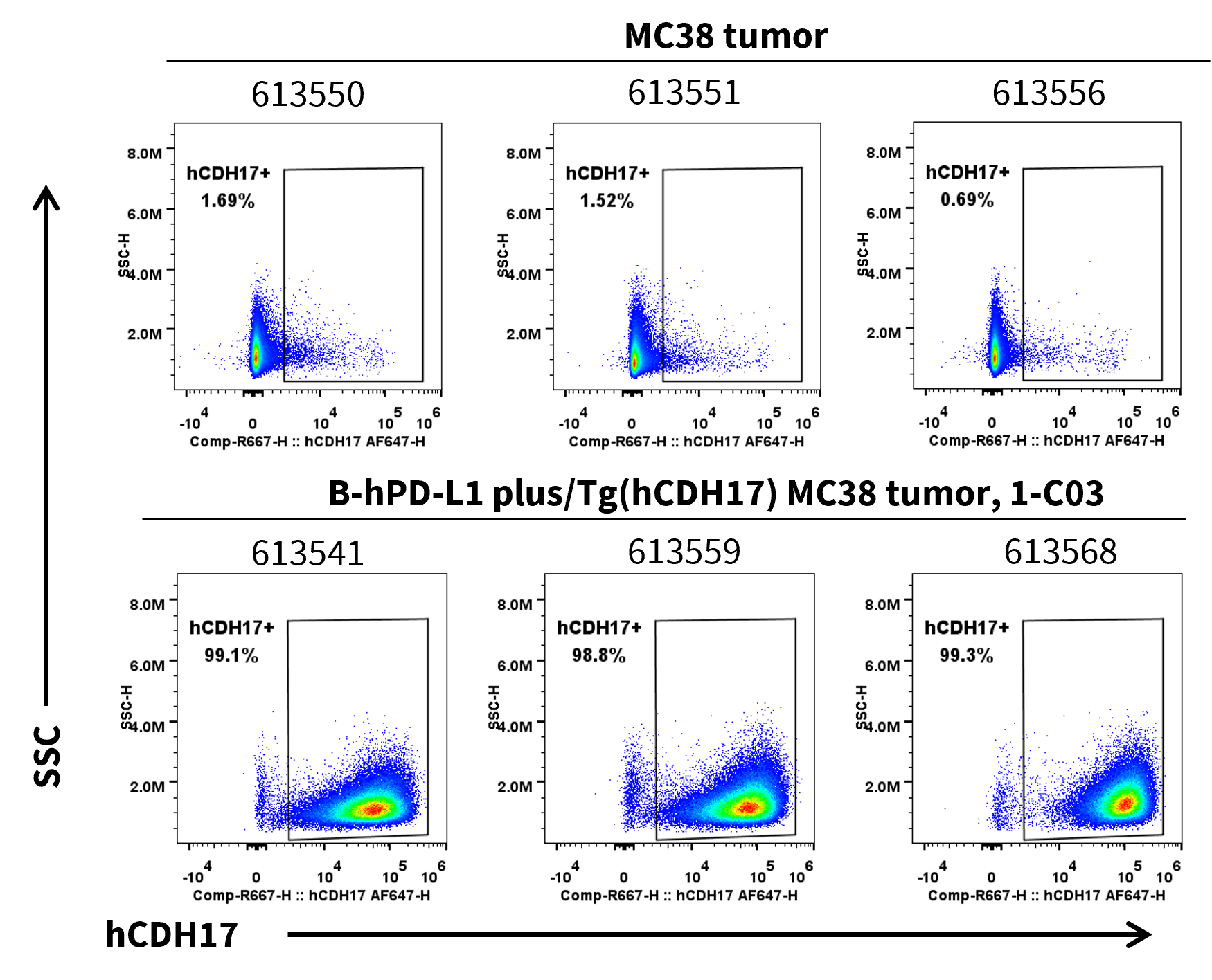
      • Human CDH17 was detected on the surface of B-hPD-L1 plus/Tg(hCDH17) MC38 tumors but not wild-type MC38 tumors.

      CDH17 expression was evaluated​​ on B-hPD-L1 plus/Tg(hCDH17) MC38​​ by flow cytometry. ​​These cells​​ were subcutaneously transplanted into B-hCDH17 mice (n=6). At the end of the experiment, tumor cells were harvested and ​​analyzed for human CDH17 expression​​ by flow cytometry. ​​