
C57BL/6-Ccr8tm1(CCR8)Bcgen Ccl1tm1(CCL1) Bcgen/Bcgen • 121836
Key Advantages
Validation
Applications
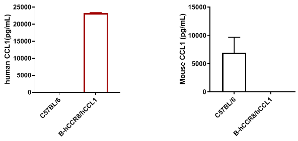
Strain-specific CCL1 expression analysis was performed in homozygous CCR8/CCL1 humanized mice by ELISA. Serum samples were collected from wild-type mice and homozygous CCR8/CCL1 humanized mice stimulated in vivo with 7.5 μg anti-mCD3ε antibody (n=2 or 3). Species-specific ELISA kits were used for analysis. Mouse CCL1 was detectable in wild-type mice, whereas human CCL1 was detectable in homozygous CCR8/CCL1 humanized mice but not in wild-type controls.
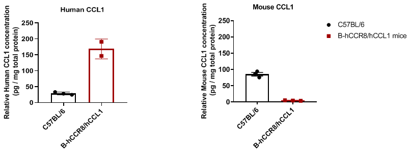
Detection of CCL1 in the tumor microenvironment was performed in C57BL/6 and CCR8/CCL1 humanized mice bearing MC38 tumors by ELISA. Murine MC38 colon cancer cells were subcutaneously implanted into C57BL/6 and CCR8/CCL1 humanized mice (n=2 or 3). Tumor tissues were harvested when tumor volume reached approximately 600 mm³ and analyzed by ELISA. Human CCL1 was detectable in homozygous CCR8/CCL1 humanized mice at approximately 150 pg per mg total protein.
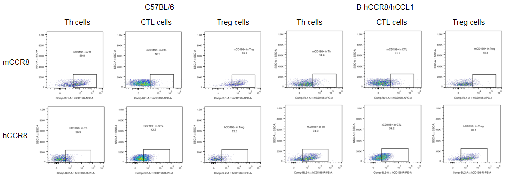
CCR8 (CD198) protein expression analysis was performed in CCR8/CCL1 humanized mice by flow cytometry. Murine MC38 colon cancer cells were subcutaneously implanted into homozygous CCR8/CCL1 humanized mice. Tumor-infiltrating lymphocytes (TILs) were analyzed when tumor volume reached approximately 600 mm³. Human CCR8 was detectable in Th cells and Treg cells of homozygous CCR8/CCL1 humanized mice. Human CCR8 signal was also observed in CTL cells of both wild-type C57BL/6 mice and humanized mice, suggesting potential nonspecific antibody binding in CTLs.
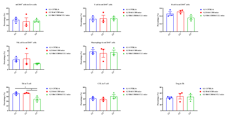
Tumor-infiltrating lymphocyte populations were compared among different mouse strains. The percentage of Treg cells within Th cells in CCR8/CCL1 humanized mice showed no significant difference compared with wild-type C57BL/6 mice and CCR8 humanized mice.
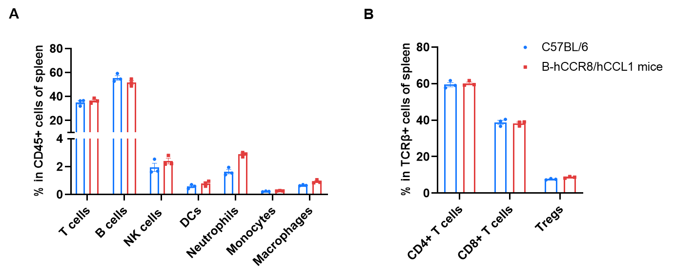
Frequency of leukocyte subpopulations in spleen was analyzed by flow cytometry. Splenocytes were isolated from wild-type C57BL/6 mice (female, n=3, 8-week-old) and homozygous CCR8/CCL1 humanized mice (female, n=3, 8-week-old).
A. Flow cytometry analysis of splenocytes was performed to assess leukocyte subpopulations.
B. Frequency analysis of T cell subpopulations.
Percentages of T cells, B cells, NK cells, dendritic cells, neutrophils, monocytes, macrophages, CD4+ T cells, CD8+ T cells, and Tregs in CCR8/CCL1 humanized mice were comparable to wild-type C57BL/6 mice, demonstrating that humanization of CCR8 and CCL1 did not alter immune cell frequency or distribution in spleen. Frequencies of leukocyte subpopulations in lymph nodes and blood were also comparable between CCR8/CCL1 humanized mice and wild-type controls (data not shown).
Values are expressed as mean ± SEM. Significance was determined by two-way ANOVA test. *P < 0.05, **P < 0.01, ***P < 0.001.

Antitumor activity of anti-human CCR8 antibody was evaluated in CCR8/CCL1 humanized mice.
A. Anti-human CCR8 antibody inhibited MC38 tumor growth in CCR8/CCL1 humanized mice. Murine MC38 colon cancer cells (5 × 10⁵) were subcutaneously implanted into homozygous CCR8/CCL1 humanized mice (female, 7-week-old, n=6). Mice were grouped when tumor volume reached approximately 100–150 mm³ and treated by intraperitoneal injection with anti-human CCR8 antibody as indicated.
B. Body weight changes during treatment.
Anti-human CCR8 antibody treatment effectively controlled tumor growth in CCR8/CCL1 humanized mice, demonstrating that this model provides a powerful preclinical platform for in vivo evaluation of anti-human CCR8 antibodies. Values are expressed as mean ± SEM.
The tumor model overage was 41.7%.

Individual MC38 tumor growth curves were analyzed in CCR8/CCL1 humanized mice. Murine MC38 colon cancer cells (5 × 10⁵) were subcutaneously implanted into homozygous CCR8/CCL1 humanized mice (female, 7-week-old, n=6). Mice were grouped when tumor volume reached approximately 100–150 mm³ and subsequently treated with anti-human CCR8 antibody as indicated.
Results demonstrated that anti-human CCR8 antibody effectively inhibited tumor growth in CCR8/CCL1 humanized mice, supporting use of this model for preclinical efficacy evaluation of anti-human CCR8 therapeutics.

Tumor-infiltrating lymphocytes were analyzed by flow cytometry at the study endpoint (n=6). Tumor tissues were harvested and analyzed to evaluate changes in immune cell number and proportion following anti-human CCR8 antibody treatment.
The proportions of CD3+ T cells, CD4+ T cells, Treg cells, and hCCR8+ Treg cells in the anti-CCR8 antibody treatment group showed no significant changes compared with the control group.
Values are expressed as mean ± SEM. Significance was determined by one-way ANOVA test. *P < 0.05, **P < 0.01, ***P < 0.001.
What are CCR8/CCL1 humanized mice?
CCR8/CCL1 humanized mice are genetically engineered mice expressing human CCR8 and human CCL1, enabling translational studies of CCR8-targeted therapeutics and tumor immunology.
Why is CCR8 an important immuno-oncology target?
CCR8 is highly expressed on tumor-infiltrating regulatory T cells and contributes to immune suppression within tumors. Targeting CCR8 may enhance antitumor immunity.
Can these mice be used for antibody efficacy studies?
Yes. CCR8/CCL1 humanized mice support in vivo efficacy evaluation of anti-human CCR8 antibodies in tumor models.
What tumor models are compatible with CCR8/CCL1 humanized mice?
MC38 colorectal tumor models have been validated, and the model may also support additional syngeneic immuno-oncology studies.
Do CCR8/CCL1 humanized mice maintain normal immune homeostasis?
Yes. Leukocyte subset frequencies in spleen, blood, and lymph nodes are comparable to wild-type C57BL/6 mice.