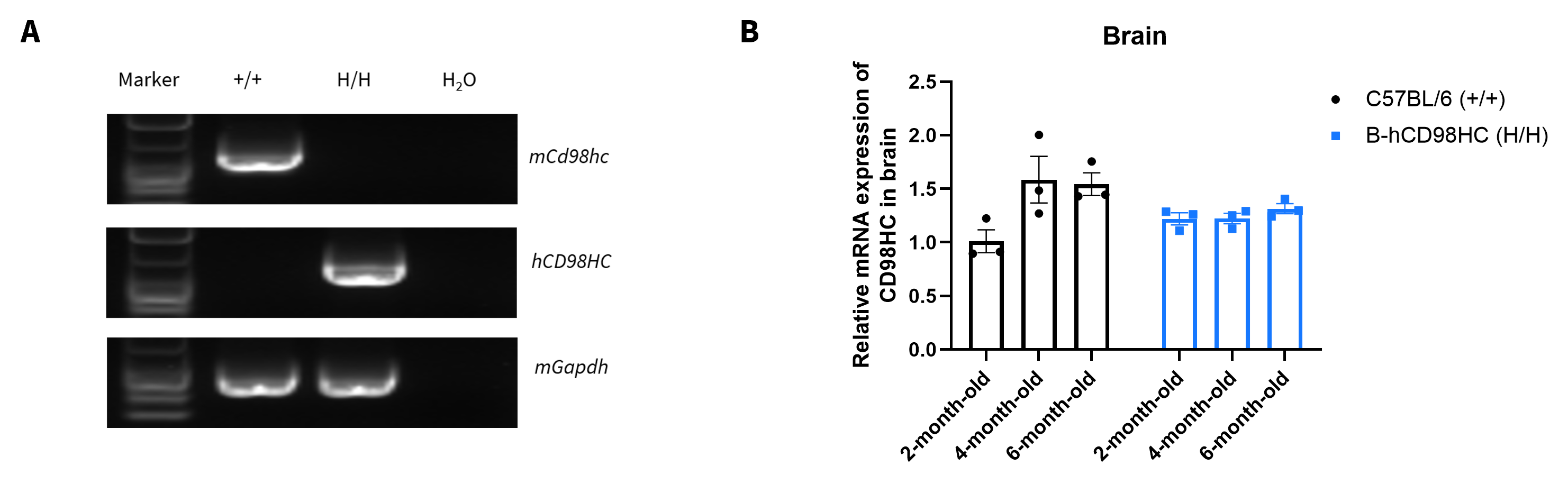
mRNA Expression Analysis
- (A) Human CD98HC mRNA is exclusively detectable in kidney of homozygous B-hCD98HC mice, but not in wild-type C57BL/6 mice.
- (B) Relative CD98HC mRNA expression levels in the brain were comparable between B-hCD98HC mice and wild-type C57BL/6 mice across 2, 4 and 6-months old.
Strain-specific CD98HC expression analysis in wild-type C57BL/6 mice and homozygous B-hCD98HC mice. (A) Kidney RNA was isolated from wild-type C57BL/6 mice (+/+) and homozygous B-hCD98HC mice (H/H), then cDNA libraries were synthesized by reverse transcription, followed by PCR with mouse or human CD98HC primers. (B) Quantitative real-time PCR (qRT-PCR) analysis of relative CD98HC mRNA expression in the brain of male wild-type C57BL/6 mice and homozygous B-hCD98HC mice at 2, 4 and 6-months of age. Expression levels were normalized to CD98HC expression in brain of 2-month-old C57BL/6 male mice. Values are expressed as mean ± SEM.
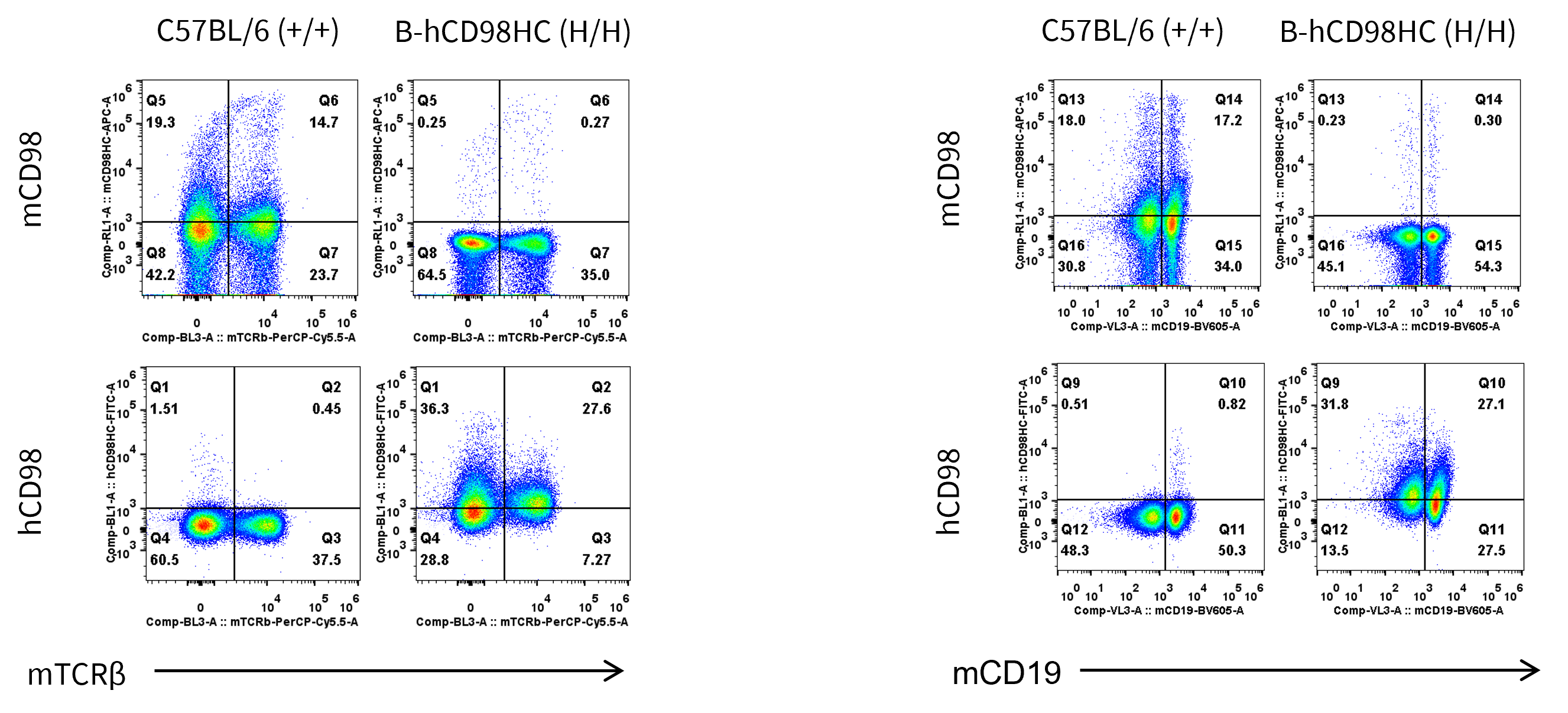
Protein Expression Analysis
- Mouse CD98hc is detectable only in wild-type C57BL/6 mice.
- Human CD98HC is detectable only in homozygous B-hCD98HC mice, but not in wild-type C57BL/6 mice.
Strain specific CD98HC expression analysis in wild-type C57BL/6 mice and homozygous humanized B-hCD98HC mice. Splenocytes were collected from wild-type C57BL/6 mice (+/+) and homozygous B-hCD98HC mice (H/H) (female, 7-week-old, n=1). Protein expression was analyzed with anti-mouse CD98HC antibody (Biolegend, 128211) and anti-human CD98HC antibody (Biolegend, 315603) by flow cytometry.
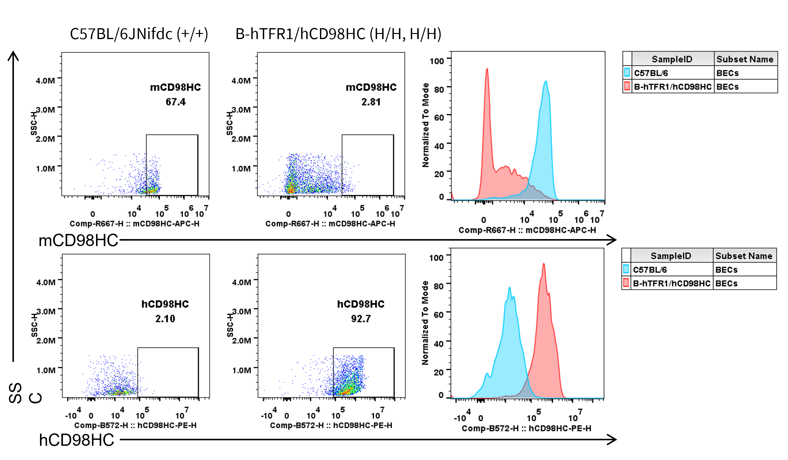
Human CD98HC Expression in Brain Endothelial Cells
- Mouse CD98HC was detectable only in brain endothelial cells of wild-type mice
- Human CD98HC was detectable only in brain endothelial cells of homozygous B-hTFR1/hCD98HC mice
Strain specific CD98HC expression in wild-type C57BL/6JNifdc and homozygous B-hTFR1/hCD98HC mice by flow cytometry. Brain cells were collected from wild-type C57BL/6JNifdc (+/+) and homozygous B-hTFR1/hCD98HC mice (H/H, H/H) and analyzed by flow cytometry with anti-mouse CD98HC antibody (Biolegend, 128211) and anti-human CD98HC antibody (231161-CD98BBBB-h1.L produced in-house).
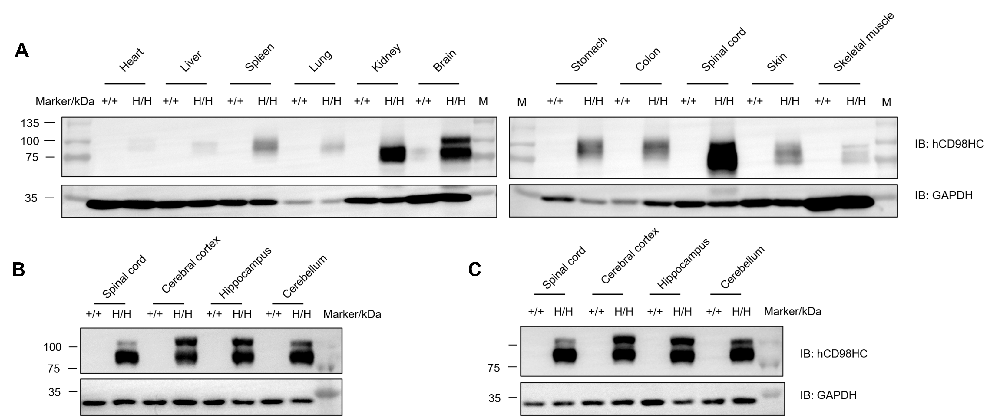
Human CD98HC Protein Expression Across Tissues
- Human CD98 is detected in the spleen, lung, kidney, stomach, colon, spinal cord, skin and brain of homozygous B-hCD98HC mice
- Human CD98HC was exclusively detected in spinal cord, cerebral cortex, hippocampus and cerebellum from B-hTFR1/hCD98HC mice
Western blot analysis of hCD98HC protein expression in homozygous B-hCD98HC mice.
Various tissue lysates were collected from wild-type C57BL/6 and homozygous B-hCD98HC mice (H/H), and then analyzed by western blot with species-specific anti-human CD98 antibody (Abcam, ab307587). (A) Male. (B) Male. (C) Female.
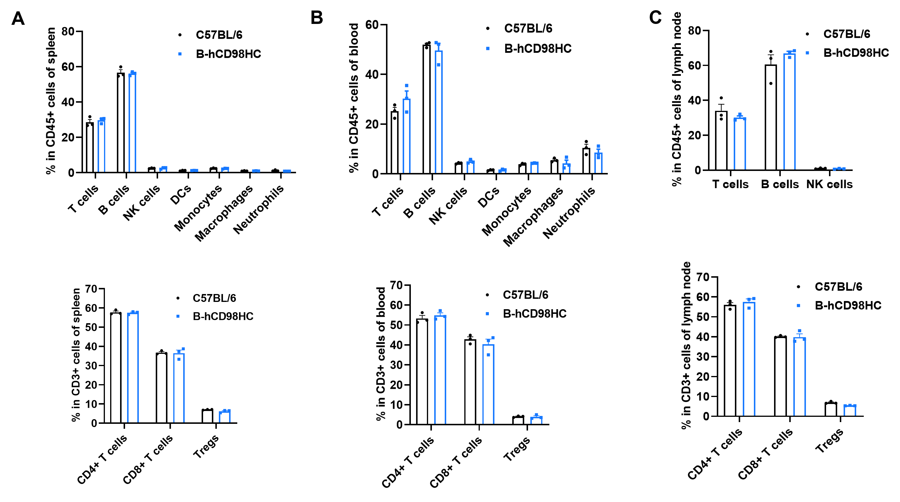
Animal State Evaluation-Leukocyte Profiling
- Humanization of CD98HC does not alter the frequency or distribution of immune cell types in spleen (A), blood (B) and lymph nodes (C).
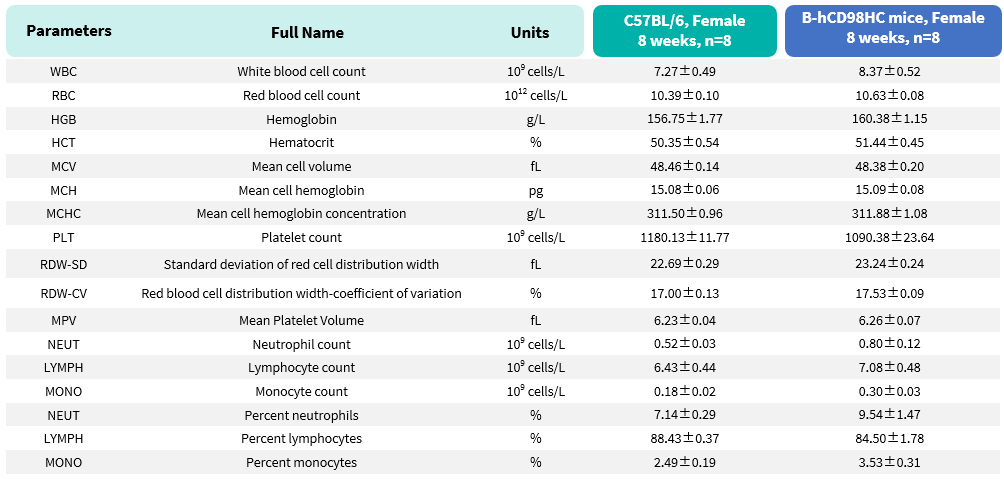
Normal Hematological Profiles
- CD98HC humanization does not disrupt hematopoiesis or immune cell composition.
Complete blood count (CBC) of B-hCD98HC mice. Values are expressed as mean ± SD.
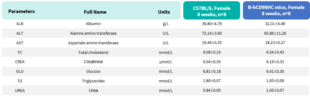
Normal Liver and Kidney Function Profiles
- CD98HC humanization does not adversely affect metabolic health or organ function.
Biochemical test of B-hCD98HC mice. Values are expressed as mean ± SD.
Active Uptake of Anti-Human CD98HC Antibody into the Brain
- Anti-human CD98HC antibody show faster serum clearance and enhanced brain exposure than control IgG
- B-hCD98HC mice enable active uptake of intravenously administered anti-human CD98HC antibody into the brain.
- B-hCD98HC mice provide a powerful preclinical platform for evaluating the in vivo drug delivery to the central nervous system.
In vivo pharmacokinetic (PK) evaluation of anti-human CD98HC antibody. B-hCD98HC mice (n=2, female, 8-week-old) were injected with control IgG (10 mpk) and anti-human CD98HC antibody (CD98BBBB-h1.L analog, monovalent, produced in house, 13.3 mpk) via tail vein. Brain and serum were taken for in vivo PK evaluation. Brain concentrations, serum concentrations, and brain-to-serum ratio of anti-human CD98HC antibody were quantified. Graphs represent mean ± SEM.
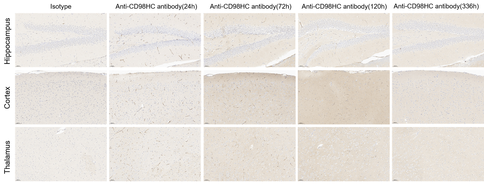
Anti-Human CH98HC Antibody Can Penetrate the Brain Parenchyma
- Anti-human CD98HC antibody penetrates the brain parenchyma following intravenous administration.
- B-hCD98HC mice enable uptake of anti-human CD98HC antibody into the brain.
- B-hCD98HC mice provide a powerful preclinical platform for evaluating the in vivo drug delivery to the central nervous system.
IHC staining of anti-CD98HC Abs penetrate brain parenchyma. B-hCD98HC mice were injected with control IgG (10 mpk) and anti-human CD98HC antibody (CD98BBBB-h1.L analog, monovalent, produced in house, 13.3 mpk) via tail vein. Brain comparts were taken for IHC staining after 120 h. Blue: nucleus; dark brown: antibodies.
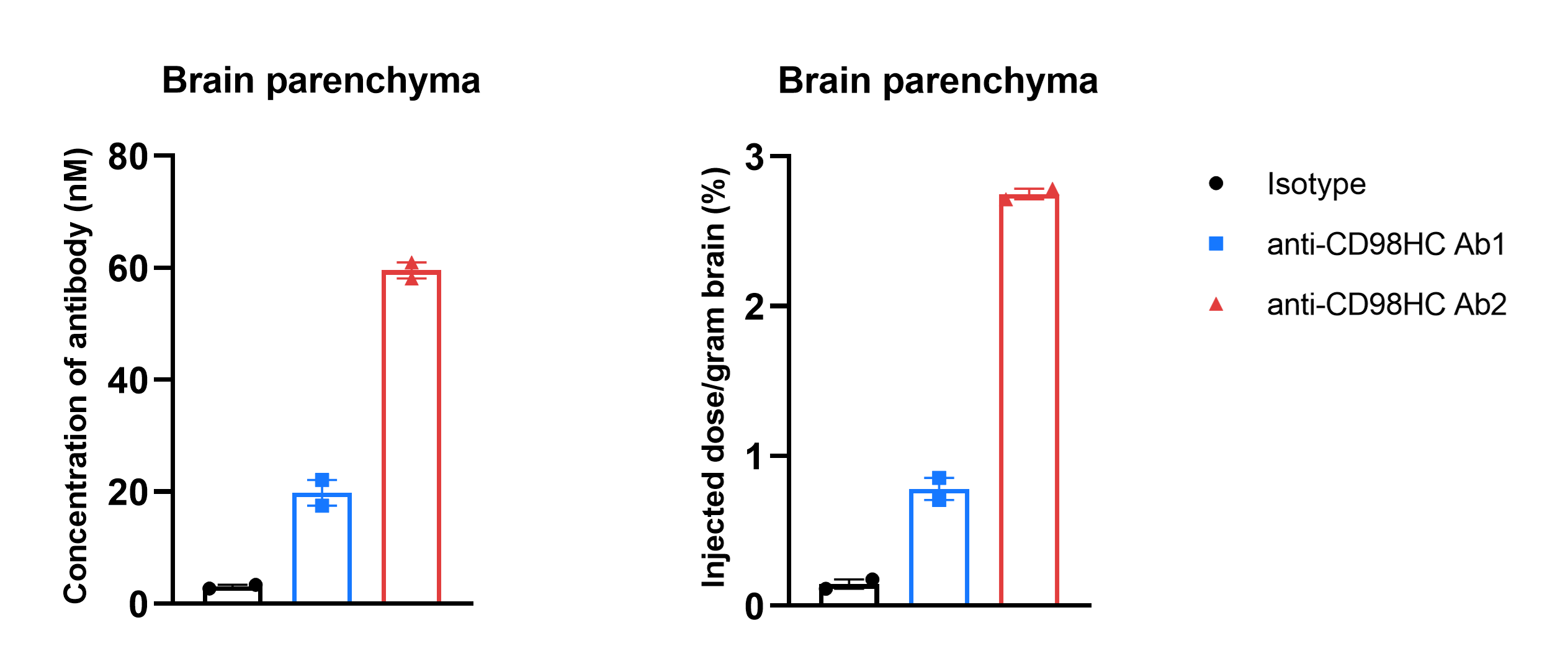
Comparative Brain Penetration Kinetics of different CD98HC antibodies
In vivo PK evaluation and comparison of different anti-CD98HC antibodies. B-hCD98HC mice (n=2, 8-week-old) were injected with Isotype IgG (20 mpk) and anti-human CD98HC antibodies (Ab1 and Ab2, produced in house according to patent) via tail vein. Brain were taken for in vivo PK evaluation after dosing 5 days. Brain concentrations (A) and % of injection/gram brain (B) were quantified. As shown in panel, anti-CD98HC Ab2 exhibited higher brain exposure. Graphs represent mean ± SEM.
Comparative Brain Penetration Kinetics of TFR1 and CD98HC
- B-hTFR1/hCD98HC mice enable uptake of intravenously administered anti-human TFR1 or anti-human CD98HC antibodies
- B-hTFR1/hCD98HC mice can be used to compare penetration efficacy of shuttle molecules targeting TFR1 vs. CD98HC
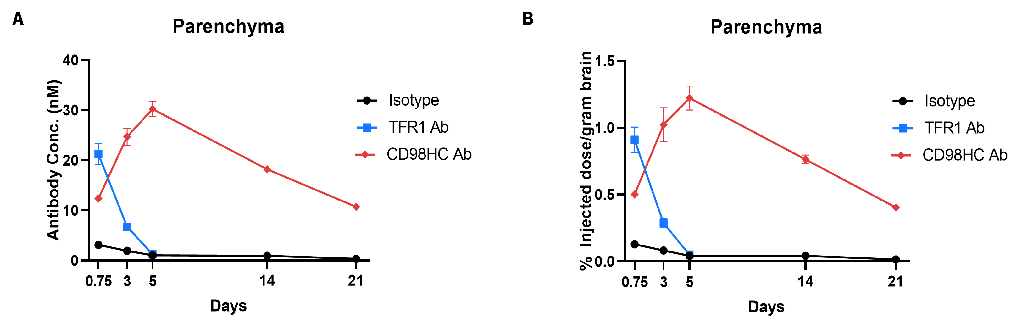
In vivo PK evaluation and comparison of anti-human TFR1 and anti-CD98HC antibody.
B-hTFR1/hCD98HC mice (n=2, female, 8-week-old) were injected with control IgG (10 mpk) anti-human TFR1 antibody (TFR1 Ab, JR-141 analog, monovalent, produced in house, 12.56 mpk) and anti-human CD98HC antibody (CD98HC Ab, CD98BBBB-h1.L analog, monovalent, produced in house, 13.3 mpk) via tail vein. Brain were taken for in vivo PK evaluation after dosing 18 h and 3, 5, 14, 21 days. Brain concentrations (A) and % of injection/gram brain (B) were quantified. As shown in panel, anti-human TFR1 antibody exhibited higher brain exposure in 24 h after dose, while anti-CD98HC antibody exhibited higher brain exposure in 72 h after dose. The results confirmed that B-hTFR1/hCD98HC mice enables uptake of an intravenously administered anti-human TFR1 antibody or anti-human CD98HC antibody, and this mice can be used for the comparison of penetration efficacy of shuttle molecules targeting TFR1 or CD98HC. Graphs represent mean ± SEM.
* When publishing results obtained using this animal model, please acknowledge the source as follows: The animal model [B-hCD98HC mice] (Cat# 110983) was purchased from Biocytogen.