

C57BL/6-Dgat2tm1(DGAT2)Bcgen/Bcgen • 113047

Diacylglycerol O-acyltransferase 2 (DGAT2) is one of two isoenzymes responsible for triglyceride (TAG) synthesis, together with DGAT1, accounting for nearly all TAG production in eukaryotic cells. Triglycerides serve as the primary form of energy storage. DGAT (acyl-CoA:diacylglycerol acyltransferase; EC 2.3.1.20) catalyzes the final step of TAG biosynthesis by covalently linking fatty acyl-CoA to diacylglycerol (DG).
In DGAT2 humanized mice (B-hDGAT2), exons 1–8 of the mouse Dgat2 gene, encoding the entire protein (ATG to STOP codon), including the promoter, 5′UTR, and 3′UTR regions, are replaced by the corresponding human DGAT2 genomic sequence. Human DGAT2 expression is driven by the endogenous human promoter sequence, while transcription and translation of the endogenous mouse Dgat2 gene are disrupted.
Validation analyses demonstrate species-specific expression in this model. Mouse Dgat2 mRNA is detectable in the liver of wild-type C57BL/6JNifdc mice, whereas human DGAT2 mRNA is exclusively detectable in the livers of homozygous DGAT2 humanized mice (B-hDGAT2). These characteristics support the use of this model for in vivo evaluation of human DGAT2-targeted therapeutics.
Key Advantages
Validation
Applications
DGAT2 humanized mice (B-hDGAT2) are used for pharmacodynamic and safety evaluation of therapeutics targeting DGAT2 in non-alcoholic steatohepatitis (NASH) and other metabolic diseases.

In DGAT2 humanized mice (B-hDGAT2), exons 1–8 of the mouse Dgat2 gene, which encode the complete protein from ATG to STOP codon and include the promoter, 5′UTR, and 3′UTR regions, are replaced by the corresponding human DGAT2 genomic sequence. Human DGAT2 expression is driven by the endogenous human DGAT2 promoter, while transcription and translation of the mouse Dgat2 gene are disrupted.

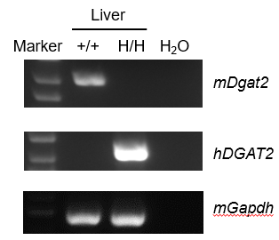
Strain-specific analysis of DGAT2 mRNA expression was performed in liver tissue from wild-type C57BL/6JNifdc mice and homozygous DGAT2 humanized mice (B-hDGAT2) by RT-PCR. Liver RNA was isolated from wild-type C57BL/6JNifdc mice (+/+) and homozygous DGAT2 humanized mice (H/H), followed by cDNA synthesis by reverse transcription and PCR amplification using mouse or human DGAT2-specific primers. Mouse Dgat2 mRNA was detectable only in wild-type C57BL/6JNifdc mice, whereas human DGAT2 mRNA was detectable only in homozygous DGAT2 humanized mice (B-hDGAT2) and not in wild-type mice.
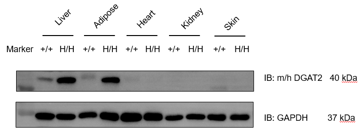
Strain-specific analysis of DGAT2 protein expression was performed in wild-type C57BL/6JNifdc mice and homozygous DGAT2 humanized mice (B-hDGAT2) by western blot. Tissue lysates from liver, adipose, heart, kidney, and skin were collected from wild-type C57BL/6JNifdc mice (+/+) and homozygous DGAT2 humanized mice (H/H) and analyzed using an anti-DGAT2 antibody (Proteintech, 17100-1-AP). DGAT2 protein bands were detected in liver and adipose tissues of both wild-type and DGAT2 humanized mice (B-hDGAT2) due to antibody cross-reactivity. GAPDH was used as a loading control.

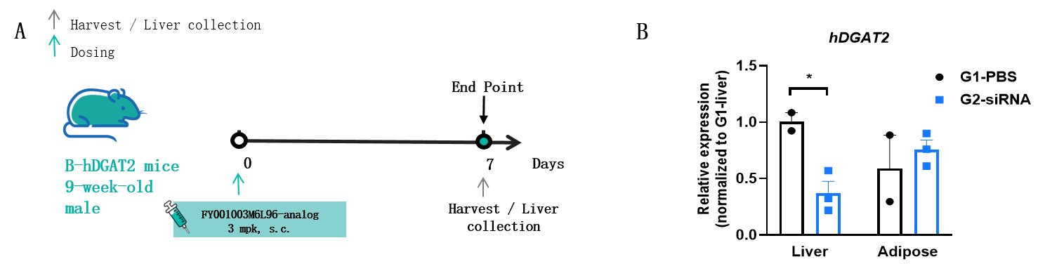
The inhibitory efficiency of the nucleic acid drugs against human DGAT2 in B-hDGAT2 mice. B-hDGAT2 mice were randomly divided into two groups (9 weeks old). The human DGAT2-targeted nucleic acid drugs (synthesized according to patents) and PBS were administered individually to the mice. The nucleic acid drugs were administered in the form of a PBS aqueous solution. The mice were sacrificed on day 7, and the liver tissue was collected to detect the expression level of human DGAT2 mRNA by qPCR and liver triglyceride (TG). (A) The schematic diagram of experimental processing. (B) The expression of human DGAT2 mRNA in the liver after treatment. Significance was determined by a t-test, *p<0.05. Values are expressed as mean ± SEM.
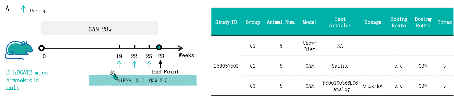
In vivo efficacy of the GAN-induced model in B-hDGAT2 mice. (A) The schematic diagram of experimental processing. Male B-hDGAT2 mice (8 weeks old) were fed either a standard chow diet (G1, n = 6) or a GAN diet (n = 20). Following a 19-week induction period, 16 mice from the GAN-fed cohort were evenly allocated into two groups (G2-G3) based on their body weight and total cholesterol (TC) levels. The mice in the siRNA treatment groups were injected subcutaneously with the saline or FY001003M6L96-analog (synthesized according to patents) from week 19 once every 3 weeks for 9 weeks.
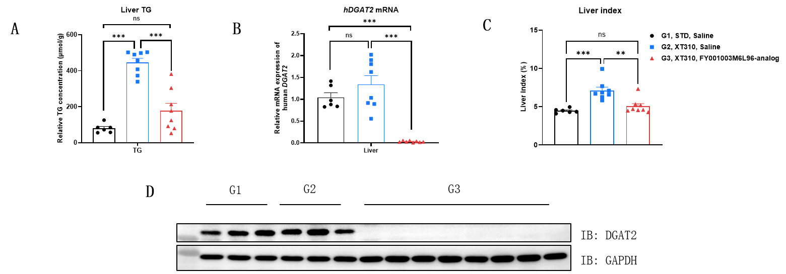
In vivo efficacy of the GAN-induced model in B-hDGAT2 mice. (A) The liver triglyceride (TG) expression after treatment. (B) The expression of human DGAT2 mRNA in the liver after treatment was measured by qPCR. (C) Liver index was calculated by the following formula: Liver index (%) = weight of liver/weight of body x 100%. (D) The expression of human DGAT2 protein in the liver after treatment was measured by Western blot with anti-DGAT2 antibody (Proteintech, 17100-1-AP). Data were shown as mean ± SEM, analyzed by one-way ANOVA (*p<0.05, **p<0.01, ***p<0.001).
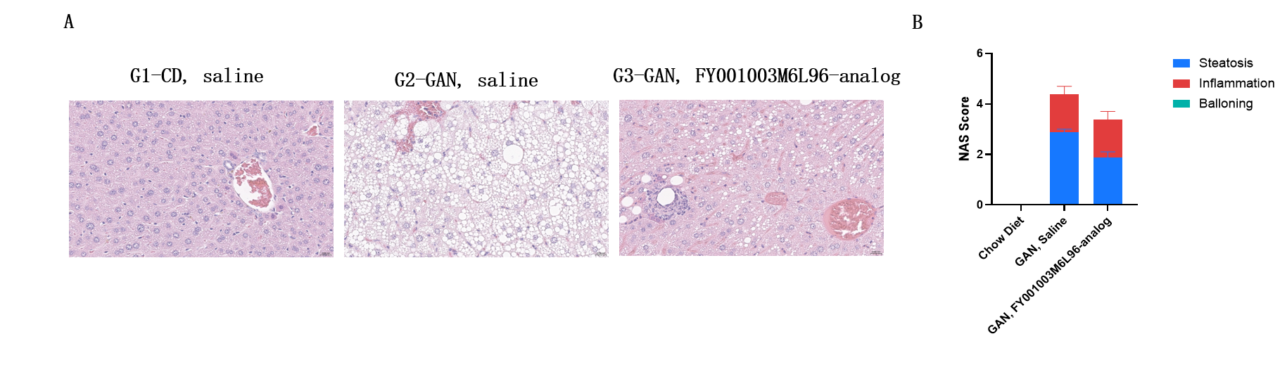
In vivo efficacy of the GAN-induced model in B-hDGAT2 mice. (A) Representative pictures of H&E staining showing the degree of NASH. (B) NAFLD activity score (NAS) in different treatment groups. Data were shown as mean ± SEM, analyzed by one-way ANOVA (*p<0.05, **p<0.01, ***p<0.001). Scale bar:20μm.
