

C57BL/6-Il33tm1(IL33)Tslptm1(TSLP)Crlf2tm2(CRLF2)/Bcgen • 111867

IL33: A key cytokine in inflammation and its therapeutic intervention
TSLP: A key cytokine in inflammation and its therapeutic intervention

IL33
TSLP
TSLPR

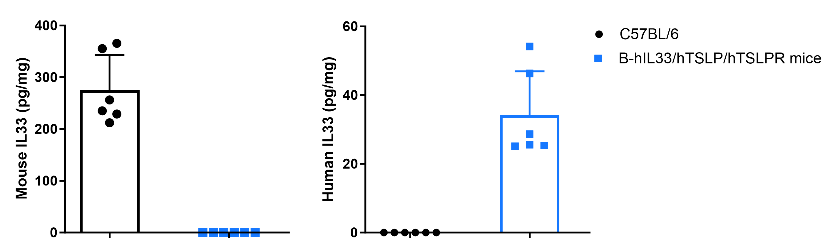
Strain-specific IL33 expression evaluated by ELISA in wild-type C57BL/6 mice and homozygous B-hIL33/hTSLP/hTSLPR mice. Calcipotriol (MC903), dissolved in ethanol, was topically applied to the ears of wild-type C57BL/6 mice and homozygous B-hIL33/hTSLP/hTSLPR mice for 7 days (male, 6-week-old, n = 3). Mouse and human IL33 levels in ear tissue homogenates were quantified by ELISA (mouse IL33, R&D Systems M3300; human IL33, R&D Systems D3300B).
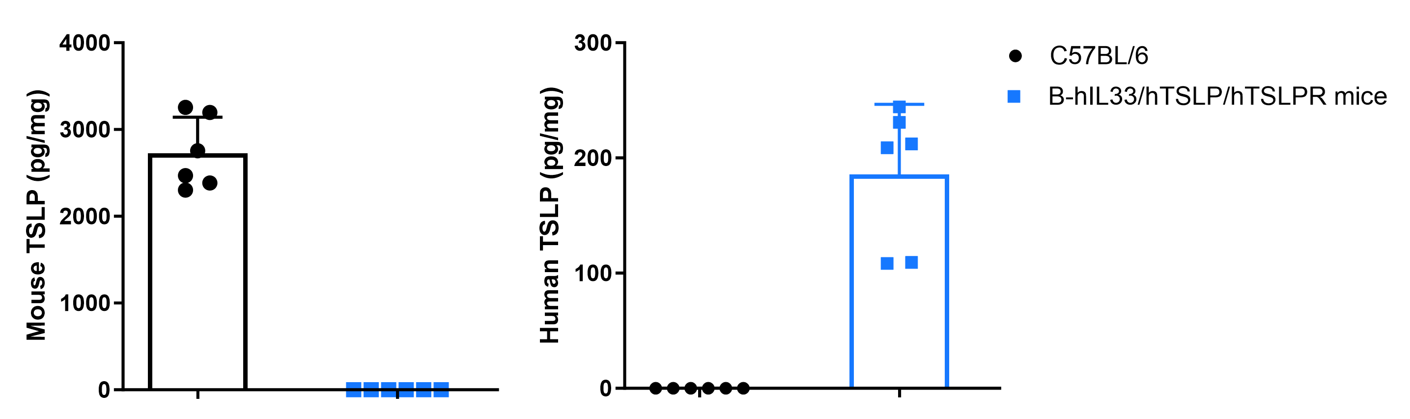
Strain-specific TSLP expression evaluated by ELISA in wild-type C57BL/6 mice and homozygous B-hIL33/hTSLP/hTSLPR mice. Calcipotriol (MC903), dissolved in ethanol, was topically applied to the ears of wild-type C57BL/6 mice and homozygous B-hIL33/hTSLP/hTSLPR mice for 7 days (male, 6-week-old, n = 3). Mouse and human TSLP levels in ear tissue homogenates were quantified by ELISA (mouse TSLP, BioLegend 434107; human TSLP, BioLegend 434207).
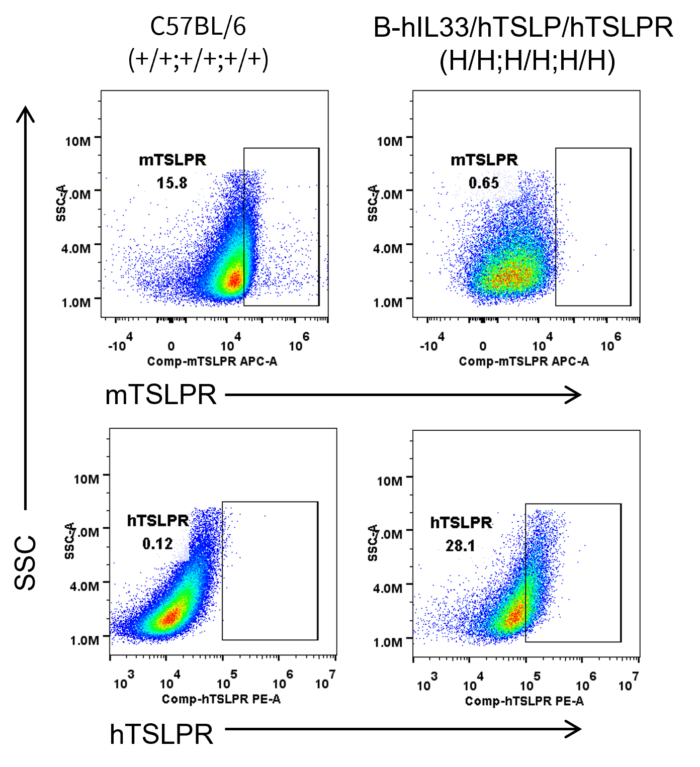
Mouse and human TSLPR expression analysis in bone marrow. Bone marrow cells were collected from wild-type C57BL/6 mice and homozygous B-hIL33/hTSLP/hTSLPR mice. TSLPR expression on bone marrow cells was analyzed by flow cytometry using anti-mouse TSLPR antibody (Biolegend, 300312) and anti-human TSLPR antibody (Biolegend, 100312).
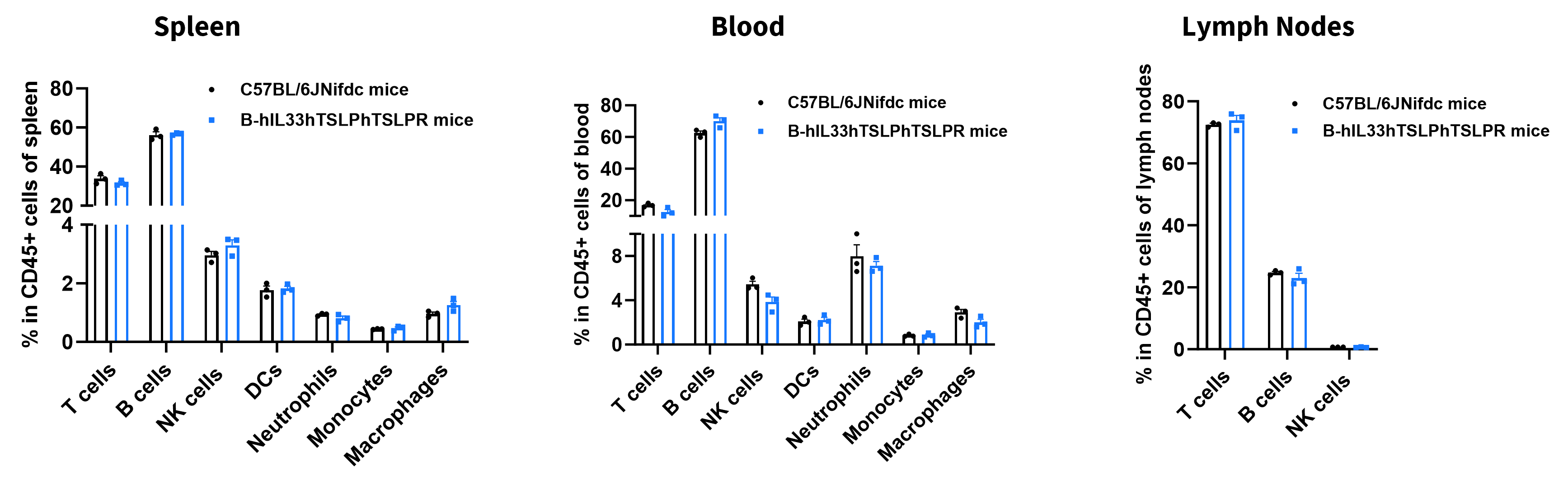
Analysis of leukocyte subpopulations by flow cytometry in immune organs and blood. Splenocytes, peripheral blood, and lymph nodes were isolated from female C57BL/6JNifdc and B-hIL33/hTSLP/hTSLPR mice (female, 9-week-old, n = 3). Single live cells were gated on the CD45⁺ population and analyzed by flow cytometry as indicated. Values are expressed as mean ± SEM.
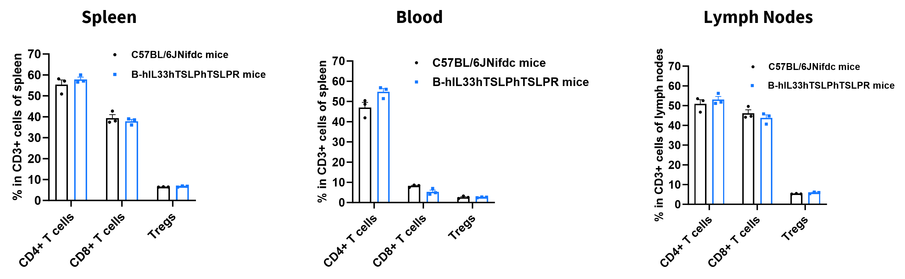
Analysis of T-cell subpopulations by flow cytometry in immune organs and blood. Splenocytes, peripheral blood, and lymph nodes were isolated from female C57BL/6JNifdc and B-hIL33/hTSLP/hTSLPR mice (female, 9-week-old, n = 3). Single live cells were gated on the CD3⁺ T-cell population and analyzed by flow cytometry as indicated. Values are expressed as mean ± SEM.
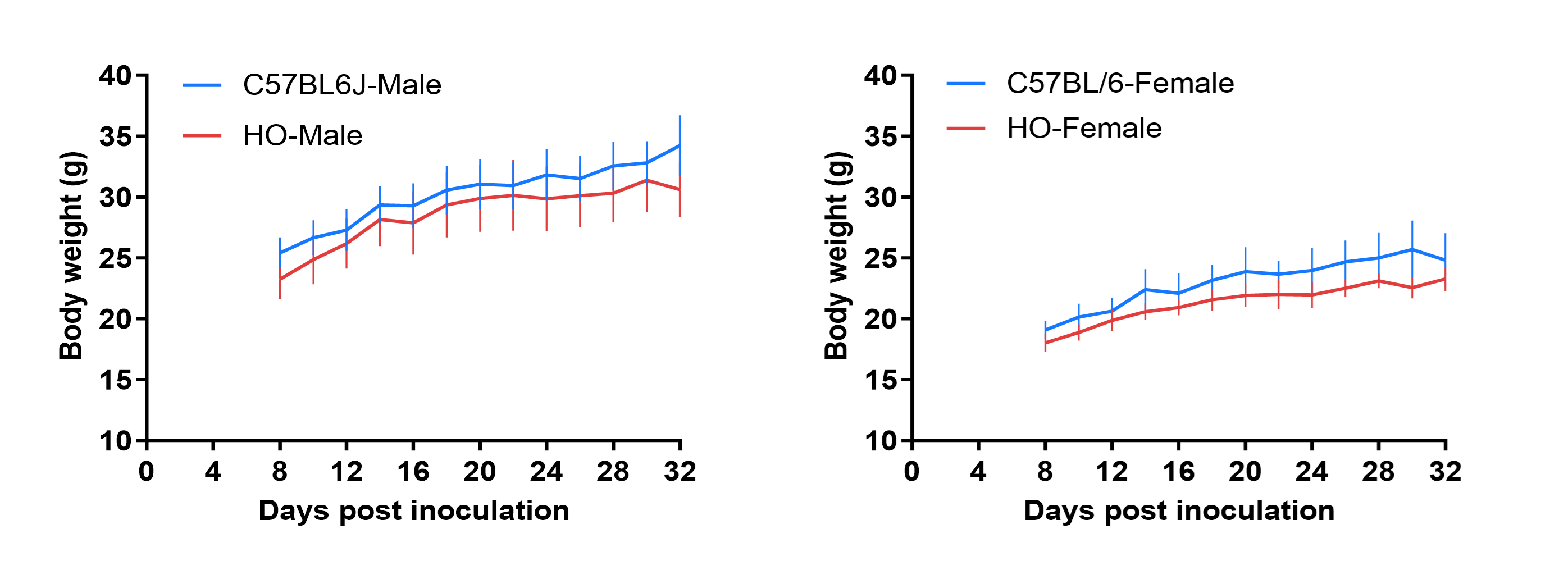
Growth curve of B-hIL33/hTSLP/hTSLPR mice. Eight-week-old mice were grouped by sex (10 males and 10 females). Body weight was measured weekly for 12 weeks on the same day each week. The minimum and maximum body weights shown in the table were calculated from the mean ± SD.
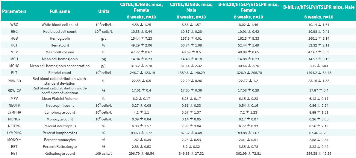
Complete blood count (CBC) of B-hIL33/hTSLP/hTSLPR mice. Values are expressed as mean ± SD.
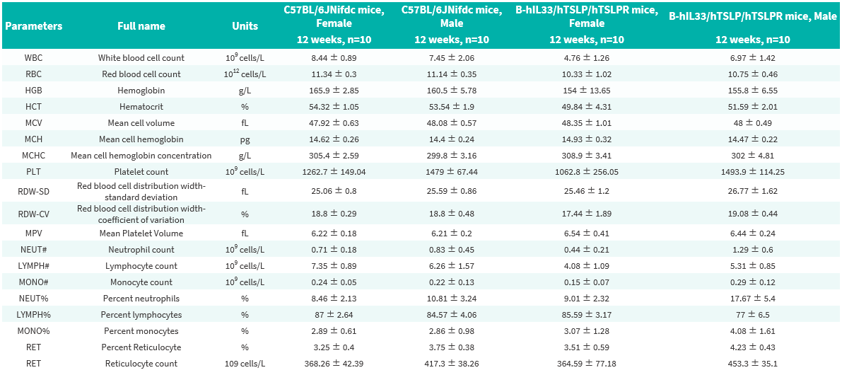
Complete blood count (CBC) of B-hIL33/hTSLP/hTSLPR mice. Values are expressed as mean ± SD.
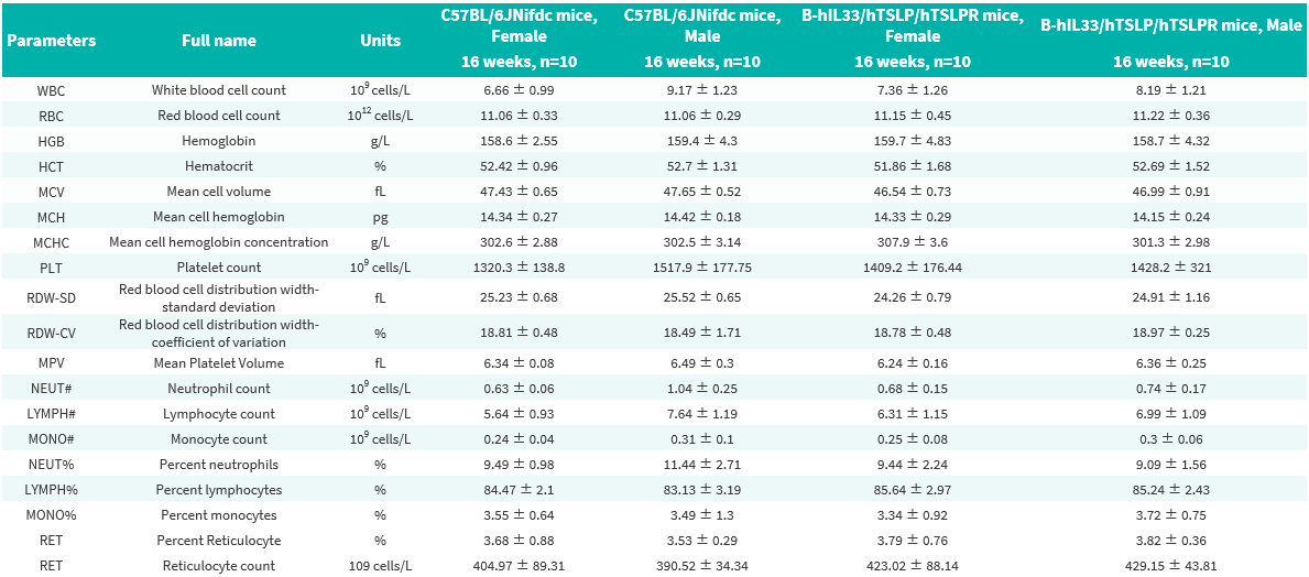
Complete blood count (CBC) of B-hIL33/hTSLP/hTSLPR mice. Values are expressed as mean ± SD.
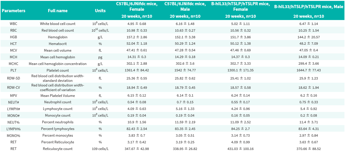
Complete blood count (CBC) of B-hIL33/hTSLP/hTSLPR mice. Values are expressed as mean ± SD.
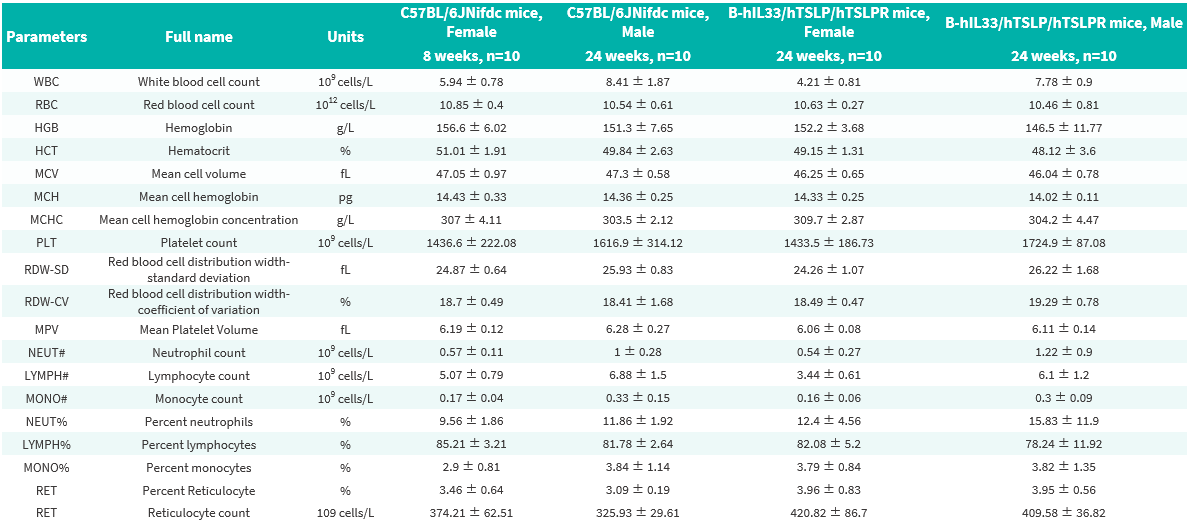
Complete blood count (CBC) of B-hIL33/hTSLP/hTSLPR mice. Values are expressed as mean ± SD.
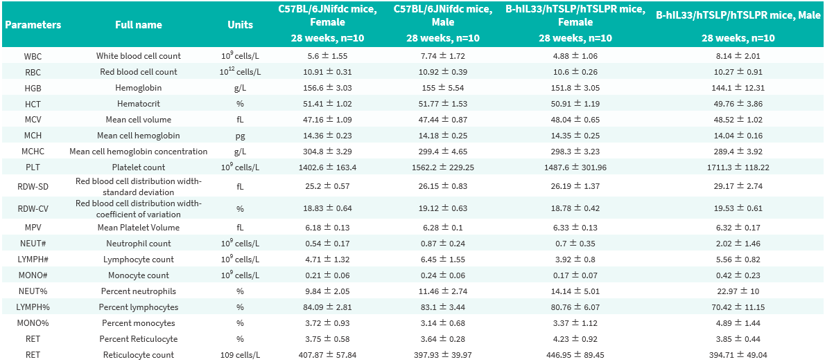
Complete blood count (CBC) of B-hIL33/hTSLP/hTSLPR mice. Values are expressed as mean ± SD.
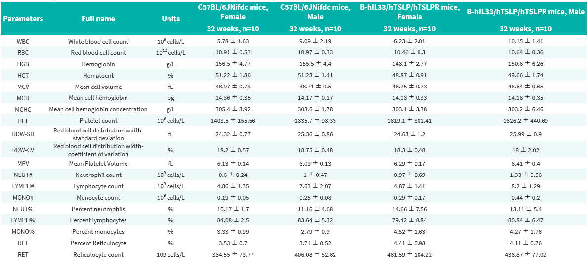
Complete blood count (CBC) of B-hIL33/hTSLP/hTSLPR mice. Values are expressed as mean ± SD.
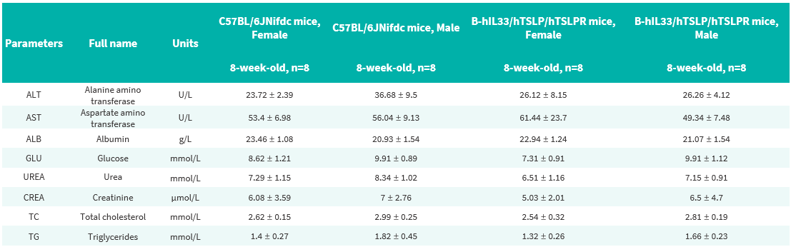
Blood biochemical parameters of B-hIL33/hTSLP/hTSLPR mice are shown. Values are expressed as mean ± SD.
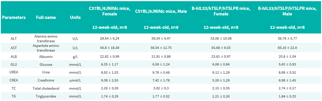
Blood biochemical parameters of B-hIL33/hTSLP/hTSLPR mice are shown. Values are expressed as mean ± SD.
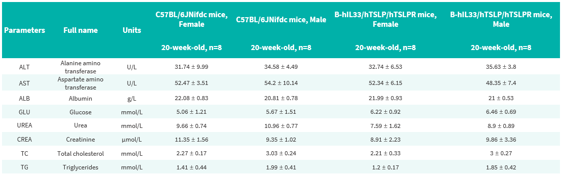
Blood biochemical parameters of B-hIL33/hTSLP/hTSLPR mice are shown. Values are expressed as mean ± SD.
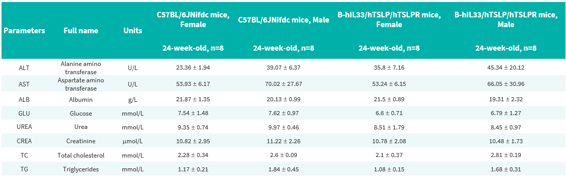
Blood biochemical parameters of B-hIL33/hTSLP/hTSLPR mice are shown. Values are expressed as mean ± SD.
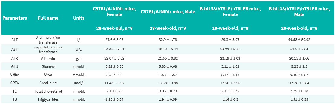
Blood biochemical parameters of B-hIL33/hTSLP/hTSLPR mice are shown. Values are expressed as mean ± SD.
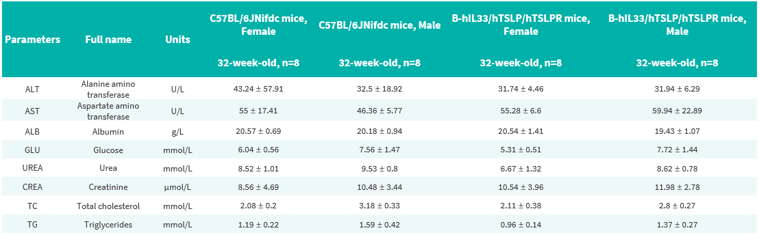
Blood biochemical parameters of B-hIL33/hTSLP/hTSLPR mice are shown. Values are expressed as mean ± SD.
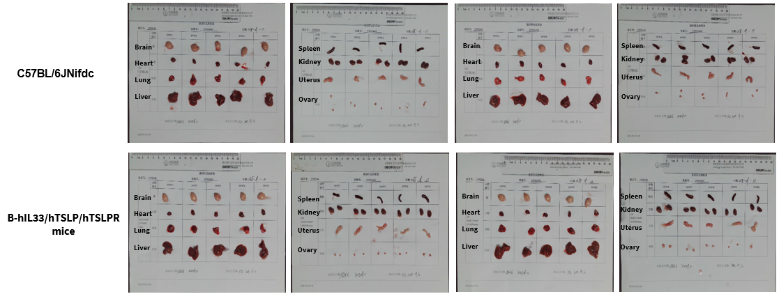
The organs of female C57BL/6JNifdc and B-hIL33/hTSLP/hTSLPR mice (32-week-old, n=10).
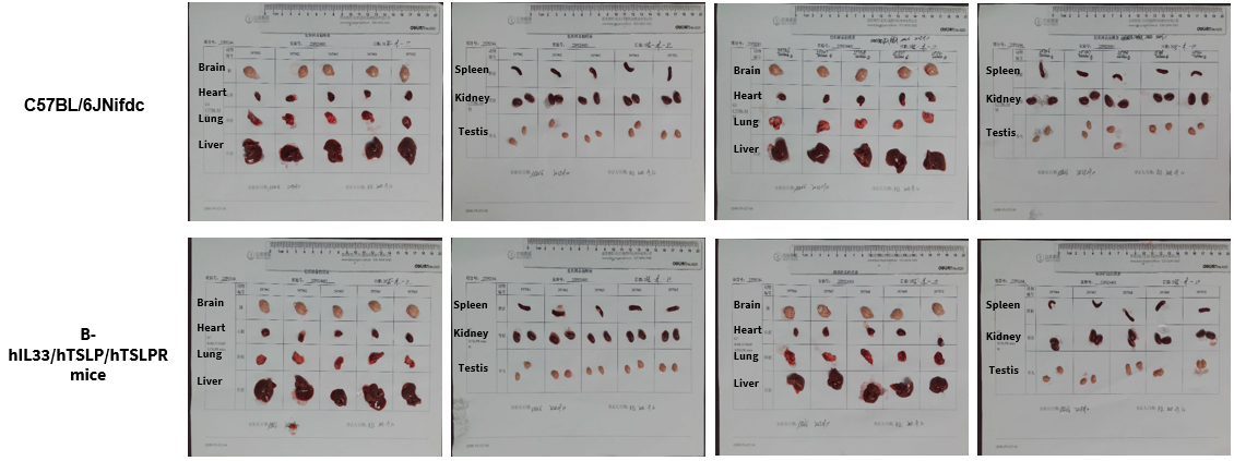
The organs of male C57BL/6JNifdc and B-hIL33/hTSLP/hTSLPR mice (32-week-old, n=10).
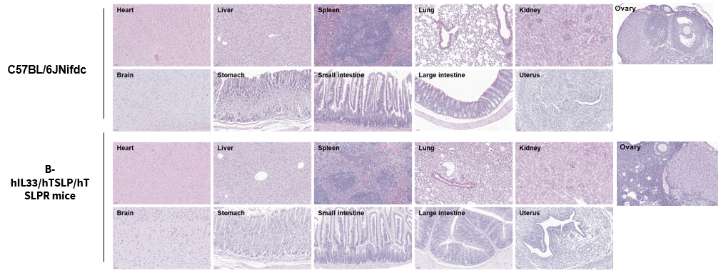
Histopathological analysis of organs in B-hIL33/hTSLP/hTSLPR mice. Major organs from B-hIL33/hTSLP/hTSLPR mice were collected at 32 weeks of age and analyzed by H&E staining (female, n = 10).
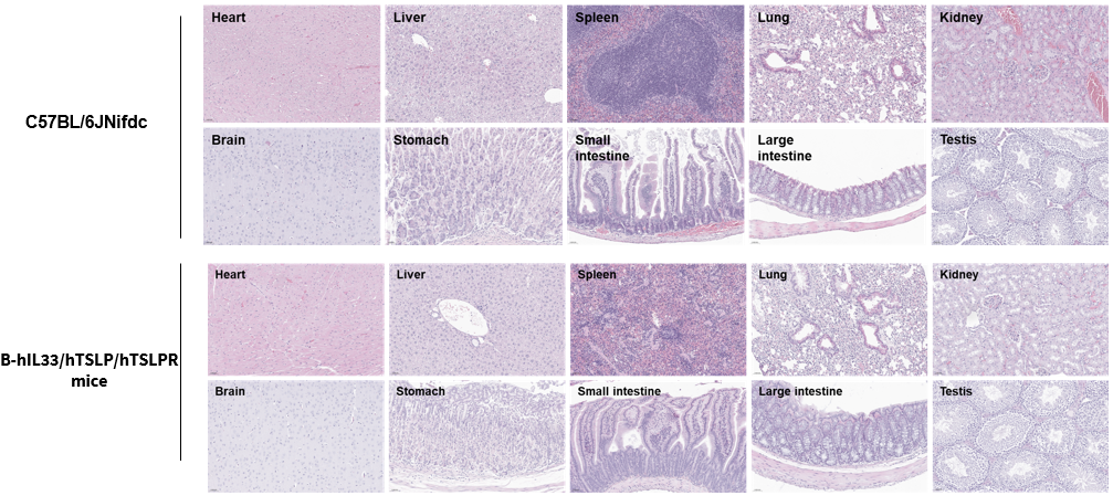
Histopathological analysis of organs in B-hIL33/hTSLP/hTSLPR mice. Major organs from B-hIL33/hTSLP/hTSLPR mice were collected at 32 weeks of age and analyzed by H&E staining (male, n = 10).

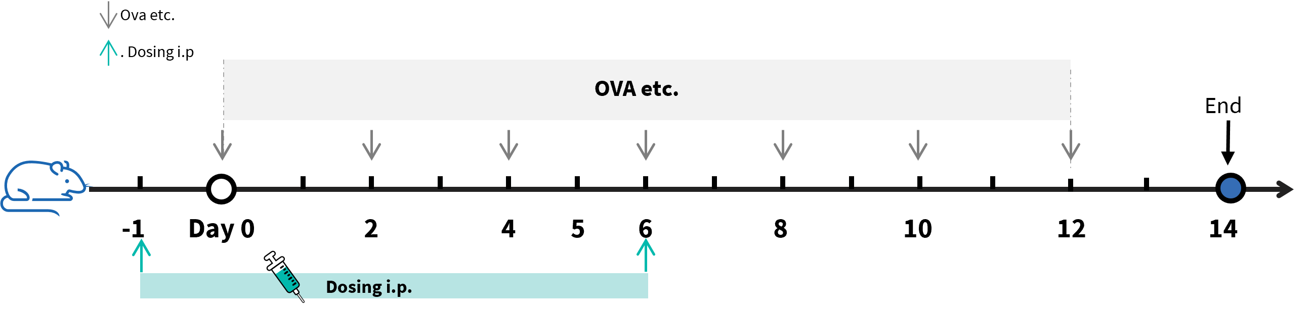
Experimental schedule for the induction of asthma and in vivo efficacy of anti-human IL33 antibody and anti-human TSLP antibody in B-hIL33/hTSLP/hTSLPR mice. In the OVA etc.-induced model, animals are administered OVA etc. intranasally to induce asthma models. The anti-human IL33 antibody itepekimab analog and anti-human TSLP antibody Tezepelumab analog (in-house) was administered by intraperitoneal injection (n=8).
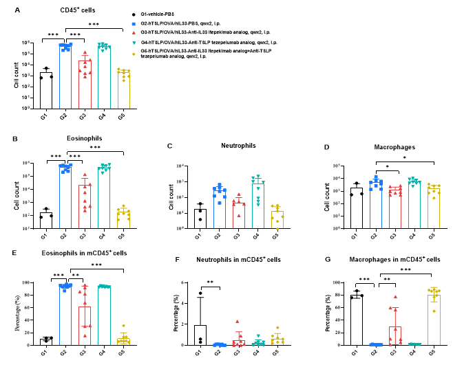
Analysis of immune cells in BALF. B-hIL33/hTSLP/hTSLPR mice (male, 11-week-old, n=8) were immunized with OVA etc. to induce asthma. Anti-human IL33 antibody (Itepekimab analog, synthesized in-house) and anti-human TSLP antibody (Tezepelumab analog, synthesized in-house) were intraperitoneally injected to B-hIL33/hTSLP/hTSLPR mice. (A&B) The number of CD45+ cells and eosinophils of BALF in the Itepekimab and the combination therapy group of Itepekimab and Tezepelumab treated groups decreased significantly compared with the OVA etc.-induced PBS treated group. Values were expressed as mean ± SEM. Significance was determined by two-way ANOVA test. *P < 0.05, **P < 0.01, ***P < 0.001.
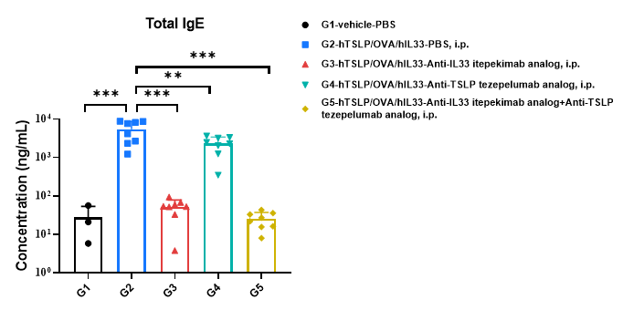
Analysis of mouse total IgE in serum. B-hIL33/hTSLP/hTSLPR mice (male, 11-week-old, n=8) were immunized with OVA etc.to induce asthma. Anti-human IL33 antibody (Itepekimab analog, synthesized in-house) and anti-human TSLP antiboday (Tezepelumab analog, synthesized in-house) were intraperitoneally injected to B-hIL33/hTSLP/hTSLPR mice. Serum was collected at the study endpoint. IgE level was analyzed by ELISA. The results showed that the levels of total IgE in mice treated with Itepekimab, Tezepelumab or the combination therapy group of Itepekimab and Tezepelumab treated groups showed a significant reduction compared with untreated mice. Values were expressed as mean ± SEM. Significance was determined by two-way ANOVA test. *P < 0.05, **P < 0.01, ***P < 0.001.
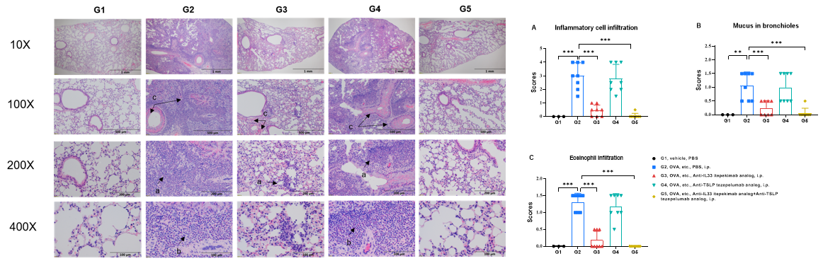
H&E staining of asthma-like model in B-hIL33/hTSLP/hTSLPR mice. Lung tissues were collected at the study endpoint and analyzed with H&E staining. The results showed that the group of mice treated with Itepekimab and the combination therapy group of Itepekimab and Tezepelumab in inflammatory infiltration and mucus secretion in lung tissue was lower than that in untreated mice, indicating that B-hIL33/hTSLP/hTSLPR mice provided a powerful preclinical model for in vivo evaluation of anti-human IL33 antibodies and anti-human TSLP antibodies. Values are expressed as mean ± SEM. Significance was determined by unpaired t-test. *P < 0.05, **P < 0.01, ***P < 0.001.
