


B-hHER2 4T1

NA • 322560

B-hHER2 4T1

Catalog Number: 322560
Strain Name: NA
Strain Background: BALB/cfC3H
---
ライセンスオプション提供可能
B-hHER2 4T1

このページで

  • Description
  • Phenotypic analysis
  • Tumorigenicity

ポスター

すべて表示

    出版物

      Description
      •  Origin: The 4T1 cell line is derived from BALB/c murine mammary carcinoma cells and is a widely used syngeneic model of stage IV human breast cancer,characterized by spontaneous tumor growth and metastatic spread.
      •  Background Information: The human epidermal growth factor receptor 2 (HER2), known as erbB-2, or proto-oncogene Neu, is a receptor tyrosine-protein kinaseencoded by the ERBB2 (HER2) gene on chromosome 17q12. ErbB2 does not bind ligand, yet appears to be the major signaling partner for other ERBB receptors byforming heteromeric complexes with ErbB1, ErbB3, or ErbB4. Upon ligand-induced dimerization, the receptors auto phosphorylate on specific tyrosine residues intheir cytoplasmic tails. These residues provide docking sites for phosphotyrosine binding, cytoplasmic signaling molecules that activate numerous intracellularsignaling pathways. HER2 overexpresses in a broad number of cancer types, such as bladder, breast, cervical, cholangio, colorectal, endometrial, esophageal, gastric,head and neck, liver, lung, ovarian, and salivary gland cancers. Especially, amplification and overexpression of HER2 occurs in 25% to 30% of the instances of human breast cancer and are associated with a poor prognosis. Several HER2-targeting therapies such as trastuzumab, pertuzumab, T-DM1, DS8201 and RC48 have been approved worldwidefor patients with HER2-positive tumors.
      •  Gene targeting strategy: The exogenous CAG promoter and human HER2 coding sequence were inserted to replace part of murine exon 2 and all of exons 3-7. Theinsertion disrupts the endogenous murine Her2 gene, resulting in a non-functional transcript.
      •  Tumorigenicity: Confirmed in B-hHER2 mice(C).
      •  Application: The B-hHER2 4T1 tumor models can be used for preclinicalevaluation of ADC and monoclonal antibody drugs targeting human HER2.
      •  Notes:

      ➢ Inoculated cell lines can be suspended with RPMI-1640 stock solution.
      ➢ Before implementing the project, it is recommended to perform tumor growth experiments. The recommended cell inoculation amount is between 1E6-3E6.
      ➢ In the experiment, it is necessary to ensure that the number of animals inoculated subcutaneously is at least 1.6 times the actual grouping number.

      HER2 Protein Expression Analysis

      • Human HER2 was detected on the surface of B-hHER2 4T1 #1-B10 cells, but not on the surface of wild-type 4T1 cells. 
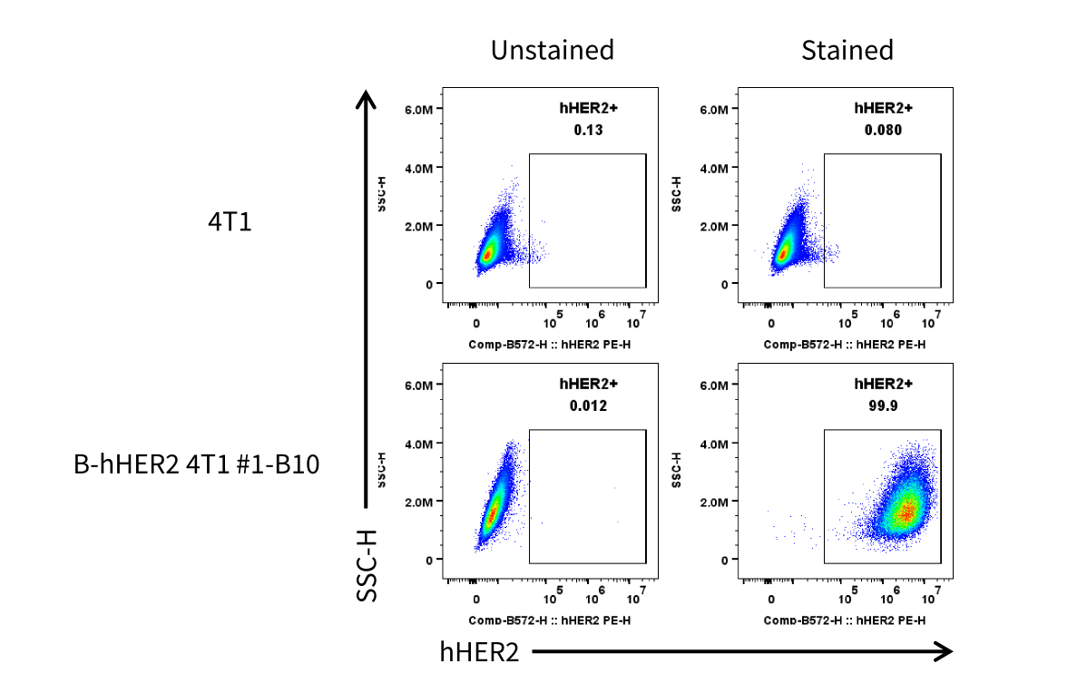
      HER2 expression analysis in B-hHER2 4T1 #1-B10 by flow cytometry. Single cell suspensions from wild-type 4T1 and B-hHER2 4T1 #1-B10 cultures were stained withhuman HER2 Antibody (BioLegend, 324406). Human HER2 was detected on the surface of B-hHER2 4T1 #1-B10 cells, but not on the surface of wild-type 4T1 cells. Theclones of B-hHER2 4T1 #1-B10 cell was used for in vivo tumor growth assays.

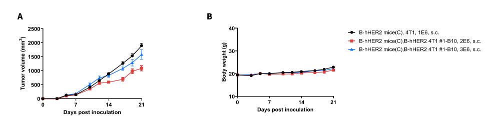
      Tumor Growth Curve & Body Weight Changes

      Subcutaneous tumor growth of B-hHER2 4T1. B-hHER2 4T1 (2×10⁶, 3×10⁶) and wild-type 4T1 cells (1×10⁶) were subcutaneously implanted into B-hHER2mice(C) (Female, 6-week-old, n=8). Tumor volume and body weight were measured twice a week. (A) Average tumor volume. (B) Body weight. Volume was expressed in mm3 using the formula: V=0.5 × long diameter × short diameter2. Results indicate that B-hHER2 4T1 cells were able to establish tumors in vivo and can be used for efficacy studies. Values are expressed as mean ± SEM.
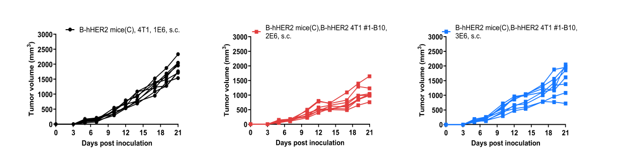
      Tumor Growth Curves of Individual Mouse

      B-hHER2 4T1 tumor growth curves from individual mouse. B-hHER2 4T1 (2×10⁶, 3×10⁶) and wild-type 4T1 cells (1×10⁶) were subcutaneously implanted into B-hHER2 mice(C) (Female, 6-week-old, n=8). Results indicate that B-hHER2 4T1 #1-B10 cells were able to establish tumors in vivo and can be used for efficacy studies. Values are expressed as mean ± SEM.

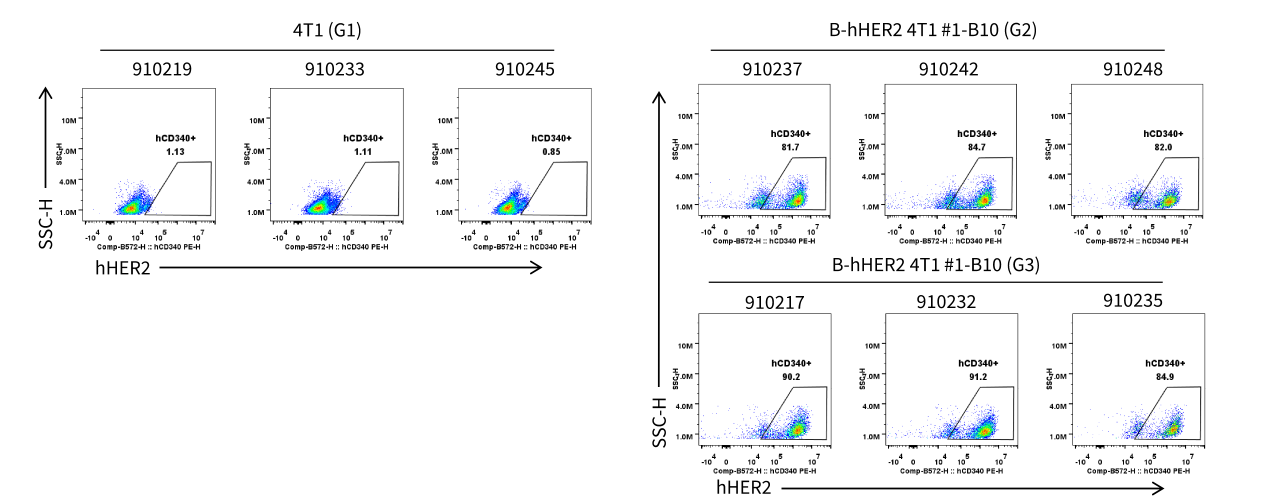
      HER2 Protein Expression Analysis of Tumor Tissue

      • Human HER2 was detected on the surface of B-hHER2 4T1 tumors cells.
      HER2 expression was evaluated on B-hHER2 4T1 by flow cytometry. These cells were subcutaneously transplanted into B-hHER2 mice(C) (n=8). At the end of the experiment, tumor cells were harvested and analyzed with species-specific anti-HER2 antibody (Biolegend, 324406) expression by flow cytometry.
