B-hCD3EDG/hHER2 mice

C57BL/6-Cd3etm1(CD3E)Bcgen Cd3dtm1(CD3D)Bcgen Cd3gtm1(CD3G)Bcgen Erbb2tm1(ERBB2)Bcgen/Bcgen • 114367

B-hCD3EDG/hHER2 mice

Catalog Number: 114367
Strain Name: C57BL/6-Cd3etm1(CD3E)Bcgen Cd3dtm1(CD3D)Bcgen Cd3gtm1(CD3G)Bcgen Erbb2tm1(ERBB2)Bcgen/Bcgen
Strain Background: C57BL/6
NCBI gene ID: 916,915,917,2064 (Human)
Aliases: CD3epsilon, IMD18, T3E, TCRE; CD3-DELTA, CD3DELTA, IMD19, T3D; CD3-GAMMA, CD3GAMMA, IMD17, T3G; CD340, HER-2, HER-2/neu, HER2, MLN 19, MLN-19, NEU, NGL, TKR1, VSCN2, c-ERB-2, c-ERB2, p185(erbB2)
---
ライセンスオプション提供可能
B-hCD3EDG/hHER2 mice

このページで

  • Description
  • Phenotypic analysis
  • Efficacy

ポスター

すべて表示

    出版物

      Description

      CD3×HER2 Bispecific Antibody-Based Therapy

      • Gene Information: CD3 is encoded by CD3E, CD3D, and CD3G; it is part of the Ig superfamily and forms the essential signaling backbone of the T cell receptor (TCR) complex. HER2 is a transmembrane receptor tyrosine kinase located on human chromosome 17q12. It belongs to the epidermal growth factor receptor (EGFR) family and plays a critical role in cell growth, differentiation, and survival across various epithelial tissues.
      • Protein Expression: CD3 is a constitutive and universal marker for all mature T cells (CD4+ and CD8+) and is always present on the cell surface. HER2 is expressed in a wide range of epithelial tissues, including the breast, gastrointestinal tract, respiratory tract, and reproductive systems. Its expression is typically low to moderate under normal physiological conditions but is markedly overexpressed or amplified in specific human cancers (such as HER2-positive breast and gastric cancers).
      • Signaling Pathway: CD3 operates via ITAM phosphorylation and ZAP-70. It triggers the initial "on" switch, Ca2+ flux, and immediate cytotoxicity. HER2 is a key member of the EGFR family. By forming homodimers or heterodimers (preferably with HER3 or EGFR), it auto-phosphorylates intracellular tyrosine residues and activates downstream signaling cascades, including the PI3K/AKT and MAPK/ERK pathways.
      • Therapeutic Inhibition: CD3×HER2 bispecific antibodies recruit and activate T cells by simultaneously binding CD3 on T cells and the extracellular domain of HER2 on tumor cells, leading to the targeted killing of HER2-positive malignant cells. This approach is actively being evaluated in HER2-overexpressing solid tumors, with multiple candidates currently undergoing early-to-mid-stage clinical trials. Ongoing efforts focus on optimizing tumor selectivity, mitigating on-target off-tumor toxicity, and enhancing therapeutic durability.
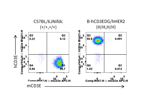
      CD3E Protein Expression in Spleen

      Mouse and human CD3E expression analysis in splenocytes by flow cytometry. Splenocytes were collected from wild-type C57BL/6JNifdc mice (+/+, +/+) and homozygous B-hCD3EDG/hHER2 mice (H/H, H/H) (female, 6-week-old, n=1). CD3E expression on T cells was analyzed by flow cytometry using species-specific anti-CD3E antibodies (anti-human CD3E antibody, BD Horizon, 562426; anti-mouse CD3E antibody, Biolegend, 100312).
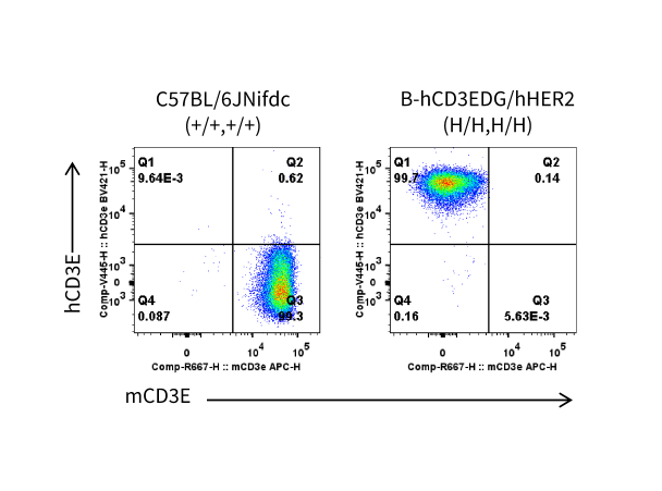
      CD3E Protein Expression in Blood

      Mouse and human CD3E expression analysis in blood by flow cytometry. Blood was collected from wild-type C57BL/6JNifdc mice (+/+, +/+) and homozygous B-hCD3EDG/hHER2 mice (H/H, H/H) (female, 6-week-old, n=1). CD3E expression on T cells was analyzed by flow cytometry using species-specific anti-CD3E antibodies (anti-human CD3E antibody, BD Horizon, 562426; anti-mouse CD3E antibody, Biolegend, 100312).
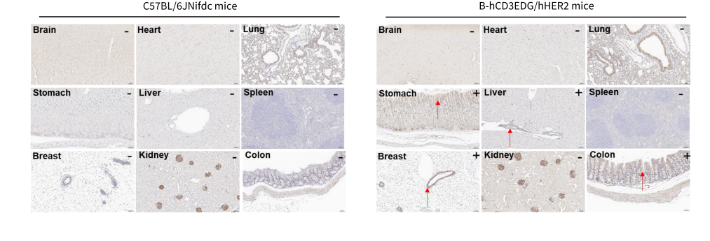
      HER2 Protein Expression

      Immunohistochemical (IHC) analysis of HER2 protein expression in wild-type mice and homozygous B-hCD3EDG/hHER2 mice. Major tissues were collected from wild-type C57BL/6JNifdc mice and homozygous B-hCD3EDG/hHER2 mice and analyzed by IHC with an anti-human HER2 antibody (HUABIO, HA721178). This antibody exhibits significant non-specific binding in the kidney and lung. The arrow indicates tissue cells with positive HER2 staining (brown). "+" indicates that the tissue is positive, and "-" indicates that the tissue is negative.

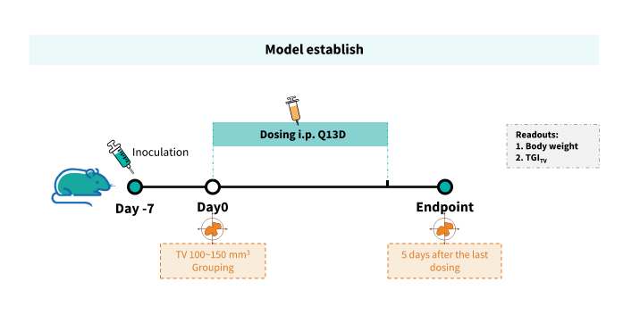
      In Vivo Efficacy of Anti-hCD3/hHER2 BsAb

      Establishment of a B-hHER2 MC38 model and in vivo efficacy study of an anti-human CD3/HER2 bispecific antibody. B-hHER2 MC38 plus cells were implanted subcutaneously into homozygous B-hCD3EDG/hHER2 mice (female, 8-weeks-old, n=6). When the average tumor volume reached  approximately 100~150 mm³, mice were randomized and subsequently administered the anti-hCD3/hHER2 BsAb (in-house) via intraperitoneal injection.
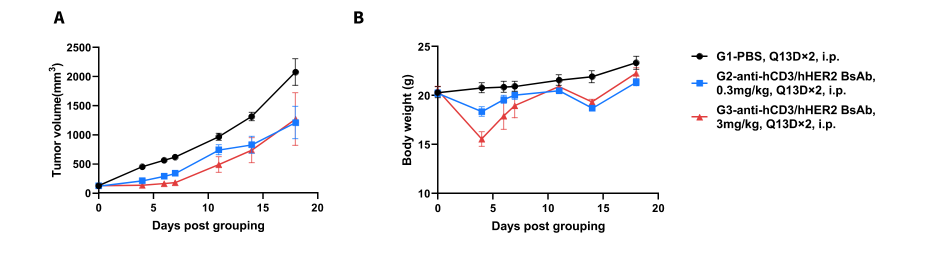
      Efficacy Evaluation of anti-hCD3/hHER2 BsAb in the Treatment of the Subcutaneous B-hHER2 MC38 Model in B-hCD3EDG/hHER2 Mice

      Establishment of a B-hHER2 MC38 model and in vivo efficacy study of an anti-human CD3/HER2 bispecific antibody. (A) Tumor growth curves. (B) Body weight changes during treatment. As shown in panel A, the anti-hCD3/hHER2 BsAb (in-house) inhibited B-hHER2 MC38 plus tumor growth in B-hCD3EDG/hHER2 mice, demonstrating that B-hCD3EDG/hHER2 mice provide a powerful preclinical model for the in vivo evaluation of anti-hCD3/hHER2 BsAbs. In group 3, one mouse died on day 4, and another mouse died on day 5.

      * When publishing results obtained using this animal model, please acknowledge the source as follows: The animal model [B-hCD3EDG/hHER2 mice] (Cat# 114367) was purchased from Biocytogen.