

C57BL/6-Ifngr1tm1(IFNGR1)BcgenIfngtm1(IFNG)BcgenIfngr2tm1(IFNGR2)Bcgen/Bcgen • 130949

IFNG
•The full coding sequence of mouse Ifng gene was replaced by counterpart of human IFNG gene in B-hIFNGR1/hIFNG/hIFNGR2 mice.
•Expression of the human IFNG protein is driven by the native mouse Ifng promoter, while endogenous mouse Ifng transcription and translation are disrupted.
IFNGR1
•The sequence that encoding the extracellular region of mouse Ifngr1 gene was replaced by counterpart human IFNGR1 gene in B-hIFNGR1/hIFNG/hIFNGR2 mice.
•Expression of the human IFNGR1 protein is driven by the native mouse Ifngr1 promoter, while endogenous mouse Ifngr1 transcription and translation are disrupted.
•
IFNGR2
•The sequence that encoding the extracellular region of mouse Ifngr2 gene was replaced by counterpart human IFNGR2 gene in B-hIFNGR1/hIFNG/hIFNGR2 mice.
•Expression of the human IFNGR2 protein is driven by the native mouse Ifngr2 promoter, while endogenous mouse Ifngr2 transcription and translation are disrupted.
B-hIFNGR1/hIFNG/hIFNGR2 mice (130949) was obtained by cross breeding B-hIFNGR1/hIFNG mice (120819) with B-hIFNGR2 mice (110803).

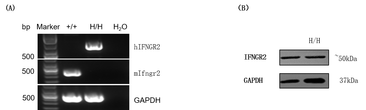
Strain specific analysis of IFNGR2 gene expression in wild-type mice and B-hIFNGR2 mice by RT-PCR and western blotting. (A) mRNA expression. Mouse Ifngr2 mRNA was exclusively detectable in the small intestine of wild-type mice (+/+). Human IFNGR2 mRNA was exclusively detectable in homozygous B-hIFNGR2 mice (H/H). (B) Protein expression. Skeletal muscle was collected from wild-type C57BL/6 mice (+/+) and homozygous B-hIFNGR2 mice (H/H), and analyzed by western blotting with anti-IFNGR2 antibody which can recognize both human and mouse IFNGR2. The results show that IFNGR2 protein can be detected in homozygous B-hIFNGR2 mice.
•Human IFNγ was detected in homozygous B-hIFNGR1/hIFNG/hIFNGR2 mice, but not in wild-type mice.

Analysis of hIFNγ production in activated splenocytes from homozygous B-hIFNGR1/hIFNG/hIFNGR2 mice (Triple-KI).
Cells were seeded in a 96-well plate for 5 × 10^5 cells/well. The plate was precoated with 8μg/mL anti-mCD3 mAb at 4 ℃ for overnight. Then incubated the cells at 37℃ 5% CO2 for 5 days. Anti-CD28 at 1μg/mL and 100U hIL2 were added 16 hours after culture. Get the supernatant for ELISA analysis. hIFNγ was detected in triple-KI mice, but not in wild-type mice.
*The experiment is verified by the cooperation partner.
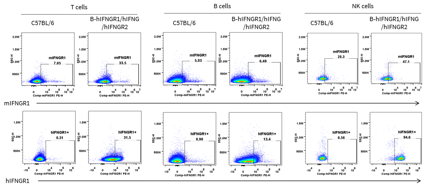
IFNGR1 expression analysis in wild-type C57BL/6 mice, homozygous B-hIFNGR1/IFNG/hIFNGR2 mice by flow cytometry.
Splenocytes were collected from wild-type C57BL/6 mice and homozygous B-hIFNGR1/hIFNG/hIFNGR2 mice, and analyzed by flow cytometry with anti-IFNGR1 antibody. Human IFNGR1 was detectable on T cells, B cells and NK cells of B-hIFNGR1/hIFNG/hIFNGR2 mice.
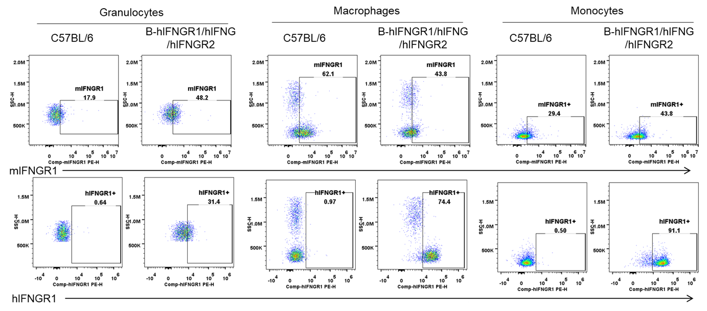
IFNGR1 expression analysis in wild-type C57BL/6 mice, homozygous B-hIFNGR1/IFNG/hIFNGR2 mice by flow cytometry.
Splenocytes were collected from wild-type C57BL/6 mice and homozygous B-hIFNGR1/hIFNG/hIFNGR2 mice, and analyzed by flow cytometry with anti-IFNGR1 antibody. Human IFNGR1 was detectable on granulocytes, macrophages and monocytes of B-hIFNGR1/hIFNG/hIFNGR2 mice.
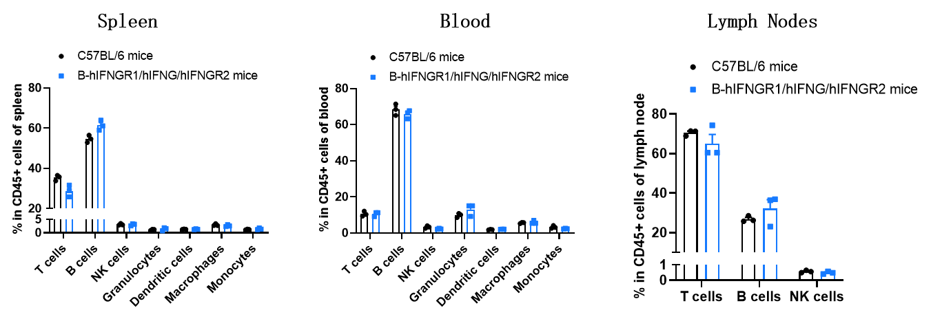
Analysis of leukocyte subpopulations by flow cytometry in immune organs and blood. Splenocytes, peripheral blood, and lymph nodes were isolated from female C57BL/6 and B-hIFNGR1/hIFNG/hIFNGR2 mice (female, 9-week-old, n = 3). Single live cells were gated on the CD45⁺ population and analyzed by flow cytometry as indicated. Values are expressed as mean ± SEM.
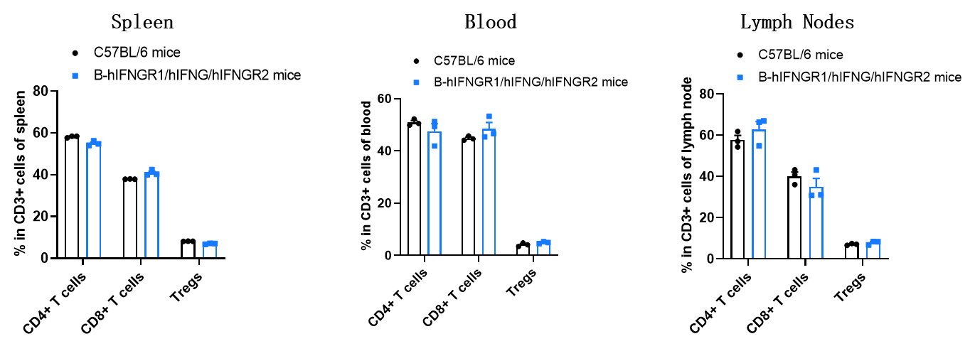
Analysis of T-cell subpopulations by flow cytometry in immune organs and blood. Splenocytes, peripheral blood, and lymph nodes were isolated from female C57BL/6 and B-hIFNGR1/hIFNG/hIFNGR2 mice (female, 9-week-old, n = 3). Single live cells were gated on the CD3⁺ T-cell population and analyzed by flow cytometry as indicated. Values are expressed as mean ± SEM.
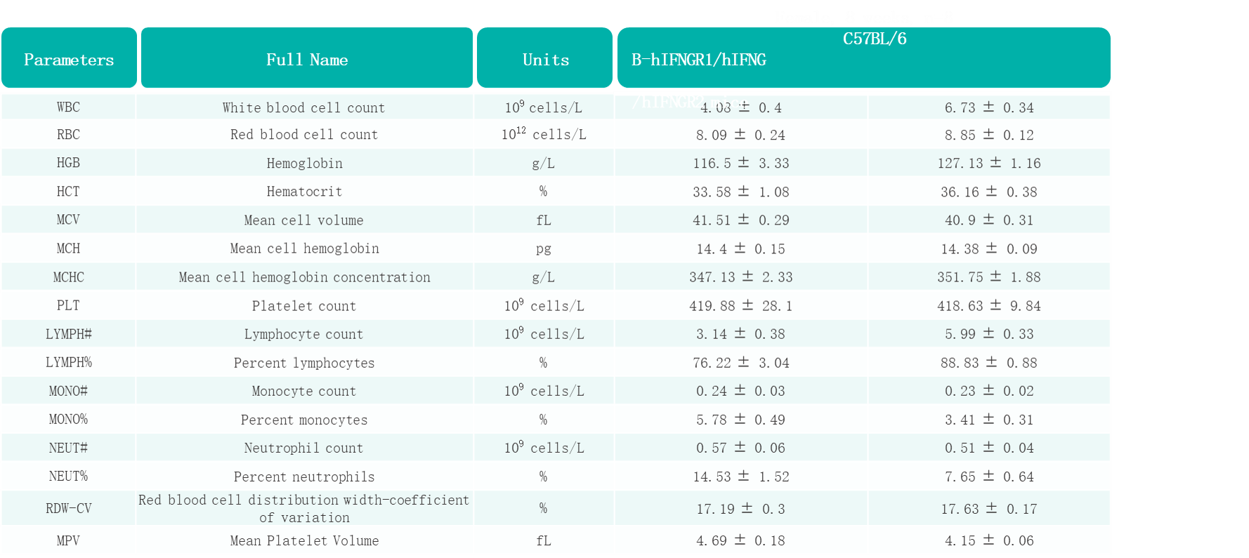
Complete blood count (CBC) of B-hIFNGR1/hIFNG/hIFNGR2 mice. Values are expressed as mean ± SD.
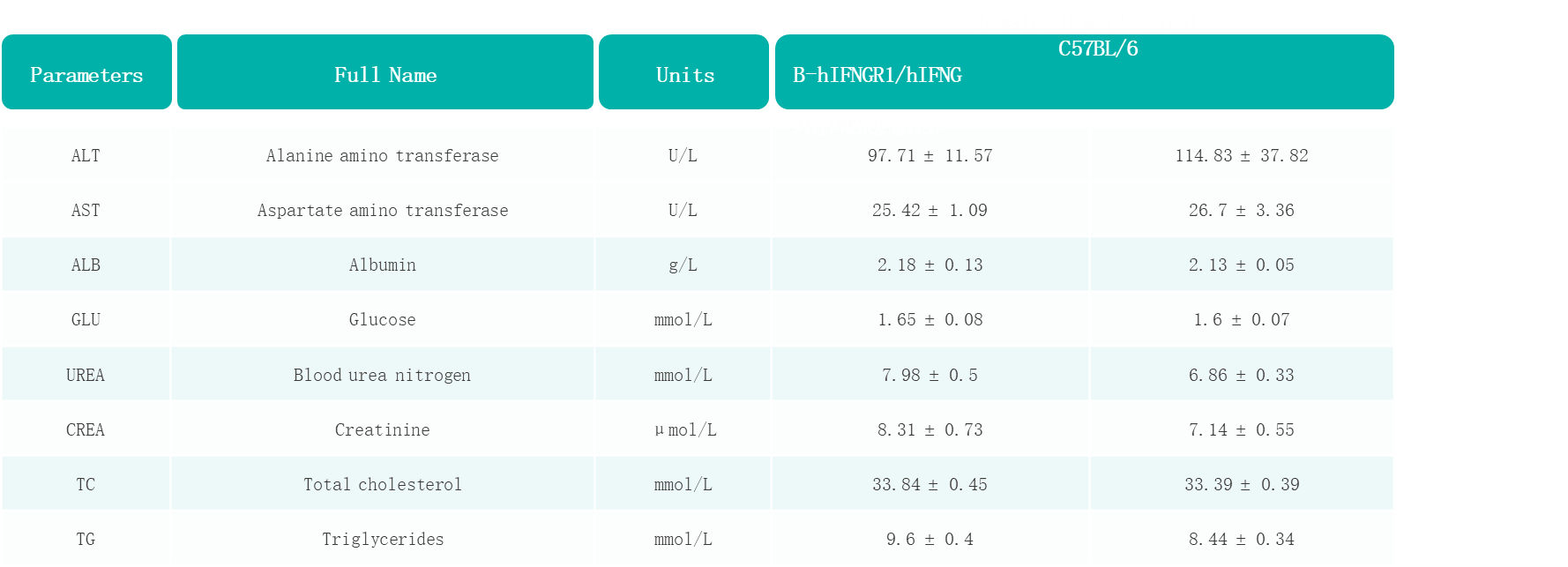
Blood biochemical parameters of B-hIFNGR1/hIFNG/hIFNGR2 mice are shown. Values are expressed as mean ± SD.
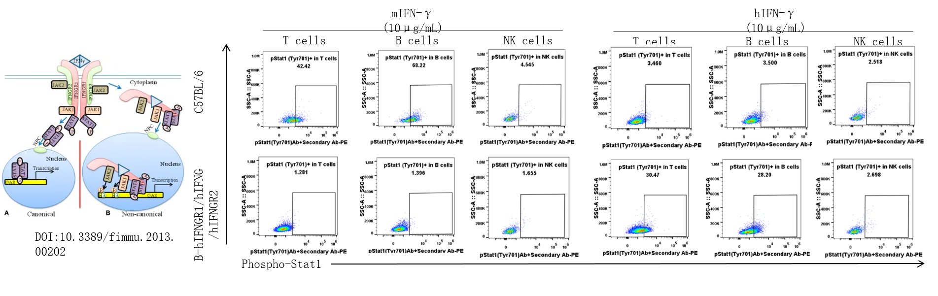
Analysis of the IFNγ-induced pSTAT1(Tyr701) in wild-type C57BL/6 mice and homozygous B-hIFNGR1/hIFNG/hIFNGR2 mice by flow cytometry.
Splenocytes from wild-type mice and homozygous B-hIFNGR1/hIFNG/hIFNGR2 mice (female, 7-week-old) were treated with IFNγ for ex vivo, and then flow cytometry was performed to analyze the phosphorylation of STAT1. Mouse IFN-γ induced the expression of pSTAT1 in T and B cells of wild-type mice but not humanized mice. Human IFN-γ induced the expression of pSTAT1 in T and B cells of humanized mice but not wild-type mice. Among NK cells, IFN-γ did not induce the STAT1 phosphorylation in both wild-type mice and homozygous B-hIFNGR1/hIFNG/hIFNGR2 mice.
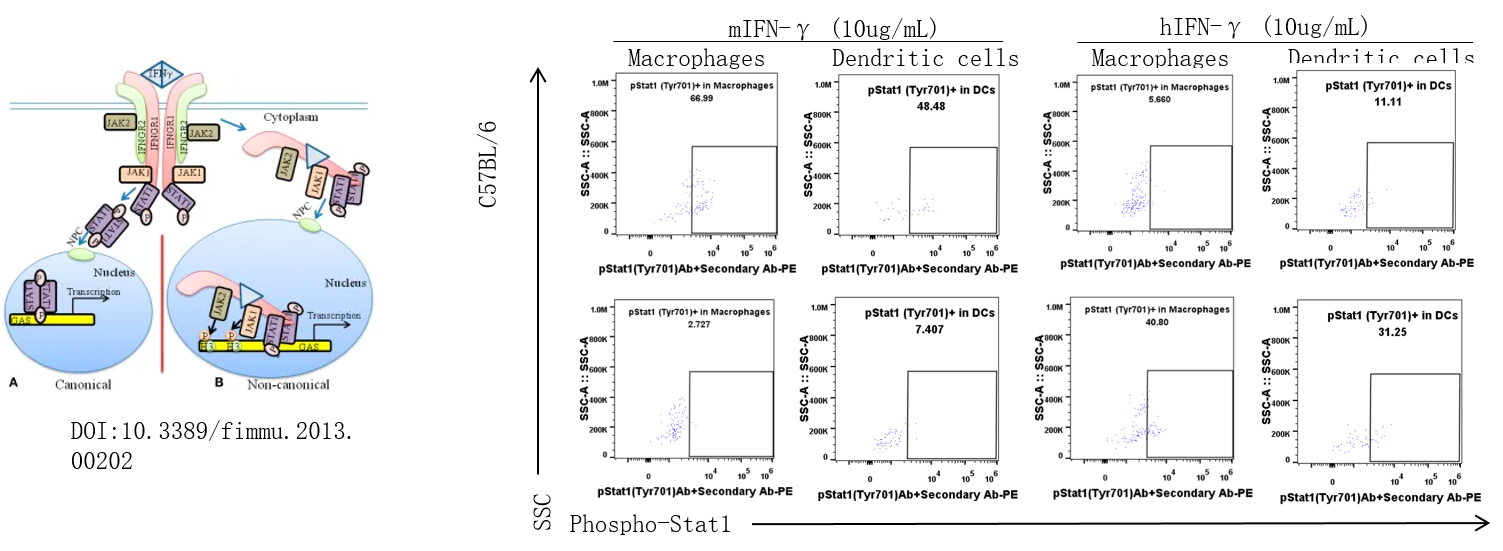
Analysis of the IFNγ induced pSTAT1(Tyr701) in wild-type C57BL/6 mice and homozygous B-hIFNGR1/hIFNG/hIFNGR2 mice by flow cytometry.
Splenocytes from wild-type mice and homozygous B-hIFNGR1/hIFNG/hIFNGR2 mice (female, 7-week-old) were treated with IFNγ for ex vivo, and then flow cytometry was performed to analyze the phosphorylation of STAT1. Mouse IFN-γ induced the expression of pSTAT1 in macrophages and dendritic cells of wild-type mice but not humanized mice. Human IFN-γ induced the expression of pSTAT1 in macrophages and dendritic cells of humanized mice but not wild-type mice.
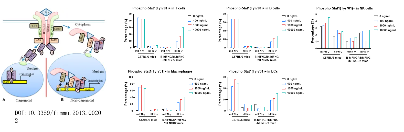
Analysis of the phosphorylation of stat1 in B-hIFNGR1/hIFNG/hIFNGR2 mice by flow cytometry. Splenocytes from wild-type mice and homozygous B-hIFNGR1/hIFNG/hIFNGR2 mice (female, 7-week-old) were treated with IFNγ for ex vivo, and then flow cytometry was performed to analyze the phosphorylation of STAT1. Mouse IFN-γ induced the expression of pSTAT1 in T cells, B cells, macrophages, and dendritic cells of wild-type mice but not humanized mice. Human IFN-γ induced the expression of pSTAT1 in T cells, B cells, macrophages, and dendritic cells of humanized mice but not wild-type mice. Among NK cells, IFN-γ did not induce the STAT1 phosphorylation in both wild-type mice and homozygous B-hIFNGR1/hIFNG/hIFNGR2 mice.
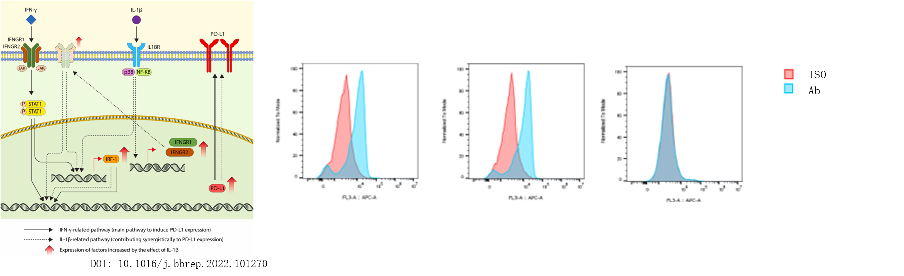
hIFNγ upregulated mPD-L1 in splenocytes from homozygous B-hIFNGR1/hIFNG/hIFNGR2 mice (Triple-KI).
Splenocytes at 0.5x10⁶/well (96-well plate) were treated with 100ng/mL hIFNγ for 24 hours at 37℃. Cells were collected and assessed for mPD-L1 expression by flow cytometry. As shown, hIFNγ induced the expression of mPD-L1 in triple-KI mice but not in wild-type mice.
*The experiment is verified by the cooperation partner.
