


B-hIL15/hIL15RA mice

C57BL/6-Il15tm1(IL15)BcgenIl15ratm1(IL15RA)Bcgen/Bcgen • 121217

B-hIL15/hIL15RA mice

Catalog Number: 121217
Strain Name: C57BL/6-Il15tm1(IL15)BcgenIl15ratm1(IL15RA)Bcgen/Bcgen
Strain Background: C57BL/6
NCBI gene ID: 3600,3601 (Human)
Aliases: IL-15; CD215
---
ライセンスオプション提供可能
B-hIL15/hIL15RA mice

このページで

  • Description
  • Targeting strategy
  • Phenotypic analysis
  • Efficacy

ポスター

すべて表示

    出版物

      Description

      IL15/IL15RA: A cytokine-chaperone complex driving lymphocyte survival and activation.

      • Gene Information: IL15 (chromosome 4q31) encodes a four-helix bundle cytokine, while IL15RA (chromosome 10p15) encodes its high-affinity alpha-receptor subunit, which structurally stabilizes and facilitates interleukin-15 signaling.
      • Protein Expression: Both proteins are co-expressed predominantly by monocytes, macrophages, and dendritic cells, where they physically associate intracellularly before being presented as a complex on the cell surface.
      • Signaling Pathway: The IL-15/IL15RA complex binds to IL2RB/IL2RG heterodimers on NK and CD8+ T cells in trans, activating JAK1/JAK3 and downstream STAT3/STAT5 pathways to promote proliferation.
      • Therapeutic Inhibition: Blocking IL15 axis mitigates tissue destruction in celiac disease and rheumatoid arthritis, whereas administering the intact complex or engineered agonists serves to enhance anti-tumor immunity.
      Targeting strategy

      IL15

      • Exons 3–8 of the mouse Il15 gene that encode the entire protein (from ATG to stop codon) were replaced with the corresponding human sequences in B-hIL15/hIL15RA mice.
      • The endogenous mouse promoter, 5′ UTR, and 3′ UTR regions were retained, allowing human IL15 expression to be driven by the native mouse Il15 promoter, while endogenous mouse Il15 transcription and translation are abolished.

      IL15RA

      • Exons 2-6 of mouse IL15RA gene that encode extracellular domain were replaced by human counterparts in B-hIL15/hIL15RA mice.
      • The genomic region of mouse Il15ra gene that encodes signal peptide, transmembrane domain and cytoplasmic portion was retained. The promoter, 5’UTR and 3’UTR region of the mouse gene were also retained. The chimeric IL15RA expression was driven by endogenous mouse Il15ra promoter, while mouse Il15ra gene transcription and translation will be disrupted.

      B-hIL15/hIL15RA mice (121217) were obtained by mating B-hIL15 mice (110762) with B-hIL15RA mice (110112).

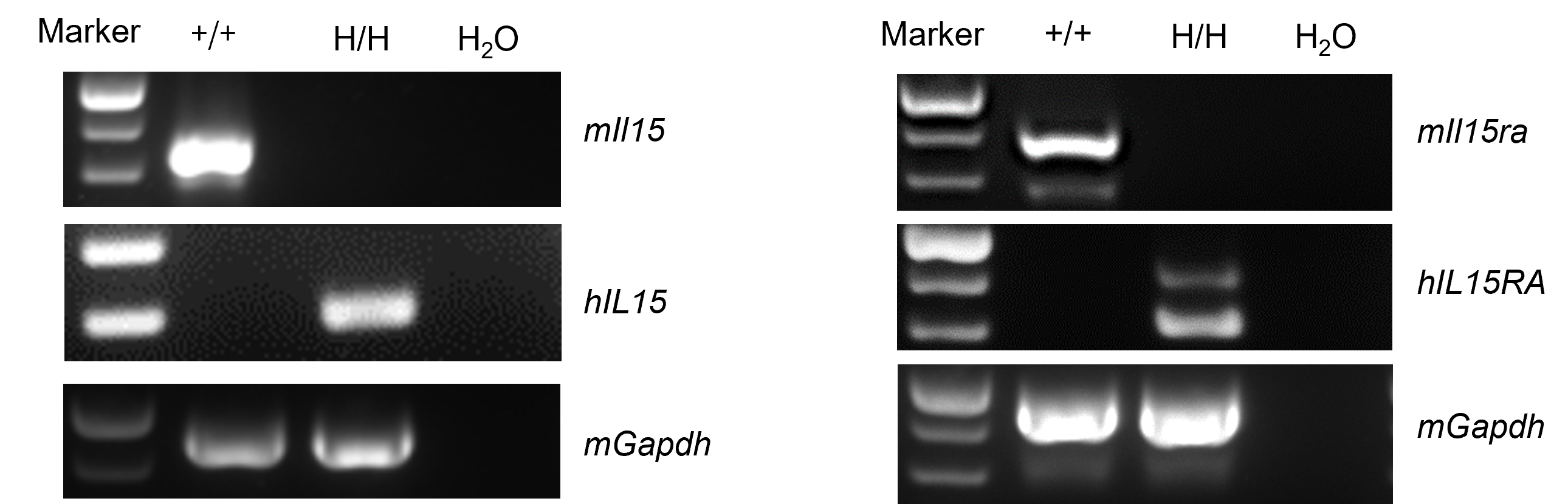
      mRNA Expression Analysis
      • Mouse Il15 and Il15ra mRNA were detected in spleen of wild-type C57BL/6 mice.
      • Human IL15 and IL15RA mRNA were detected in spleen of homozygous B-hIL15/hIL15RA mice, but not in wild-type C57BL/6 mice.

      Species specific analysis of IL15 and IL15RA gene expression in wild-type C57BL/6 mice and homozygous humanized B-hIL15/hIL15RA mice by RT-PCR. Spleen RNA was were isolated from wild-type C57BL/6 mice (+/+) and homozygous B-hIL15/hIL15RA mice (H/H), and then cDNA libraries were synthesized by reverse transcription, followed by PCR with mouse Il15 primers, human IL15 primers, mouse Il15ra primers and human IL15RA primers.
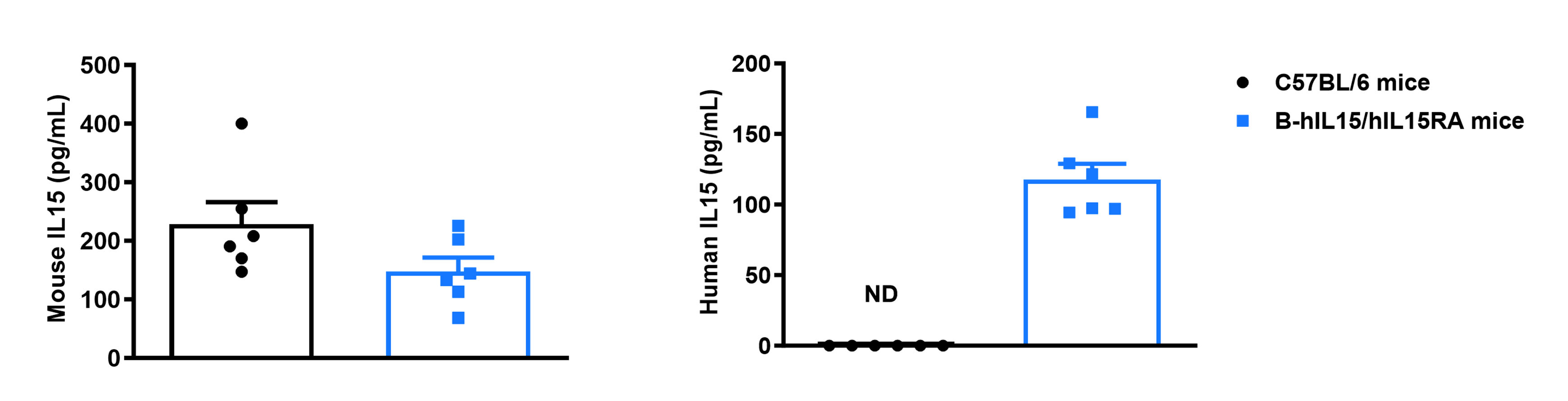
      IL15 Protein Expression Analysis
      • Mouse IL15 was detected in serum of wild-type C57BL/6 mice and homozygous B-hIL15/hIL15RA mice due to the cross-reactivity of antibodies.
      • Human IL15 was detected in serum of homozygous B-hIL15/hIL15RA mice, but not in wild-type C57BL/6 mice.

      Strain specific IL15 expression analysis in wild-type C57BL/6 mice and homozygous humanized B-hIL15/hIL15RA mice by ELISA. Serum was collected from wild-type C57BL/6 mice and homozygous B-hIL2RB/hIL2RG/hIL15/hIL15RA mice (female, 8-week-old, n = 3) stimulated with APAP‌ for 24 hours. Expression level of mouse and human IL15 were analyzed by ELISA (anti-mouse IL15 kit: Abcam, ab275898; anti-human IL15 kit: RD, D1500). Values are expressed as mean ± SEM. ND: not detectable.
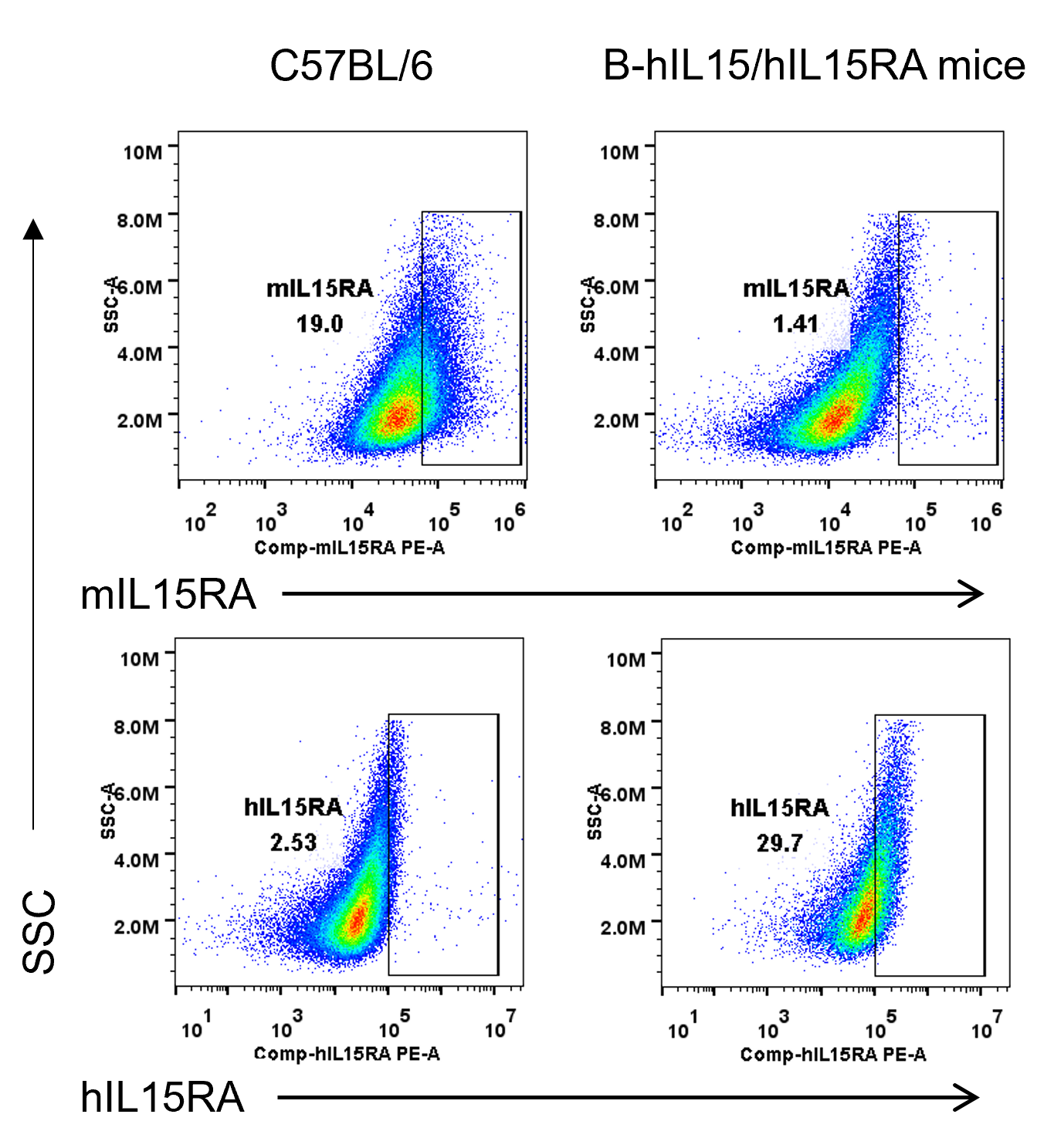
      IL15RA Protein Expression
      • Mouse IL15RA was detectable on BMDCs of wild-type C57BL/6 mice.
      • Human IL15RA was detectable on BMDCs of homozygous B-hIL15/hIL15RA mice, but not in wild-type C57BL/6 mice.

      Strain specific IL15RA expression analysis in wild-type C57BL/6 mice and homozygous humanized B-hIL15/hIL15RA mice by flow cytometry. Bone marrow derived dendritic cells (BMDCs) were produced by culturing the bone marrow from wild-type C57BL/6 mice and homozygous B-hIL15/hIL15RA mice , which were stimulated with LPS in vitro. Protein expression was analyzed with anti-mouse IL15RA antibody (BD, 568235) and anti-human IL15RA antibody (Biolegend, 330207) by flow cytometry.
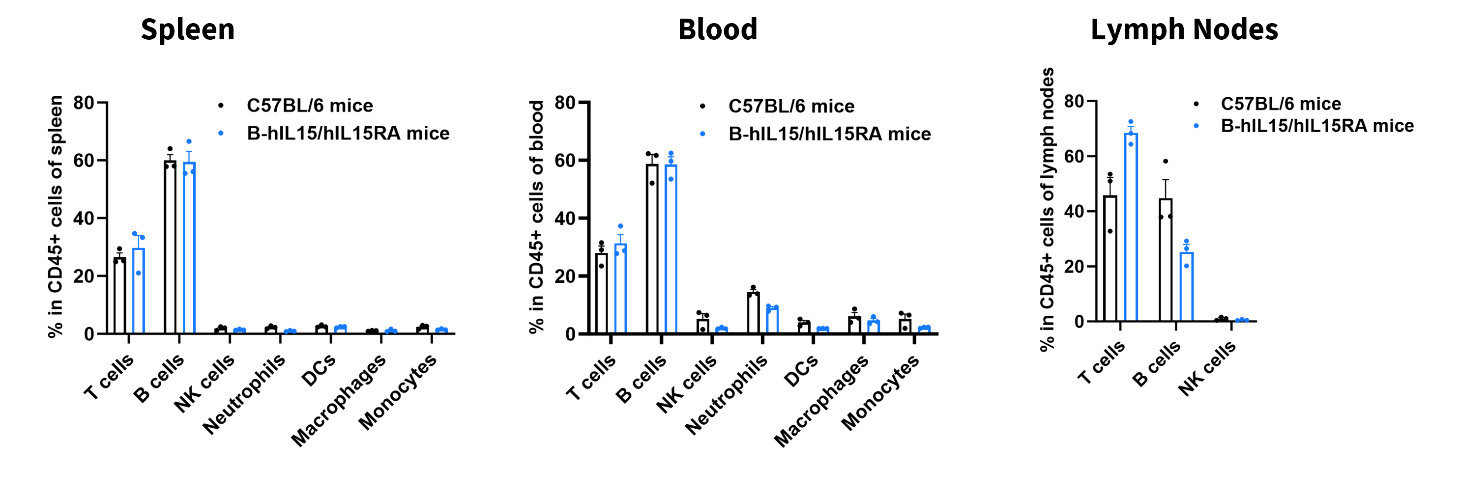
      Analysis of Leukocyte Subpopulations
      • The percentages of T cells, B cells, NK cells, DCs, neutrophils, monocytes, and macrophages in homozygous B-hIL15/hIL15RA mice were similar to those in C57BL/6 mice.
      • Humanization of IL15 and IL15RA does not affect normal immune cell development or distribution.

      Analysis of leukocyte subpopulations by flow cytometry in immune organs and blood.

      Splenocytes, peripheral blood, and lymph nodes were isolated from C57BL/6 mice and homozygous B-hIL15/hIL15RA mice (female, 8-week-old, n = 3). Single live cells were gated on the CD45⁺ population and analyzed by flow cytometry as indicated. Values are expressed as mean ± SEM.
      Analysis of T Cell Subpopulations
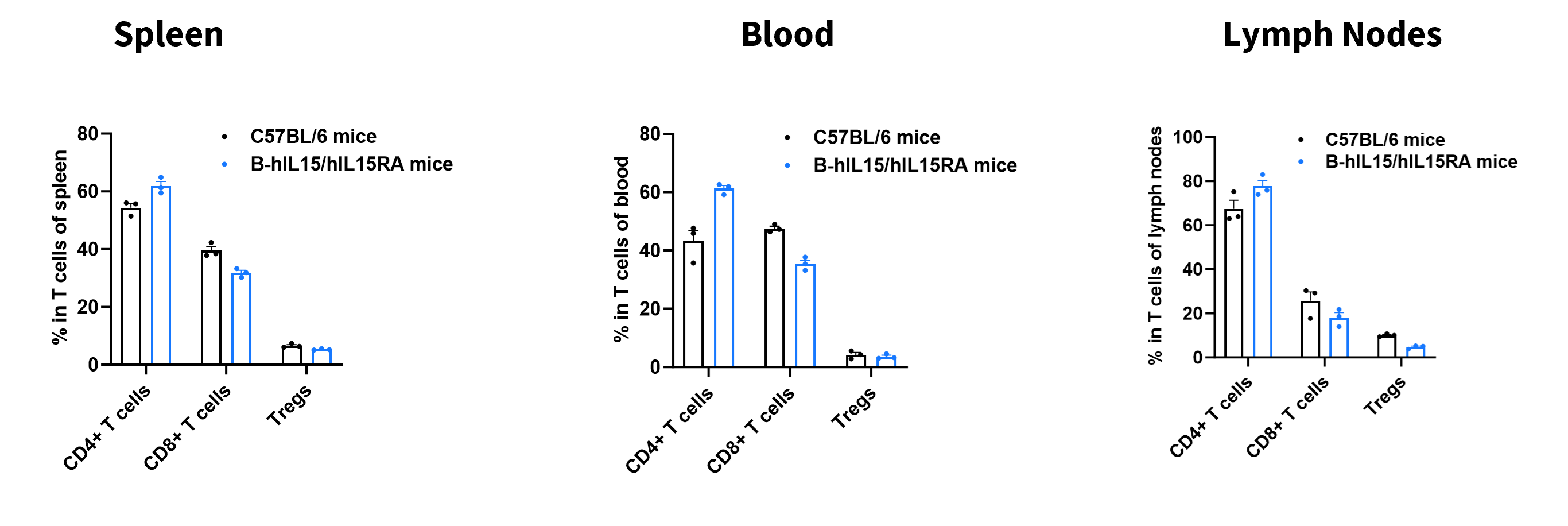
      • The proportions of CD4⁺ T cells, CD8⁺ T cells, and Tregs in homozygous B-hIL15/hIL15RA mice were comparable to those in C57BL/6 mice.
      • Humanization of IL15 and IL15RA does not affect normal T cell development, differentiation, or distribution.

      Analysis of T-cell subpopulations by flow cytometry in immune organs and blood.

      Splenocytes, peripheral blood, and lymph nodes were isolated from C57BL/6 mice and homozygous B-hIL15/hIL15RA mice (female, 8-week-old, n = 3). Single live cells were gated on the CD3⁺ T-cell population and analyzed by flow cytometry as indicated. Values are expressed as mean ± SEM.
      Phosphorylation of STAT5 Induced by Soluble IL15 and IL15RA Complex
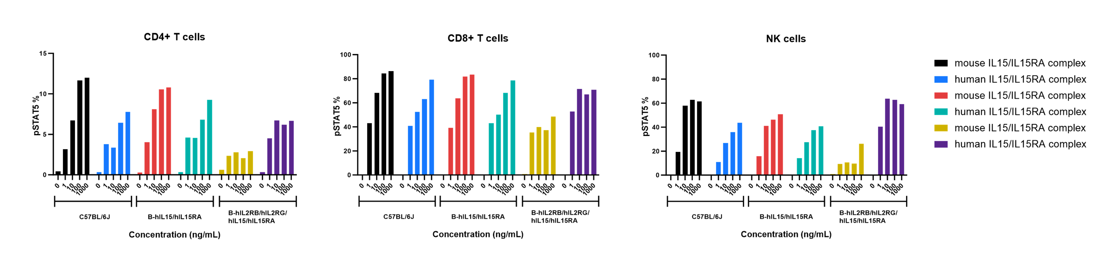
      • Mouse IL-15/IL-15RA complex and human IL-15/IL-15RA complex induced pSTAT5 in CD4+ T cells, CD8+ T cells and NK cells of wild-type C57BL/6 mice, homozygous B-hIL15/hIL15RA mice and homozygous B-hIL2RB/hIL2RG/hIL15/hIL15RA mice.

      Intracellular phosphorylation of STAT5 induction by human and mouse IL15/hIL15RA in wild-type C57BL/6 mice, homozygous B-hIL15/hIL15RA mice and homozygous B-hIL2RB/hIL2RG/hIL15/hIL15RA mice. Splenocytes were harvested from wild-type C57BL/6JNifdc mice, homozygous B-hIL15/hIL15RA mice and homozygous B-hIL2RB/hIL2RG/hIL15/hIL15RA mice, and stimulated with mouse or human IL15 and IL15RA  complex A in vitro for 30 min. Then cells were analyzed by flow cytometry with anti-pSTAT5 antibodies.

      Effects of Anti-human IL15 Antibody in Psoriasis Model

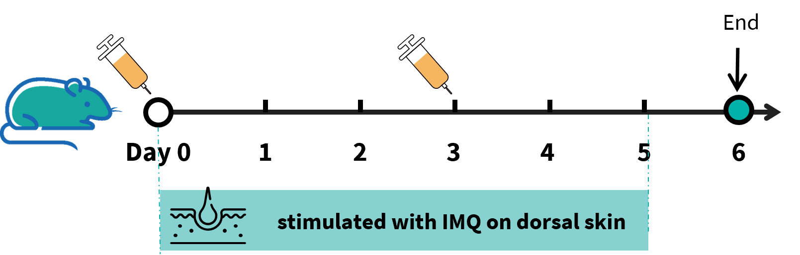
      In vivo efficacy of anti-human IL15 antibody AMG-714 analog (in house) in B-hIL15/hIL15RA mice. B-hIL15/hIL15RA mice (female, 6-8-week-old, n=5) were scored daily for up to 7 days for body weight and clinical signs of skin inflammation following treatment with imiquimod (IMQ) cream, Control mice were treated similarly with Vaseline cream. Mice in group 3 were treated anti-human IL15 antibody AMG-714 analog (in house).
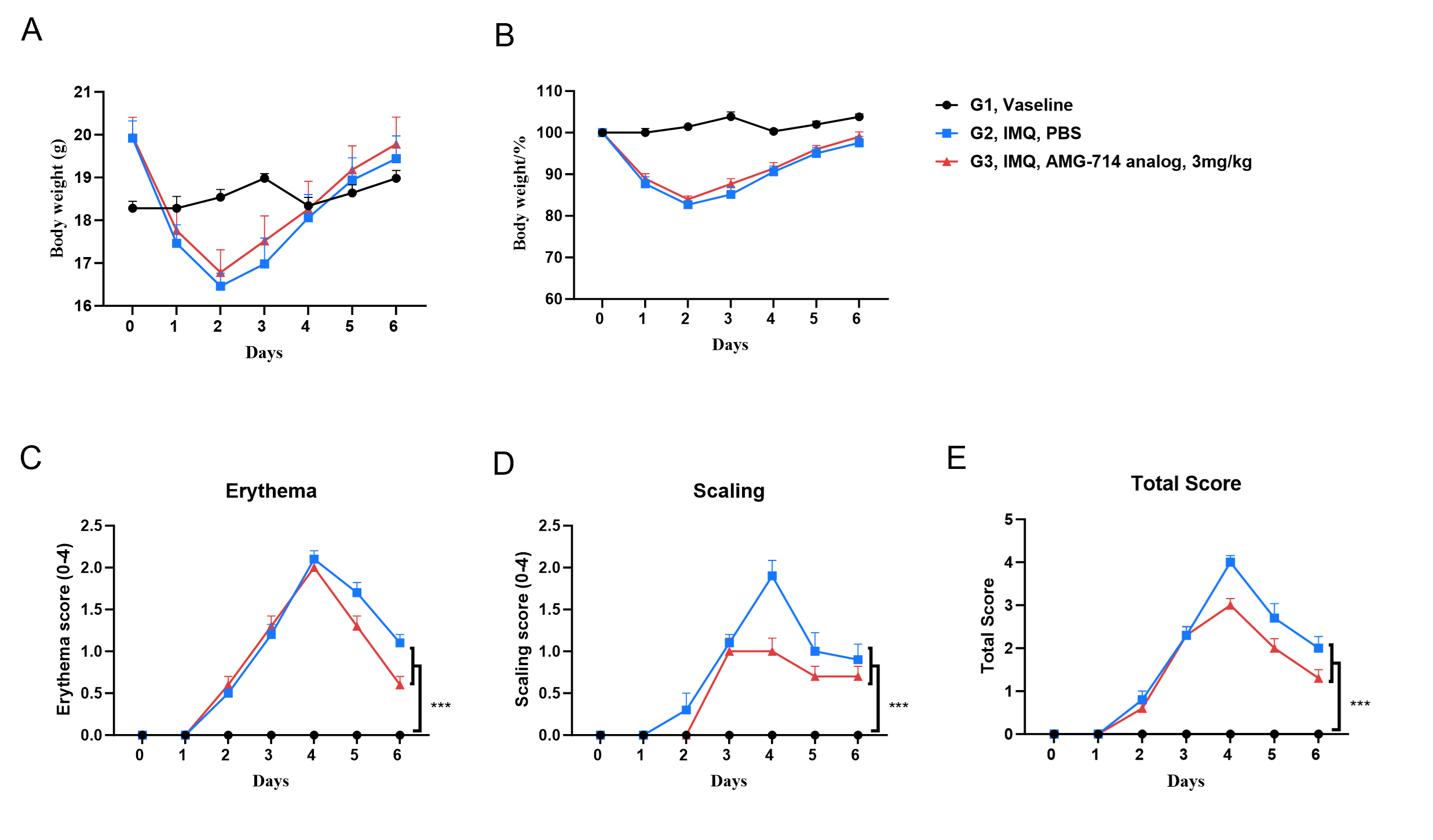
      In vivo efficacy of anti-human IL15 antibody AMG-714 analog (in house) in B-hIL15/hIL15RA mice. (A-B) Body weight changes during treatment. (C-D) Erythema and the scaling score of the back were scored daily from 0 to 6. Additionally, the total score (erythema plus scaling) is depicted (E). Results indicated that structural features of IMQ-induced skin inflammation is visible and increased in severity up to day 4.  Anti-human IL15 antibody AMG-714 analog (in house) improve the disease score of erythema and scaling, but had no effect on body weight.

      * When publishing results obtained using this animal model, please acknowledge the source as follows: The animal model [B-hIL15/hIL15RA mice] (Cat# 121217) was purchased from Biocytogen.