

C57BL/6JNifdc-Il23rtm2(IL23R)Bcgen Il12rb1tm2(IL12RB1)Bcgen/Bcgen • 113296

IL23R and IL12RB1: Key Receptors Driving the Pathogenic Th17 Axis

IL23R
IL12RB1

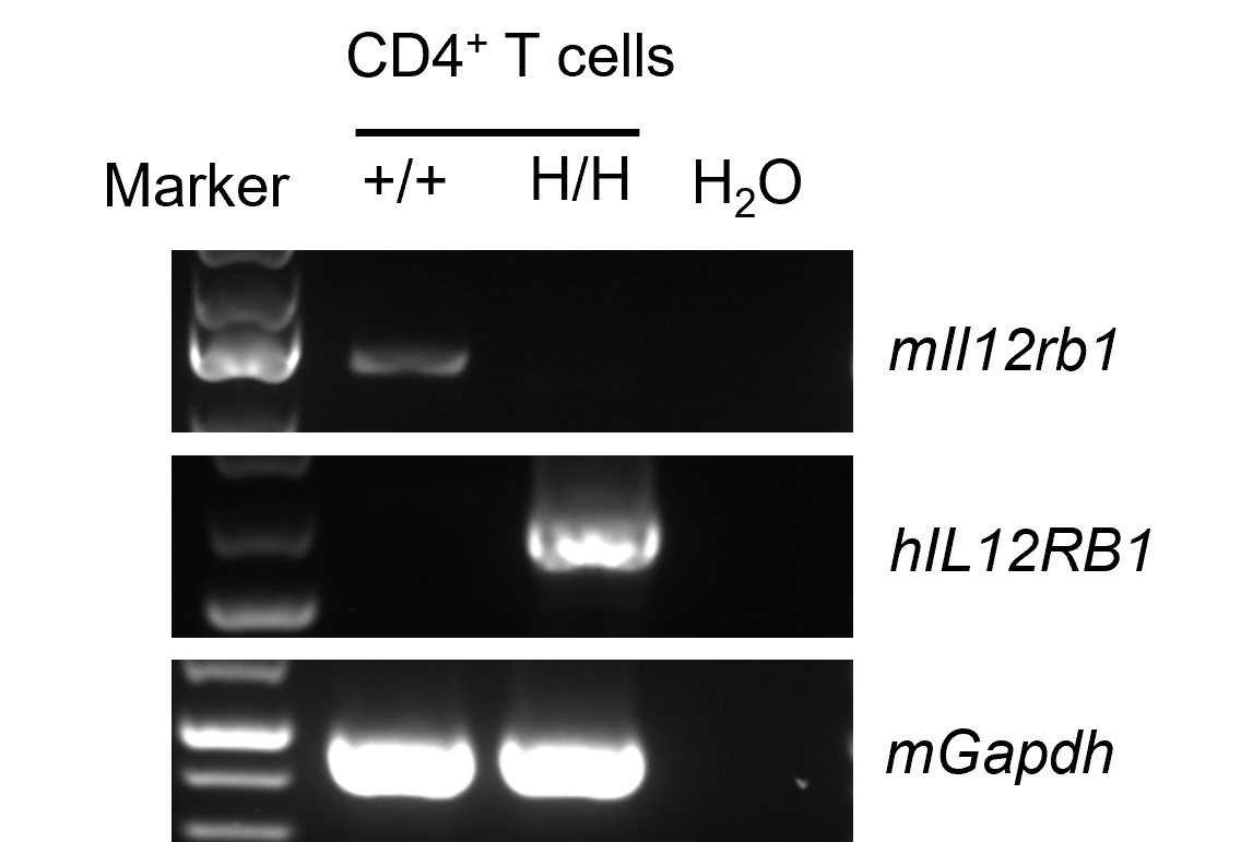
Strain specific analysis of IL12RB1 mRNA expression in wild-type C57BL/6JNifdc mice and B-hIL23R/hIL12RB1 plus mice by RT-PCR. Spleen separated CD4+ T cells RNA were isolated from wild-type C57BL/6JNifdc mice (+/+) and B-hIL23R/hIL12RB1 plus mice (H/H, H/H), and then cDNA libraries were synthesized by reverse transcription, followed by PCR with mouse or human IL12RB1 primers. Mouse Il12rb1 mRNA was only detectable in wild-type mice. Human IL12RB1 mRNA was exclusively detectable in B-hIL23R/hIL12RB1 plus mice.
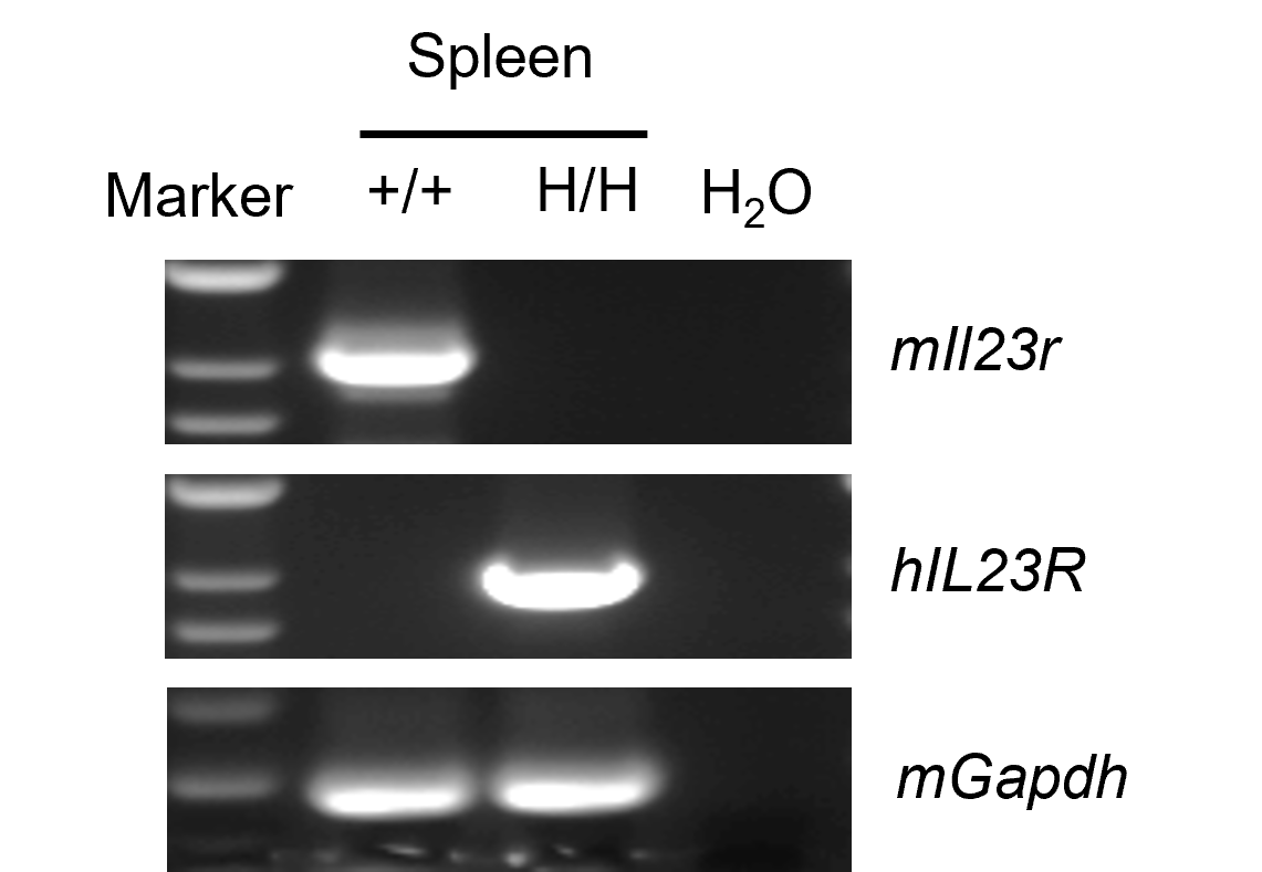
Strain specific analysis of IL23R mRNA expression in wild-type C57BL/6JNifdc mice and B-hIL23R/hIL12RB1 plus mice by RT-PCR. Splenocytes RNA were isolated from wild-type C57BL/6JNifdc mice (+/+) and homozygous B-hIL23R/hIL12RB1 plus mice (H/H, H/H), and then cDNA libraries were synthesized by reverse transcription, followed by PCR with mouse or human IL23R primers. Mouse Il23r mRNA was detectable in wild-type mice. Human IL23R mRNA was exclusively detectable in homozygous B-hIL23R/hIL12RB1 plus mice.

Function analysis of IL23R in wild-type C57BL/6JNifdc mice and homozygous B-hIL23R/hIL12RB1 plus mice by ELISA. Splenocytes were collected from wild-type C57BL/6JNifdc (+/+) mice and homozygous B-hIL23R/hIL12RB1 plus mice (H/H), then CD4+ T cells were isolated and cultured on anti-mCD3ε antibody and anti-mCD28 antibody pre-coated plates, and stimulated with indicated concentrations of mouse IL23 recombinant protein (10 ng/mL) or human IL23 recombinant protein (10 ng/mL), and different concentrations of positive drug (0 nM, 0.2 nM, 1 nM) at 37℃ for 48 h. Cell supernatants were collected for ELISA analysis of mouse IL17A (BioLegend, 432504). Mouse IL23 can induced mouse IL17A production in wild-type C57BL/6JNifdc mice and homozygous B-hIL23R/hIL12RB1 plus mice. Human IL23 can induced mouse IL17A production in wild-type C57BL/6JNifdc mice and homozygous B-hIL23R/hIL12RB1 plus mice. The positive drug can inhibit the expression of mouse IL17A in homozygous B-hIL23R/hIL12RB1 plus mice with hIL23 stimulated. The positive drug is provided by the client.

Analysis of subpopulation of leukocytes by FACS. Splenocytes were isolated from female C57BL/6 and B-hIL23R/hIL12RB1 plus mice (n=3, 6-week-old). Flow cytometry analysis of the spleen were performed to assess leukocyte subpopulations. Percent of T cells, B cells, NK cells, dendritic cells, granulocytes, monocytes and macrophages in homozygous B-hIL23R/hIL12RB1 plus mice were similar to those in the C57BL/6 mice, demonstrating that introduction of hIL23R and hIL12RB1 in place of its mouse counterpart does not change the overall development, differentiation or distribution of these cell types. Values are expressed as mean ± SEM.

Analysis of spleen T cell subpopulations by FACS. Splenocytes were isolated from female C57BL/6 and B-hIL23R/hIL12RB1 plus mice (n=3, 6-week-old). Flow cytometry analysis of the spleen were performed to assess leukocyte subpopulations. Percent of CD4+ T cells, CD8+ T cells and Treg cells in homozygous B-hIL23R/hIL12RB1 plus mice were similar to those in the C57BL/6 mice, demonstrating that introduction of hIL23R and hIL12RB1 in place of its mouse counterpart does not change the overall development, differentiation or distribution of these T cell subtypes. Values are expressed as mean ± SEM.

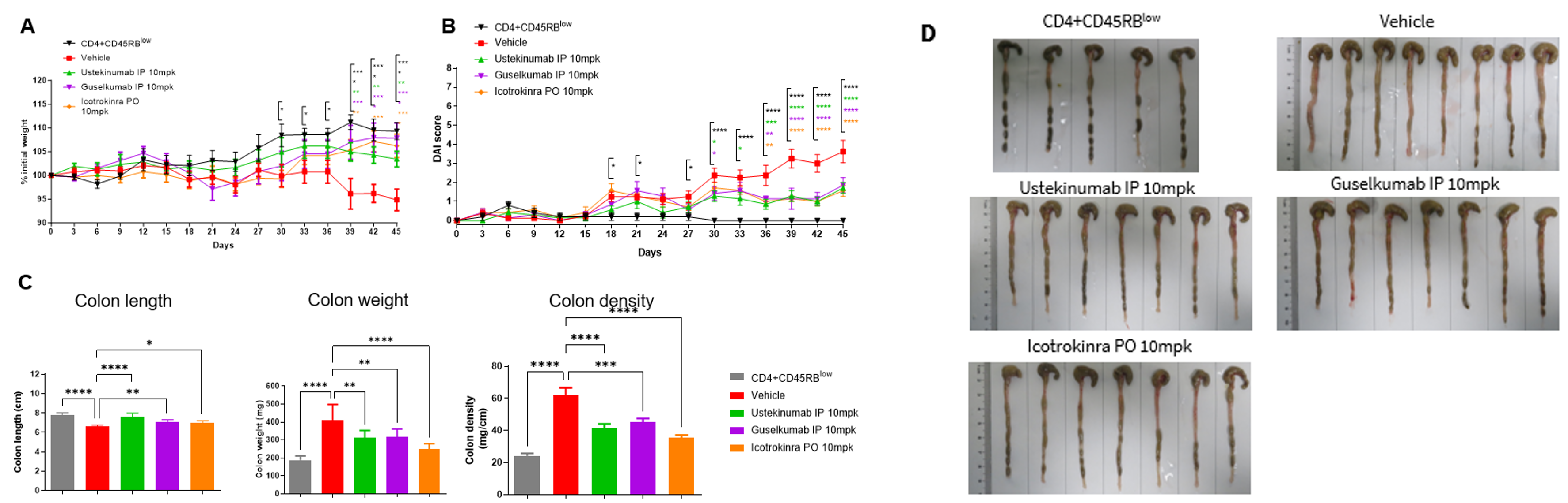
The therapeutic efficacy of Ustekinuma, Guselkumab and Icotrokinra on T cells transfer induced colitis model in B-hIL23R/hIL12RB1 plus mice. CD4+CD45RBlow T and CD4+CD45RBhigh T cells were isolated from the spleens of B-hIL23R/hIL12RB1 plus mice. B-hIL23A/hIL12B, Rag2 KO mice in group G1 were injected with CD4+CD45RBlow T cells, while B-hIL23A/hIL12B, Rag2 KO mice in groups G2-G5 were injected with CD4+CD45RBhigh T cells. The treatment groups received anti-human IL12/IL23p40 antibody Ustekinumab (10 mpk, provided by WuXi AppTec), anti-human IL23p19 antibody Guselkumab (10 mpk, provided by WuXi AppTec) and anti-human IL23R oral peptide Icotrokinra (10 mpk, provided by WuXi AppTec). (A) Body weight change. (B) DAI score. (C) Colon index. (D) Colon photo. Administration of anti-human IL12/IL23p40 antibody Ustekinumab, anti-human IL23p19 antibody Guselkumab and anti-human IL23R oral peptide Icotrokinra effectively improved T cells transfer induced colitis. Two-way ANOVA or one-way ANOVA was used for multiple comparisons, with each group compared to Vehicle. Values are expressed as mean ± SEM. *p<0.05, **p<0.01, ***p<0.001, ****p<0.0001.
Note: This experiment was conducted by WuXi AppTec, T cell transfer induced chronic colitis in B-hIL23A/hIL12B, Rag2 KO mice (Donor: B-hIL23R/hIL12RB1 plus mice).
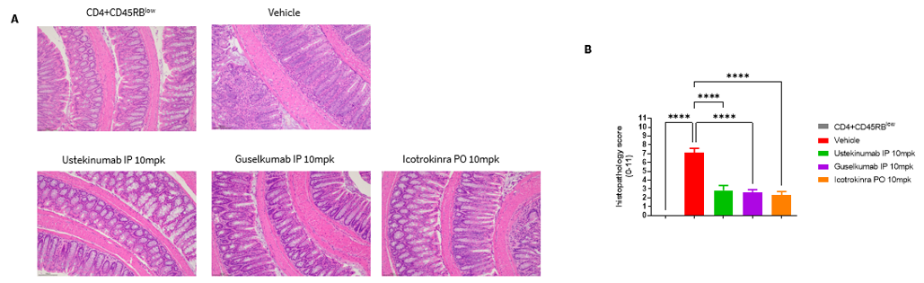
The therapeutic efficacy of Ustekinuma, Guselkumab and Icotrokinra on T cells transfer induced colitis model in B-hIL23R/hIL12RB1 plus mice. CD4+CD45RBlow T and CD4+CD45RBhigh T cells were isolated from the spleens of B-hIL23R/hIL12RB1 plus mice. B-hIL23A/hIL12B, Rag2 KO mice in group G1 were injected with CD4+CD45RBlow T cells, while B-hIL23A/hIL12B, Rag2 KO mice in groups G2-G5 were injected with CD4+CD45RBhigh T cells. The treatment groups received anti-human IL12/IL23p40 antibody Ustekinumab (10 mpk, provided by WuXi AppTec), anti-human IL23p19 antibody Guselkumab (10 mpk, provided by WuXi AppTec) and anti-human IL23R oral peptide Icotrokinra (10 mpk, provided by WuXi AppTec). (A) H&E staining of colon tissue. (B) Pathological score. Administration of anti-human IL12/IL23p40 antibody Ustekinumab, anti-human IL23p19 antibody Guselkumab and anti-human IL23R oral peptide Icotrokinra effectively improved T cells transfer induced colitis. Two-way ANOVA or one-way ANOVA was used for multiple comparisons, with each group compared to Vehicle. Values are expressed as mean ± SEM. *p<0.05, **p<0.01, ***p<0.001, ****p<0.0001.
Note: This experiment was conducted by WuXi AppTec, T cell transfer induced chronic colitis in B-hIL23A/hIL12B, Rag2 KO mice (Donor: B-hIL23R/hIL12RB1 plus mice).
