Description
TNFR2: Signaling in Tregs for Mechanism and Therapeutic Potential
- Gene Information: The TNFRSF1B gene, located on chromosome 1p36, encodes TNFR2 (CD120b), a 75 kDa type I transmembrane glycoprotein that lacks a death domain and belongs to the tumor necrosis factor receptor superfamily.
- Protein Expression: TNFR2 expression is highly restricted to specific immune cell subsets—particularly regulatory T cells (Tregs), myeloid-derived suppressor cells (MDSCs), and certain effector T cells—as well as some oncogenic cells within the tumor microenvironment.
- Signaling Pathway: Upon binding to membrane-bound TNF (mTNF), TNFR2 recruits TRAF1, TRAF2, and TRAF3 to initiate the canonical and non-canonical NF-κB pathways and the PI3K/Akt pathway, which collectively promote cell survival, proliferation, and tissue regeneration.
- Therapeutic Inhibition: Therapeutic strategies primarily utilize antagonistic antibodies to selectively deplete or inhibit immunosuppressive Tregs and MDSCs, thereby "dropping the brakes" on the immune system to enhance anti-tumor activity.
Targeting strategy
TNFR2
- Exons 2–6 of the mouse Tnfr2 gene, which encode the extracellular domain, are replaced with the corresponding human sequences.
- The endogenous mouse promoter, 5′ UTR, and 3′ UTR regions are retained, allowing human TNFR2 expression to be driven by the native mouse Tnfr2 promoter, while endogenous mouse Tnfr2 transcription and translation are abolished.
IL2RA
- Exons 2–6 of the mouse Il2ra gene, which encode the extracellular domain, are replaced with the corresponding human sequences.
- The endogenous mouse promoter, 5′ UTR, and 3′ UTR regions are retained, allowing human IL2RA expression to be driven by the native mouse Il2ra promoter, while endogenous mouse Il2ra transcription and translation are abolished.
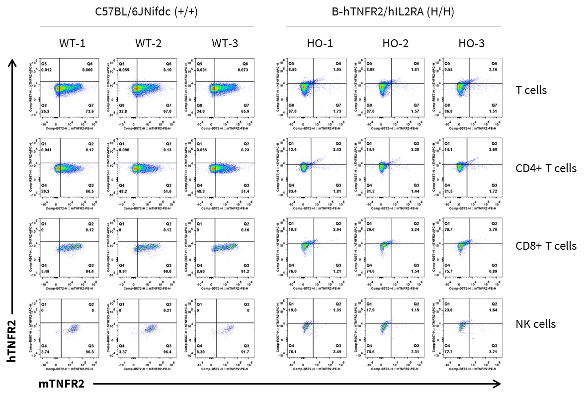
TNFR2 Protein Expression Analysis
- Mouse TNFR2 was detected in wild-type (WT) C57BL/6JNifdc mice, but not in B-hTNFR2/hIL2RA mice.
- Human TNFR2 was exclusively detected in homozygous (HO) B-hTNFR2/hIL2RA mice.
Mouse and human TNFR2 expression analysis in splenocytes. Splenocytes were collected from wild-type (WT) C57BL/6JNifdc mice, homozygous (HO) B-hTNFR2/hIL2RA mice. IL2RA expression on T cells, CD4+ T cells, CD8+ T cells and NK cells was analyzed by flow cytometry using species-specific anti-mouse TNFR2 antibody (Biolegend, 113405) and anti-human TNFR2 antibody (BioLegend, 358406).
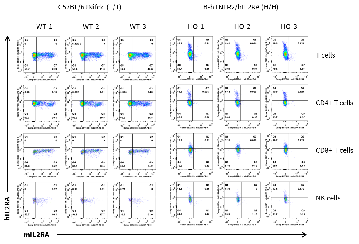
IL2RA Protein Expression Analysis
- Mouse IL2RA was detected in wild-type (WT) C57BL/6JNifdc mice, but not in B-hTNFR2/hIL2RA mice.
- Human IL2RA was exclusively detected in homozygous (HO) B-hTNFR2/hIL2RA mice.
Mouse and human IL2RA expression analysis in splenocytes. Splenocytes were collected from wild-type (WT) C57BL/6JNifdc mice, homozygous (HO) B-hTNFR2/hIL2RA mice. IL2RA expression on T cells, CD4+ T cells, CD8+ T cells and NK cells was analyzed by flow cytometry using species-specific anti-mouse IL2RA antibody (Biolegend, 102008) and anti-human IL2RA antibody (BioLegend, 302610).
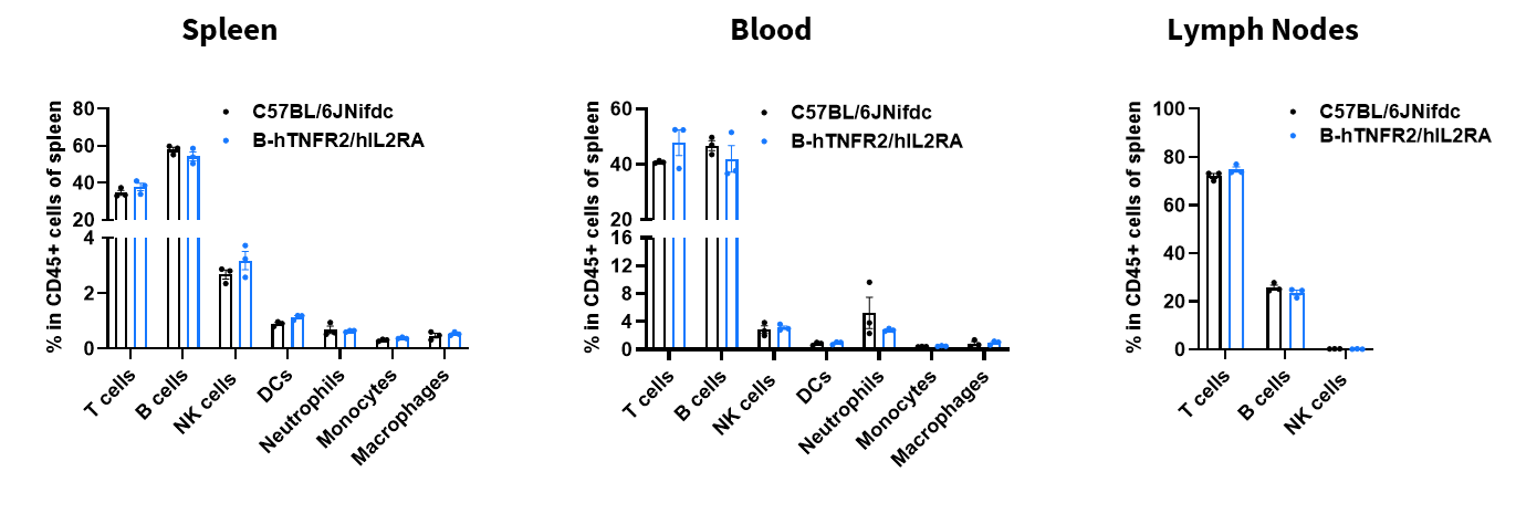
Analysis of Leukocyte Subpopulations
- The percentages of T cells, B cells, NK cells, DCs, neutrophils, monocytes, and macrophages in homozygous B-hTNFR2/hIL2RA mice were similar to those in C57BL/6JNifdc mice.
- Humanization of TNFR2, and IL2RA does not affect normal immune cell development or splenic distribution.
Analysis of leukocyte subpopulations by flow cytometry in immune organs and blood. Splenocytes, peripheral blood, and lymph nodes were isolated from female C57BL/6JNifdc mice and B-hTNFR2/hIL2RA mice (female, 9-week-old, n = 3). Single live cells were gated on the CD45⁺ population and analyzed by flow cytometry as indicated. Values are expressed as mean ± SEM.
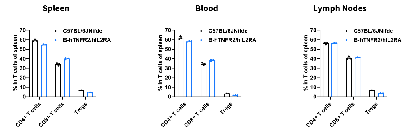
Analysis of T Cell Subpopulations
- The proportions of CD4⁺ T cells, CD8⁺ T cells, and Tregs in homozygous B-hTNFR2/hIL2RA mice were comparable to those in C57BL/6JNifdc mice.
- Humanization of TNFR2, and IL2RA does not affect normal T cell development, differentiation, or splenic distribution.
Analysis of T-cell subpopulations by flow cytometry in immune organs and blood. Splenocytes, peripheral blood, and lymph nodes were isolated from female C57BL/6JNifdc mice and B-hTNFR2/hIL2RA mice (female, 9-week-old, n = 3). Single live cells were gated on the CD3⁺ T-cell population and analyzed by flow cytometry as indicated. Values are expressed as mean ± SEM.
* When publishing results obtained using this animal model, please acknowledge the source as follows: The animal model [B-hTNFR2/hIL2RA mice] (Cat# 111846) was purchased from Biocytogen.