Description
ACVR2A: A key target for muscle growth, weight Loss, and Pulmonary Arterial Hypertension (PAH) in drug research.
- Gene Information: ACVR2A encodes a receptor that mediates the functions of activins, which are members of the transforming growth factor-beta (TGF-beta) superfamily involved in diverse biological processes.
- Protein Expression: ACVR2A is ubiquitously expressed across normal human tissues—particularly in the cytoplasm and cell membrane—and plays a vital role in cellular differentiation, embryonic development, and bone mass regulation.
- Signaling Pathway: ActRIIA/ACVR2A and ActRIIB/ACVR2B are TGF-β superfamily transmembrane proteins that serve as type II receptors for activin A, driving essential processes in cellular growth, differentiation, and tissue regeneration.
- Therapeutic Method: Currently, several preclinical and clinical studies are exploring the effects of antagonists or agonists of ACVR2A and ACVR2B receptors in disease treatment.
ACVR2B: A Receptor Involved in the Regulation of Muscle Growth
- Gene Information: The transmembrane serine/threonine kinase receptor known as ACVR2B (activin A receptor type IIB) is an essential part of the transforming growth factor-β (TGF-β) superfamily signaling pathway.
- Protein Expression: It is widely expressed in various human tissues, with particularly critical expression levels in tissues closely related to physiological functions, such as muscle, bone, and gonads.
- Signaling Pathway: ActRIIA/ACVR2A and ActRIIB/ACVR2B are TGF-β superfamily transmembrane proteins that serve as type II receptors for activin A, driving essential processes in cellular growth, differentiation, and tissue regeneration.
- Therapeutic Method: ACVR2B inhibitors cover a wide spectrum of potential clinical applications, attributable to muscle’s core involvement in whole-body wellness and disease development. Their leading promising indication targets disorders characterized by muscle depletion. Treatments that boost muscle mass and block tissue atrophy offer tangible therapeutic benefits for patients suffering from muscular dystrophy as well as cachexia triggered by tumors and long-term chronic diseases.
Targeting strategy
ACVR2A
- The exons 1-11 of the mouse Acvr2a gene that encode the whole molecule, including promoter, 5’UTR, and part 3’UTR, were replaced by the exons 1-11 of the human counterparts, including human promoter, 5’UTR, and human 3’UTR in B-hACVR2A mice.
- The human ACVR2A expression is driven by the human ACVR2A promoter, while the mouse Acvr2a gene transcription and translation will be disrupted.
ACVR2B
- The exons 1-10 of the mouse Acvr2b gene that encode the whole molecule, including the promoter, 5’UTR, and 3’UTR, were replaced by the exons 1-11 of the human counterparts, including the human promoter, 5’UTR, and human 3’UTR, in B-hACVR2B mice.
- The human ACVR2B expression is driven by the human ACVR2B promoter, while the mouse Acvr2b gene transcription and translation will be disrupted.
- B-hACVR2A/hACVR2B mice were obtained by mating B-hACVR2A mice (111393) and B-hACVR2B mice (111351). For validation data of this mouse model, you can refer to the validation data from the related gene humanized mouse models.
ACVR2A and ACVR2B mRNA Expression Analysis
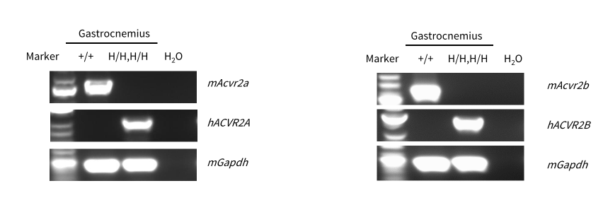
Strain specific analysis of ACVR2A and ACVR2B mRNA expression in wild-type C57BL/6JNifdc mice and homozygous B-hACVR2A/hACVR2B mice by RT-PCR. Gastrocnemius was collected from wild-type C57BL/6JNifdc mice (+/+) and homozygous B-hACVR2A/hACVR2B mice (H/H, H/H), then cDNA libraries were synthesized by reverse transcription, followed by PCR with mouse or human ACVR2A and ACVR2B primers. Mouse Acvr2a and Acvr2b mRNA was detectable in wild-type C57BL/6JNifdc mice but not in homozygous B-hACVR2A/hACVR2B mice. Human ACVR2A and ACVR2B mRNA was detectable only in homozygous B-hACVR2A/hACVR2B mice, but not in wild-type C57BL/6JNifdc mice. Human sequences were confirmed via Sanger Sequencing.
ACVR2A mRNA Expression Analysis
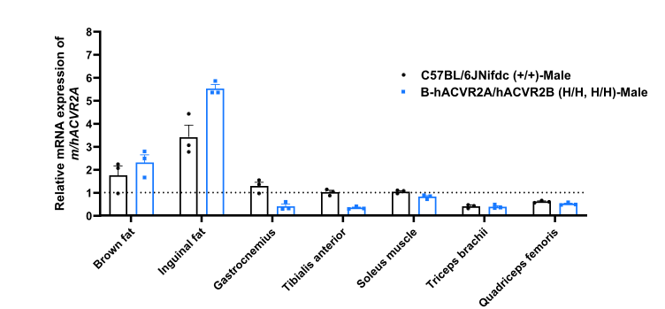
Strain specific analysis of ACVR2A mRNA expression in wild-type C57BL/6JNifdc mice and homozygous B-hACVR2A/hACVR2B mice by RT-qPCR. Various tissues was collected from wild-type C57BL/6JNifdc mice (+/+) and homozygous B-hACVR2A/hACVR2B mice (H/H, H/H), then cDNA libraries were synthesized by reverse transcription, followed by PCR with cross-reactive human-mouse ACVR2A primers. Human ACVR2A was detectable in B-hACVR2A/hACVR2B mice, and mouse Acvr2a was detectable in C57BL/6JNifdc mice. All results were normalized to the tibialis anterior. Values are expressed as mean ± SEM.
ACVR2B mRNA Expression Analysis
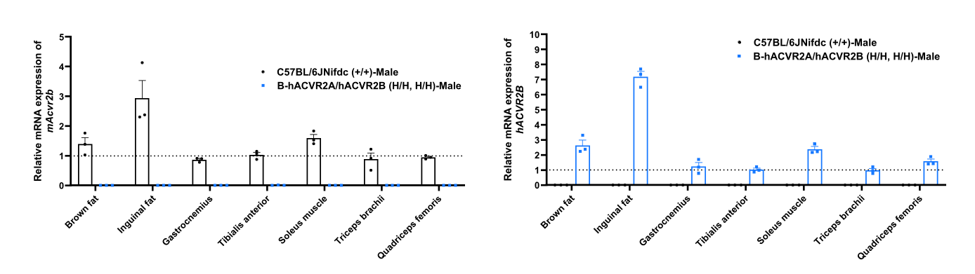
Strain specific analysis of ACVR2B mRNA expression in wild-type C57BL/6JNifdc mice and homozygous B-hACVR2A/hACVR2B mice by RT-qPCR. Various tissues was collected from wild-type C57BL/6JNifdc mice (+/+) and homozygous B-hACVR2A/hACVR2B mice (H/H, H/H), then cDNA libraries were synthesized by reverse transcription followed by RT-qPCR with mouse or human ACVR2B primers. Human ACVR2B mRNA was detectable only in homozygous B-hACVR2A/hACVR2B mice, but not in wild-type C57BL/6JNifdc mice. Mouse Acvr2b mRNA was exclusively detectable in wild-type mice. All results were normalized to the tibialis anterior. Values are expressed as mean ± SEM.
In Vivo Efficacy Study in Lean B-hACVR2A/hACVR2B Mice
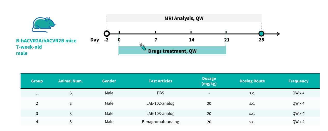
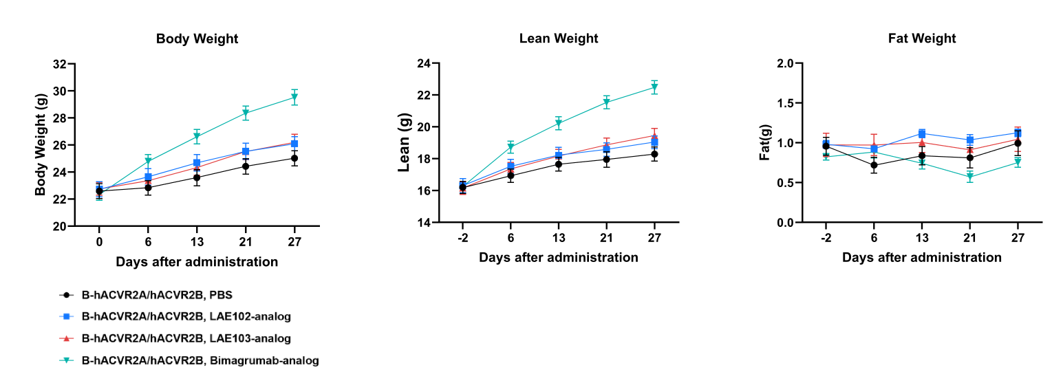
In Vivo Efficacy Study in B-hACVR2A/hACVR2B Mice. B-hACVR2A/hACVR2B mice (7-week-old, male, n=6-8) were treated with PBS, LAE-102-analog, LAE-103-analog, and Bimagrumab-analog. Whole-body MRI was performed throughout the 28-day observation period to monitor body weight, absolute lean mass, and absolute fat mass over time.
In Vivo Efficacy Study in B-hACVR2A/hACVR2B Mice. B-hACVR2A/hACVR2B mice (7-week-old, male, n=6-8) were treated with PBS, LAE-102-analog, LAE-103-analog, and Bimagrumab-analog. Whole-body MRI was performed throughout the 28-day observation period to monitor body weight, absolute lean mass, and absolute fat mass over time.
* When publishing results obtained using this animal model, please acknowledge the source as follows: The animal model [B-hACVR2A/hACVR2B mice] (Cat# 114982) was purchased from Biocytogen.