Description
CLEC7A: The potential therapeutic target of the myeloid cell engager (MCE)
- Gene Information: C-type lectin domain containing 7A (CLEC7A, also known as DECTIN1 and β-glucan receptor) is a protein-coding gene located on chromosome 12p13.2. It encodes a member of the C-type lectin/C-type lectin-like domain (CTL/CTLD) superfamily. In humans, mice and rats, Dectin-1 exists in two major isoforms which are called Dectin-1a and 1b. Dectin-1a/b are generated by alternative splicing from the same mRNA precursor with differential usage of exon 3, which is present in the mature mRNA of the Dectin-1a isoform but absent from Dectin-1b. In humans, up to six additional isoforms were reported which are, however, incapable of ligand binding.
- Protein Expression: Dectin-1 is a type II transmembrane lectin and widely expressed in the myeloid lineage, which involves macrophages/ monocytes, dendritic cells (DCs), and neutrophils, Langerhans cells, keratinocytes as well as in γδ T cells from the lymphoid lineage. Dectin-1 expression have been also detected in primary human intestinal epithelial cells from ileum and colon.
- Signaling Pathway: Dectin-1 undergoes phosphorylation at its cytoplasmic tyrosine residue within a hemi-immunoreceptor tyrosine-based activation motif to activate Syk kinase and downstream MAPK, NFκB and NFAT signaling cascades, which drive secretion of multiple pro- and anti-inflammatory cytokines to coordinate innate immune activation and modulate subsequent adaptive immunity.
- Therapeutic Inhibition: DR-0201(target CLEC7A×CD20) is a potential first-in-class CD20-directed bispecific antibody that targets and engages specific tissue-resident and trafficking myeloid cells to induce deep B-cell depletion via targeted phagocytosis.
Targeting strategy
CLEC7A
- The exons 2-5 of mouse Clec7a gene that encode extracellular domain and transmembrane domain were replaced by human counterparts in B-hCLEC7A mice(C).
- The genomic region of mouse Clec7a gene that encodes cytoplasmic portion is retained. The promoter, 5’UTR region of the mouse gene are also retained. The CLEC7A expression is driven by endogenous mouse Clec7a promoter, while mouse Clec7a gene transcription and translation will be disrupted.
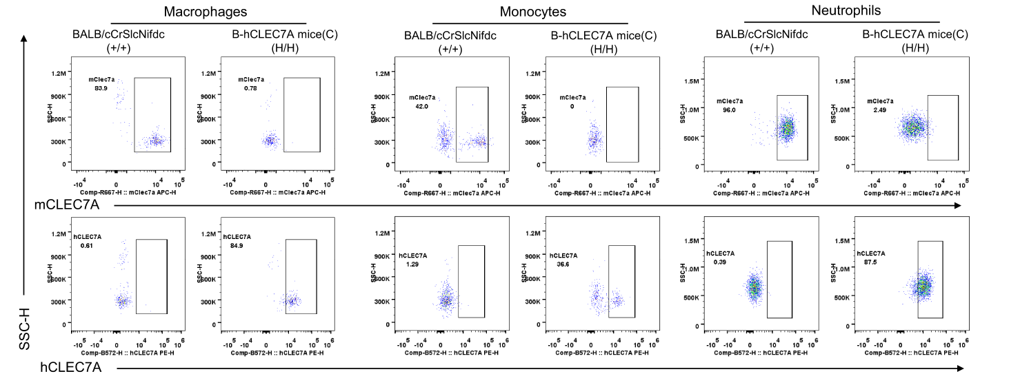
CLEC7A Protein Expression in Blood
Strain-specific CLEC7A expression analysis in homozygous B-hCLEC7A mice (C) by flow cytometry. Blood was collected from wild-type BALB/cCrSlcNifdc (+/+) and homozygous B-hCLEC7A mice (C) (H/H) and analyzed by flow cytometry with species-specific anti-mCLEC7A (APC anti-mouse CD369 Antibody, Biolegend, 144305) and anti-hCLEC7A antibody (PE anti-human CD369 Antibody, Biolegend, 355403). Mouse CLEC7A was detectable in neutrophils, macrophages, and monocytes of wild-type mice. Human CLEC7A was detectable in neutrophils, macrophages, and monocytes of B-hCLEC7A mice (C).
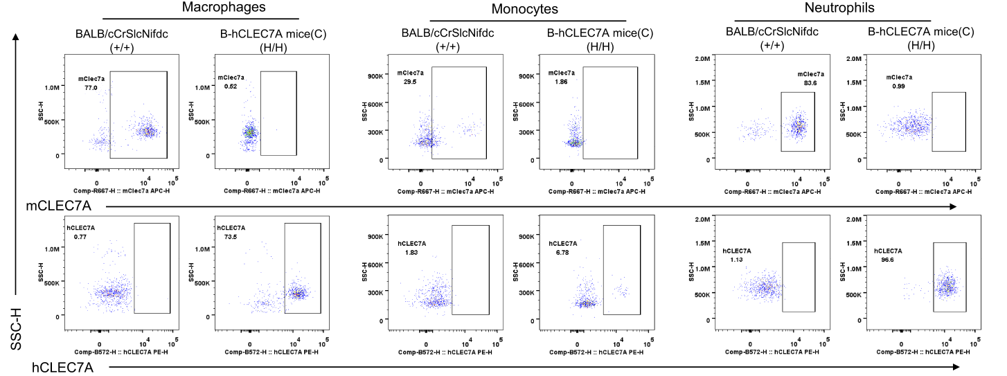
CLEC7A Protein Expression in Spleen
Strain-specific CLEC7A expression analysis in homozygous B-hCLEC7A mice (C) by flow cytometry. Splenocytes were collected from wild-type BALB/cCrSlcNifdc (+/+) and homozygous B-hCLEC7A mice (C) (H/H) and analyzed by flow cytometry with species-specific anti-mCLEC7A (APC anti-mouse CD369 Antibody, Biolegend, 144305) and anti-hCLEC7A antibody (PE anti-human CD369 Antibody, Biolegend, 355403). Mouse CLEC7A was detectable in neutrophils, macrophages, and monocytes of wild-type mice. Human CLEC7A was detectable in neutrophils, macrophages, and monocytes of B-hCLEC7A mice (C).
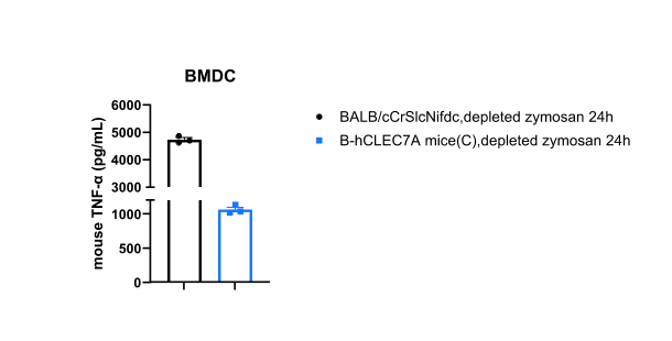
Function analysis-CLEC7A signaling pathway
Mouse TNF-α expression analysis in wild-type BALB/C mice and homozygous B-hCLEC7A mice(C) by ELISA. Bone marrow derived dendritic cells (BMDCs) were produced by culturing the bone marrow from wild-type BALB/cCrSlcNifdc mice (+/+) (male, n=1, 5-week-old) and homozygous B-hCLEC7A mice(C) (H/H) (male, n=1, 14-week-old) stimulated with depleted zymosan in vitro for 24 hours. Cell supernatant was collected for ELISA analysis. Expression level of mouse TNF-α were analyzed by ELISA (mouse TNF-α ELISA kit: Biolegend, 430907). Mouse TNF-α was detectable in bone marrow-derived dendritic cells of wild-type mice and homozygous B-hCLEC7A mice (C). Values are expressed as mean ± SEM.
* When publishing results obtained using this animal model, please acknowledge the source as follows: The animal model [B-hCLEC7A mice(C)] (Cat# 114897) was purchased from Biocytogen.