B-hPD-L1-Luc-EGFP LLC1

NA • 322483

B-hPD-L1-Luc-EGFP LLC1

Product nameB-hPD-L1-Luc-EGFP LLC1
Catalog number322483
Strain nameNA
NCBI gene ID60533 (Human)
Chromosome19
AliasesB7h1; Pdl1; Pdcd1l1; Pdcd1lg1; A530045L16Rik
TissueLung
DiseaseLewis Lung Carcinoma

このページで

  • Description
  • Phenotypic analysis
  • Tumorigenicity

ポスター

すべて表示

    出版物

      Description
      • Origin: The LLC1 (Lewis lung carcinoma) cell line is derived from a lung carcinoma that arose in C57BL/6 mice. The cell line is a widely used syngeneic mouse tumor model for studying non-small cell lung cancer (NSCLC), tumor growth, metastasis, and anti-tumor immune responses.
      • Background Information: PD-L1 (CD274), a pivotal immune checkpoint ligand of the B7 family, is broadly upregulated across many malignancies, including non-small cell lung cancer, melanoma, renal cell carcinoma, and triple-negative breast cancer. By engaging PD-1 (CD279) on activated T cells, PD-L1 delivers inhibitory signals that attenuate T cell proliferation, cytokine secretion (e.g., IFN-γ and IL-2), and cytotoxic activity, ultimately blunting anti-tumor immunity and enabling tumor immune escape. In the tumor microenvironment, PD-L1 is expressed not only on tumor cells but also on tumor-associated immune and stromal compartments—such as antigen-presenting cells (macrophages and dendritic cells), myeloid-derived suppressor cells (MDSCs), and endothelial cells—where it further reinforces T cell exhaustion and supports the establishment of an immunosuppressive niche. PD-L1 expression is dynamically regulated by oncogenic signaling pathways and inflammatory cues (notably IFN-γ–driven adaptive immune resistance), and it can be induced under microenvironmental stresses such as hypoxia, contributing to therapy resistance and disease progression. Beyond its canonical immune-inhibitory role, PD-L1 has been reported to exert tumor-intrinsic functions, including promoting epithelial–mesenchymal transition (EMT), enhancing migration and invasion, supporting cancer stemness, and facilitating metabolic adaptation, thereby accelerating tumor growth and metastasis. Accordingly, therapeutic blockade of the PD-1/PD-L1 axis—via monoclonal antibodies and next-generation modalities—has reshaped the clinical landscape, and robust, physiologically relevant human PD-L1 models (including PD-L1 humanized cell lines) provide essential tools for antibody discovery, functional potency assessment, and mechanism-of-action studies in translational immuno-oncology.
      • Gene targeting strategy: The exogenous promoter and the full coding sequences of human PD-L1 were inserted to replace part of exon 3 of mouse PD-L1 gene. The insertion disrupts the endogenous mouse PD-L1 gene, resulting in a non-functional transcript. The luciferase and EGFP genes linked by P2A were randomly inserted into the genome of B-hPD-L1 LLC1 cells.
      • Tumorigenicity: Confirmed in C57BL/6JNifdc mice.
      • Application: The B-hPD-L1-Luc-EGFP LLC1 tumor models can be used for preclinical evaluation of drugs targeting human PD-L1.
      Protein Expression Analysis

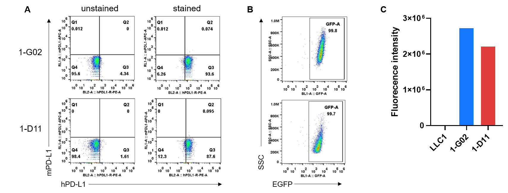
      Expression analysis of hPD-L1, EGFP and luciferase in B-hPD-L1-Luc-EGFP LLC1 cells. (A) Single cell suspensions of B-hPD-L1-Luc-EGFP LLC1 #1-G02, #1-D11 were stained with species-specific anti-mouse PD-L1 antibody (Biolegend, 124312) and anti-human PD-L1 antibody (Biolegend, 329706). Human PD-L1 was detectable on the surface of B-hPD-L1-Luc-EGFP LLC1 cells. But mouse PD-L1 was not detectable on the surface of B-hPD-L1-Luc-EGFP LLC1 cells. (B) EGFP was detectable in B-hPD-L1-Luc-EGFP LLC1 cells. (C) The activity of luciferase can be detected in the supernatant of the cell lysis solution of B-hPD-L1-Luc-EGFP LLC1 cells, but not in wild-type LLC1 cells.

      Tumor Growth Curve & Body Weight Changes

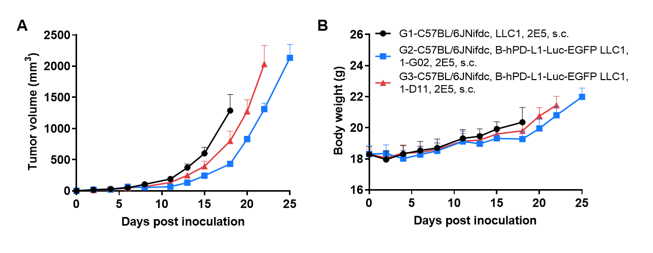
      Subcutaneous tumor growth of B-hPD-L1-Luc-EGFP LLC1 cells. B-hPD-L1-Luc-EGFP LLC1 cells (2×10⁵) and wild-type LLC1 cells (2×10⁵) were subcutaneously implanted into C57BL/6JNifdc mice (female, 6-week-old, n=6). Tumor volume and body weight were measured twice a week. (A) Average tumor volume. (B) Body weight. Volume was expressed in mm3 using the formula: V=0.5 × long diameter × short diameter2. As shown in panel A, B-hPD-L1-Luc-EGFP LLC1 cells were able to establish tumors in vivo and can be used for efficacy studies.

      Protein Expression Analysis of Tumor Tissue

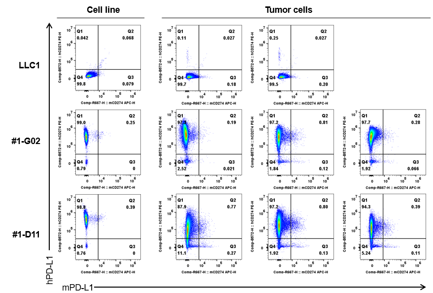
      Human PD-L1 expression evaluated in B-hPD-L1-Luc-EGFP LLC1 cells by flow cytometry. B-hPD-L1-Luc-EGFP LLC1 cells were subcutaneously transplanted into wild-type C57BL/6JNifdc mice (female, 6-week-old, n=6). Tumor cells were harvested and analyzed for mouse PD-L1 (Biolegend, 124312) and human PD-L1 (Biolegend, 329706) expression by flow cytometry. Human PD-L1 was only detectable in B-hPD-L1-Luc-EGFP LLC1 cells and tumor cells. Mouse PD-L1 was not detectable in wild-type LLC1 cells or B-hPD-L1-Luc-EGFP LLC1 cells and tumor cells.

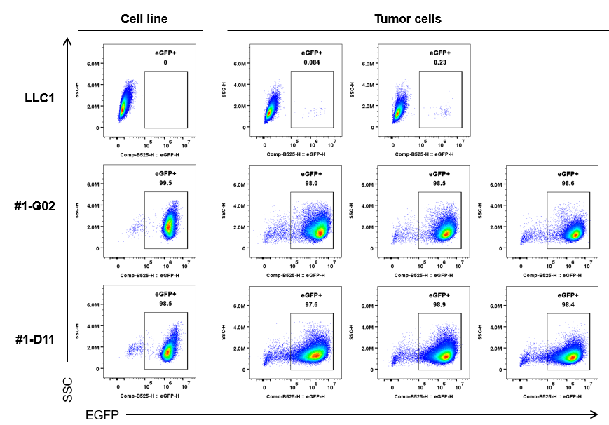
      EGFP expression evaluated in B-hPD-L1-Luc-EGFP LLC1 cells by flow cytometry. B-hPD-L1-Luc-EGFP LLC1 cells were subcutaneously transplanted into wild-type C57BL/6JNifdc mice (female, 6-week-old, n=6). EGFP was only detectable on B-hPD-L1-Luc-EGFP LLC1 cells and tumor cells, but not on the wild-type LLC1 cells or tumors.

      Luciferase Activity Analysis of Tumors

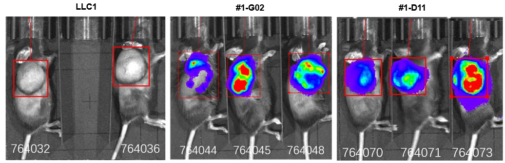
      Luciferase activity in tumors imaged by in vivo imaging system (IVIS). Wild-type LLC1 and B-hPD-L1-Luc-EGFP LLC1 cells were subcutaneously transplanted into wild-type C57BL/6JNifdc mice (female, 6-week-old, n=6). Bioluminescence signal was only detectable in the tumors from B-hPD-L1-Luc-EGFP LLC1 cells, but not in the tumors from wild-type LLC1 cells.