
NA • 322217
| Product name | B-Tg(Luc) LNCap Clone FGC |
|---|---|
| Catalog number | 322217 |
| Strain name | NA |
| Tissue | Prostate |
| Disease | Carcinoma |

Luminescence signal intensity of B-Tg(Luc) LNCap Clone FGC cells. Cell lysates of wild-type LNCap Clone FGC and B-Tg(Luc) LNCap Clone FGC were measured using the Bright-GloTM luciferase Assay (Promega, Catalog No. E4030). B-Tg(Luc) LNCap Clone FGC cells have a strong luminescence signal that is not present in wild-type LNCap Clone FGC cells.

Subcutaneous homograft tumor growth of B-Tg(Luc) LNCap Clone FGC cells. B-Tg(Luc) LNCap Clone FGC cells (2×10⁶) and LNCap Clone FGC cells (2×10⁶) were subcutaneously implanted into B-NDG mice (male, n=6). Tumor volume and body weight were measured twice a week. (A) Average tumor volume ± SEM. (B) Body weight (Mean± SEM). Volume was expressed in mm3 using the formula: V=0.5 X long diameter X short diameter2. As shown in panel A, B-Tg(Luc) LNCap Clone FGC cells were able to establish tumors in vivo and can be used for efficacy studies.
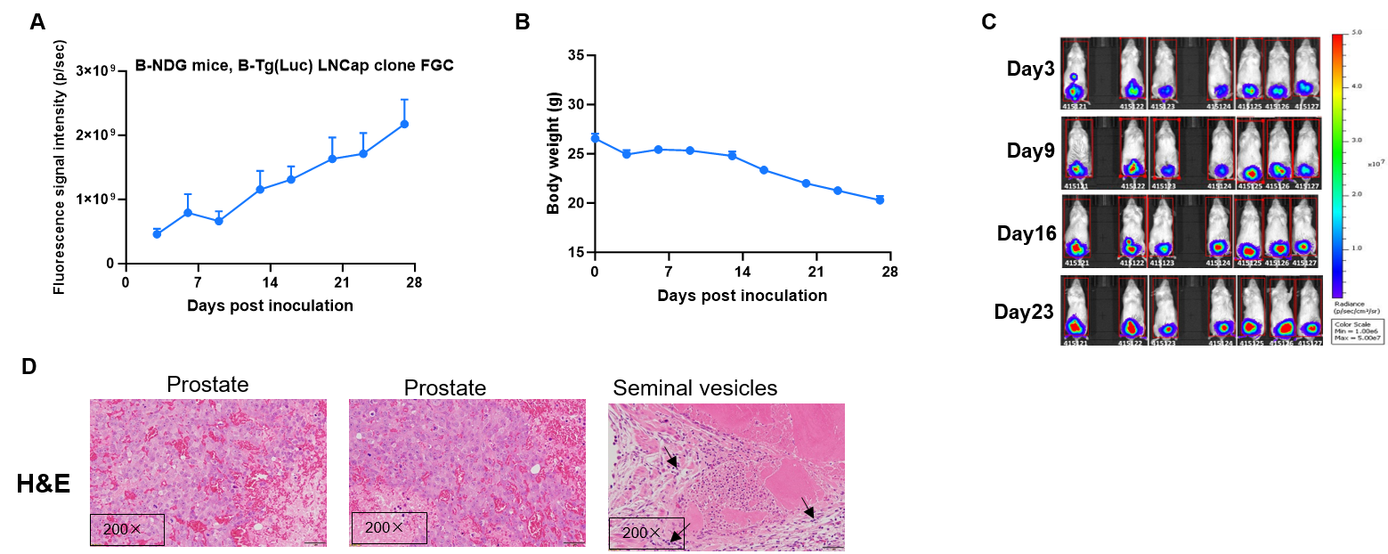
Establishment of orthotopic prostate cancer model. The mouse orthotopic prostate cancer was generated by implanting B-Tg(Luc) LNCap clone FGC cells in the dorsal prostate lobes of B-NDG mice (male, n=7), and the tumor volume, body weight and signal intensity of the mice were recorded weekly. The results showed that the tumor volume increased and body weight of mice gradually decreased during this study. This indicates that the cell line was successfully constructed as an orthotopic tumor model. H&E staining showed that necrosis and hemorrhage were seen in prostate tumors, and inflammatory cell infiltration can be seen in seminal vesicles(D). Values are expressed as mean ± SEM.