B-Tg(Luc-EGFP) Hep G2
Catalog Number: 322408
Strain Name: NA
---
ライセンスオプション提供可能

NA • 322408
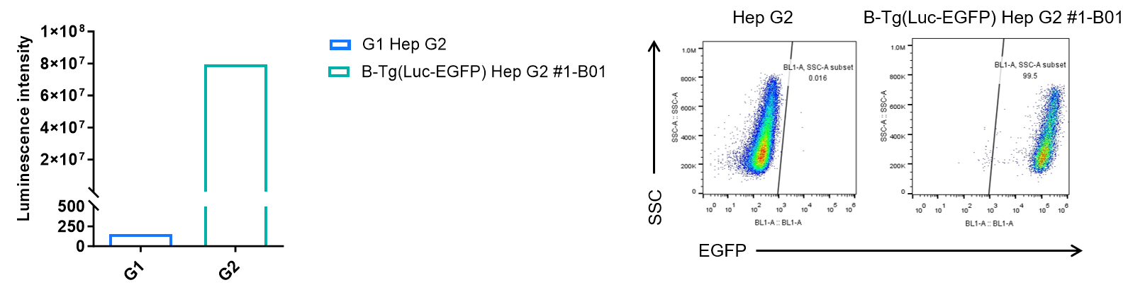
Luminescence signal intensity of B-Tg(Luc-EGFP) Hep G2 cells and EGFP expression analysis in B-Tg(Luc-EGFP) Hep G2 cells. Single cell suspensions from wild-type Hep G2 and B-Tg(Luc-EGFP) Hep G2 #1-B01 cultures were measured using the Bright-GloTM luciferase Assay (Promega, Catalog No. E4030). B-Tg(Luc-EGFP) Hep G2 cells have a strong luminescence signal that is not present in wild-type Hep G2 cells. EGFP was detected on the surface of B-Tg(Luc-EGFP) Hep G2 cells but not wild-type Hep G2 cells.
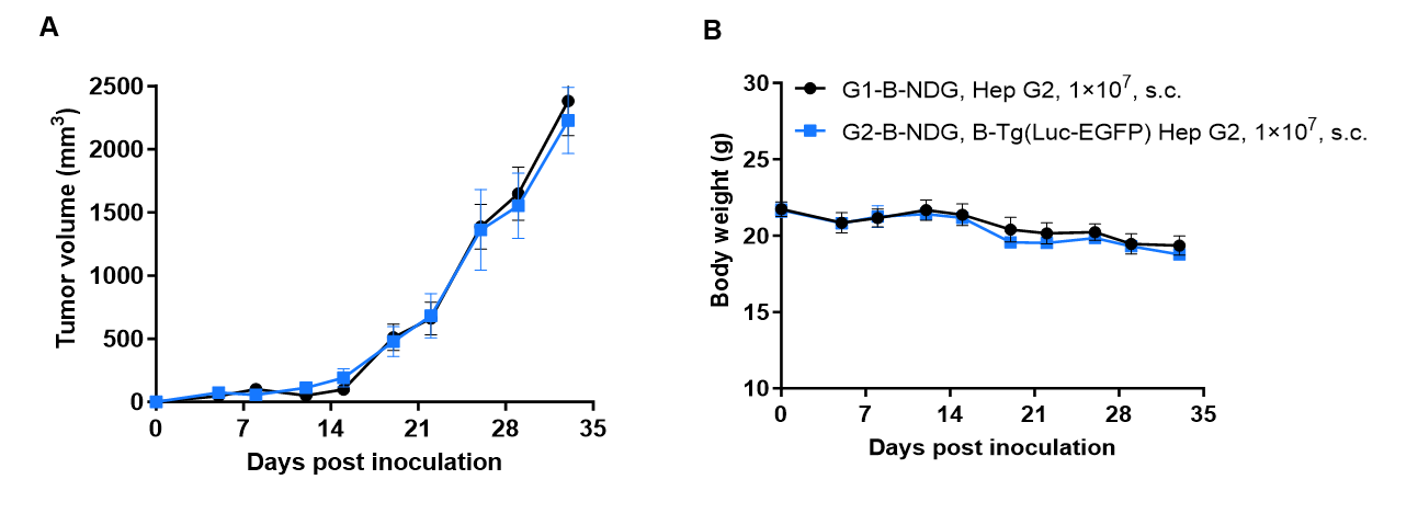
Subcutaneous tumor growth of B-Tg(Luc-EGFP) Hep G2 cells. B-Tg(Luc-EGFP) Hep G2 cells (1x10⁷) and wild-type Hep G2 cells (1x10⁷) were subcutaneously implanted into B-NDG mice (female, 7-9-week-old, n=6). Tumor volume and body weight were measured twice a week. (A) Average tumor volume. (B) Body weight. Volume was expressed in mm3 using the formula: V=0.5 X long diameter X short diameter2. Results indicate that B-Tg(Luc-EGFP) Hep G2 cells were able to establish tumors in vivo and can be used for efficacy studies. Values are expressed as mean ± SEM.
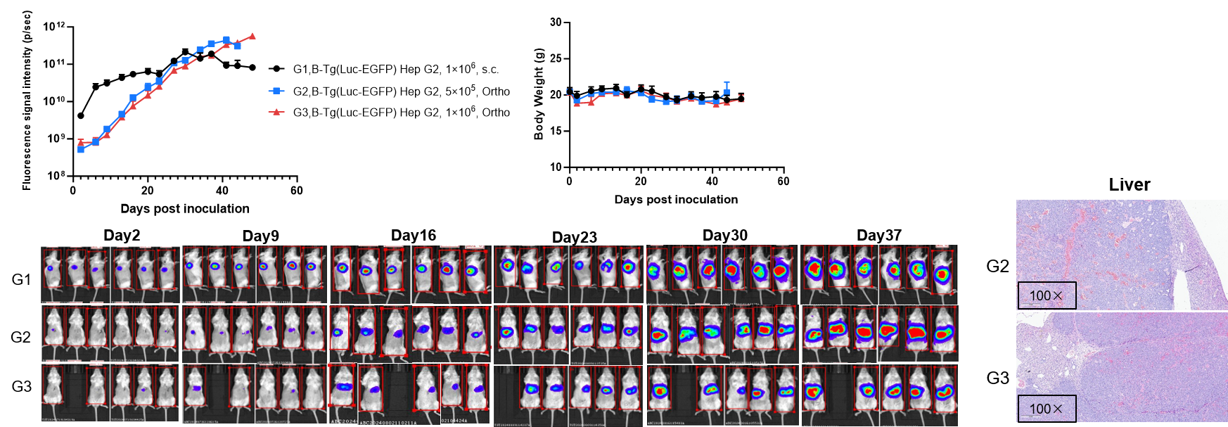
Establishment of orthotopic liver cancer model. To establish the liver cancer model, B-Tg(Luc-EGFP) Hep G2 cell suspension was injected into the left lateral lobe of the liver of B-NDG mice (femal, n=6), and the body weight and tumor fluorescence signal of the mice were recorded weekly. The results showed that the fluorescence signal gradually increased. Right panel showed H&E staining of liver tumors. This indicated that the cell line was successfully constructed as an orthotopic tumor model.
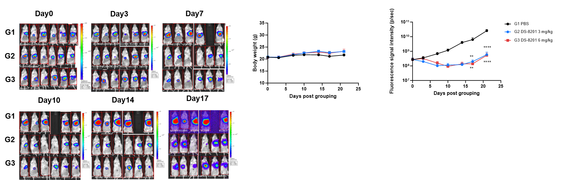
Antitumor activity of anti-HER2 antibody in B-Tg(Luc-EGFP) Hep G2 liver orthotopic model. The DS-8201 significantly B-Tg(Luc-EGFP) Hep G2 tumor growth in B-NDG mice. B-Tg(Luc-EGFP) Hep G2 cells (5×10⁵)suspension mixed in Matrigel solution (1:1) was orthotopically implanted into B-NDG mice (female, 8 week-old, n=6). Mice were grouped when Imaging signal value reached approximately 2.7×10⁸ p/sec, at which time they were treated with the DS-8201 with different doses and schedules indicated in panel (A) Body weight during treatment. As shown in panel B, this drug was efficacious, demonstrating that B-Tg(Luc-EGFP) Hep G2 liver orthotopic model could provide a powerful preclinical model for in vivo evaluation of DS-8201. Values are expressed as mean ± SEM. **p<0.01, ****p<0.0001