B-hKIT/hKITLG mice

C57BL/6-Kittm2(KIT)Bcgen Kitlgtm1(KITLG)Bcgen/Bcgen • 112090

B-hKIT/hKITLG mice

Catalog Number: 112090
Strain Name: C57BL/6-Kittm2(KIT)Bcgen Kitlgtm1(KITLG)Bcgen/Bcgen
Strain Background: C57BL/6
NCBI gene ID: 16590,17311 (Human)
Aliases: W; Bs; Fdc; Ssm; SCO1; SCO5; SOW3; CD117; c-KIT; Tr-kit; Gsfsco1; Gsfsco5; Gsfsow3; Gb; SF; Sl; Clo; Con; Mgf; SCF; SLF; blz; Kitlg; contrasted
---
ライセンスオプション提供可能
B-hKIT/hKITLG mice

このページで

  • Description
  • Targeting strategy
  • Phenotypic analysis
  • Efficacy

ポスター

すべて表示

    出版物

      Description

      SCF/KIT: A Critical Signaling Axis in Hematopoiesis, Mast Cell Biology, and Oncogenesis

      • Gene Information: Stem Cell Factor (SCF / KITLG) is a protein-coding gene on chromosome 12q21.32 that encodes a cytokine ligand. Its receptor, KIT (c-Kit / CD117), is a type III receptor tyrosine kinase proto-oncogene located on chromosome 4q12.
      • Protein Expression: SCF is expressed by bone marrow stromal cells, endothelial cells, fibroblasts, and keratinocytes as biologically active soluble or membrane-bound isoforms. KIT is highly expressed on hematopoietic stem cells (HSCs), mast cells, melanocytes, and interstitial cells of Cajal (ICCs).
      • Signaling Pathway: SCF binding to the extracellular domain of KIT induces receptor homodimerization and intracellular tyrosine autophosphorylation. This activates downstream PI3K/Akt, MAPK/ERK, and JAK/STAT pathways to regulate cell survival, proliferation, and migration.
      • Therapeutic Inhibition: Pathologic SCF/KIT signaling is targeted across two major clinical areas: Oncology: Small-molecule tyrosine kinase inhibitors (TKIs) like imatinib, sunitinib, and avapritinib block the ATP-binding site of mutated KIT, forcing tumor regression in GISTs and reducing mast cell burden in systemic mastocytosis. Autoimmunity & Allergy: In wild-type KIT diseases (e.g., Chronic Spontaneous Urticaria, asthma), biologics like barzolvolimab (anti-KIT mAb) or anti-SCF/anti-TSLP bispecific antibodies block ligand binding. This starves mast cells, inducing apoptosis to deplete tissue mast cells, lower serum tryptase, and suppress chronic inflammation.
      Targeting Strategy

      KIT

      • A chimeric CDS that encodes human KIT extracellular domain, mouse kit transmembrane and cytoplasmic domain, followed by mouse 3’UTR-STOP is inserted in exon 2 of mouse Kit gene.
      • The chimeric KIT protein expression will be driven by endogenous mouse kit promoter, while mouse Kit gene transcription and translation will be disrupted.

      KITLG

      • Exons 2-7 of mouse Kitlg gene that encode extracellular domain are replaced by human counterparts in B-hKIT/hKITLG mice.
      • The genomic region of mouse Kitlg gene that encodes transmembrane domain and cytoplasmic portion is retained. The promoter, 5’UTR and 3’UTR region of the mouse gene are also retained. The chimeric KITLG expression is driven by endogenous mouse Kitlg promoter, while mouse Kitlg gene transcription and translation will be disrupted.
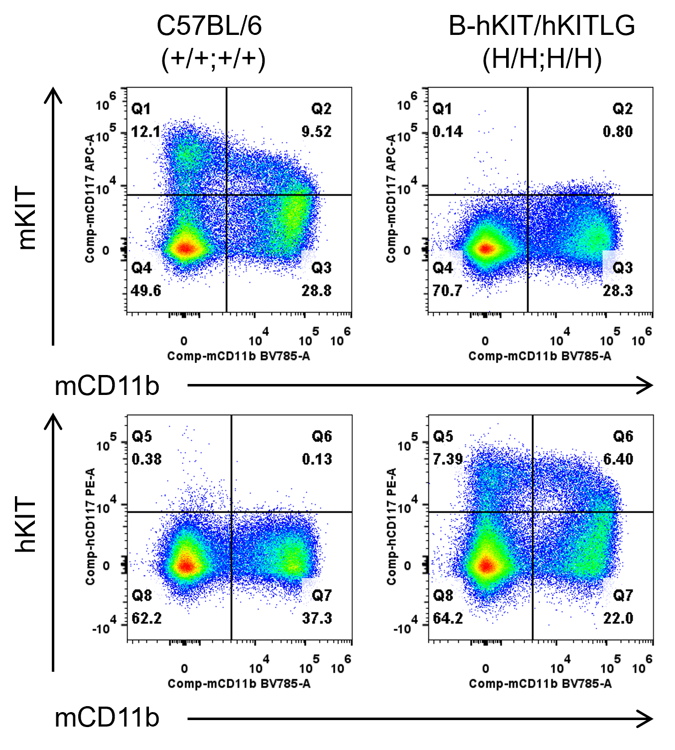
      KIT Protein Expression in Spleen
      • Mouse KIT was detectable in wild-type mice.
      • Human KIT was exclusively detectable in homozygous B-hKIT/hKITLG mice but not in wild-type mice.

      Mouse and human KIT expression analysis in Bone marrow cells. Bone marrow were collected from wild-type C57BL/6 mice and homozygous B-hKIT/hKITLG mice. KIT expression was analyzed by flow cytometry using anti-mouse KIT antibody (Biolegend, 105811) and anti-human KIT antibody (Biolegend, 313203).
      KITLG Protein Expression Analysis
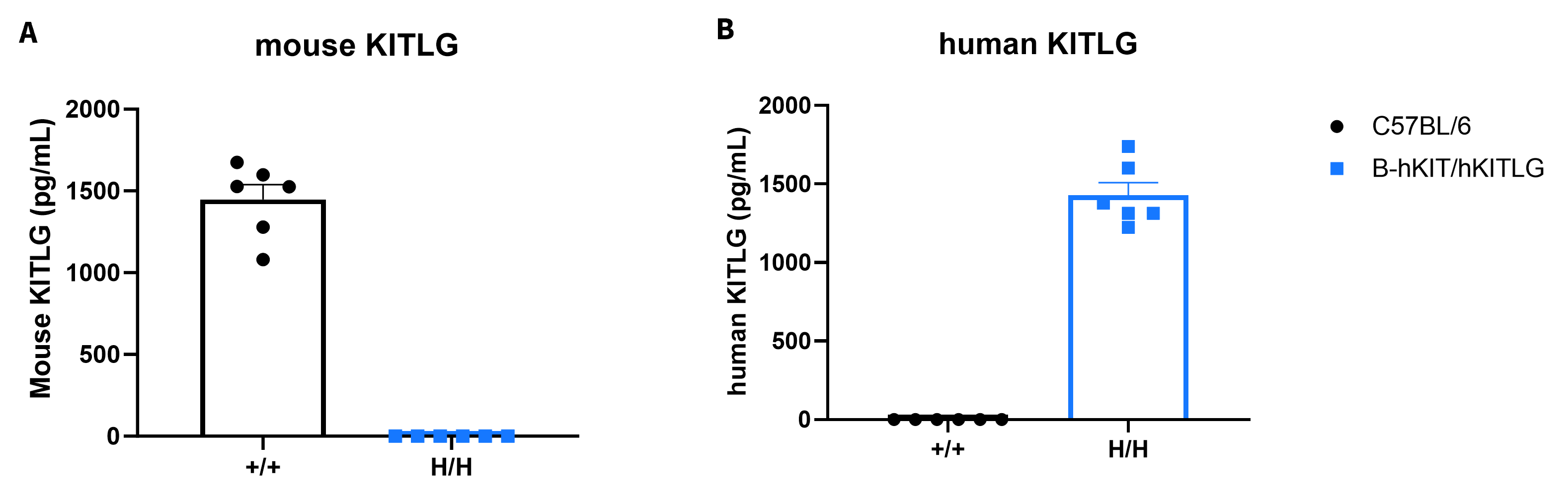
      • Mouse KITLG was detected exclusively in wild-type C57BL/6 mice.
      • Human KITLG was detected in homozygous B-hKIT/hKITLG mice, but not in wild-type mice.

      Strain specific KITLG expression analysis in homozygous B-hKITLG mice by ELISA. Serum was collected from wild-type C57BL/6 mice and homozygous B-hKITLG mice, and analyzed by ELISA with species-specific KITLG ELISA kit. Mouse KITLG was detectable in wild-type mice. Human KITLG was exclusively detectable in homozygous B-hKITLG mice but not in wild-type mice. Validation of KITLG protein expression data was not performed on double homozygous B-hKIT/hKITLG mice; theoretically, it should be consistent with the protein expression data from single-gene B-hKITLG mice (female, 7-week-old, n = 6). Mouse and human KITLG levels in serum were quantified by ELISA (mouse KITLG, abcam ab197750; human KITLG, Invitrogen EHKITLG). Values are expressed as mean ± SEM.
      mRNA expression analysis
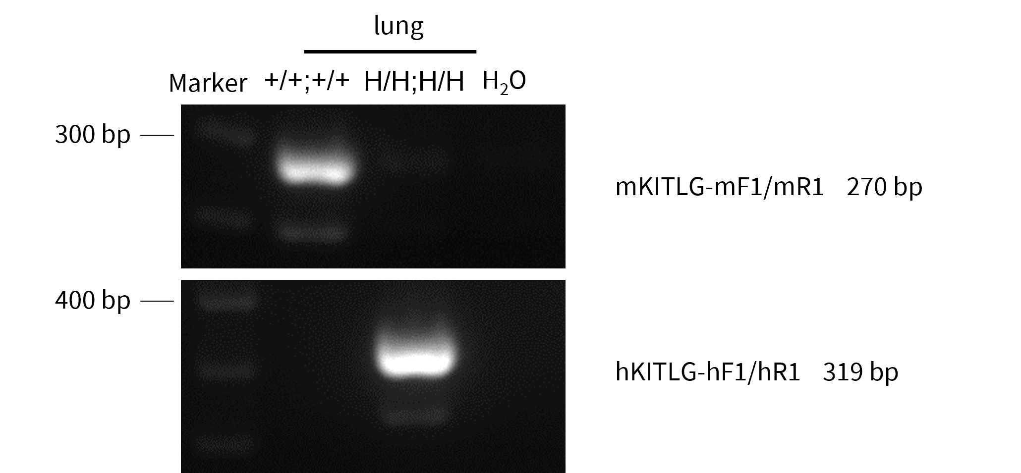
      • Both long and short human KITLG isoforms are specifically and correctly expressed in B-hKIT/hKITLG mice.

      Strain specific analysis of KITLG mRNA expression in wild-type C57BL/6JNifdc mice and B-hKIT/hKITLG mice by RT-PCR. lung RNA were isolated from wild-type C57BL/6JNifdc mice and homozygous B-hKIT/hKITLG mice, then cDNA libraries were synthesized by reverse transcription, followed by PCR with mouse and human KITLG primers. Mouse KITLG mRNA was only detectable in wild-type mice but not in homozygous B-hKIT/hKITLG mice. Human KITLG mRNA was only detectable in homozygous B-hKIT/hKITLG mice but not in wild-type mice.
      Analysis of Leukocyte Subpopulations
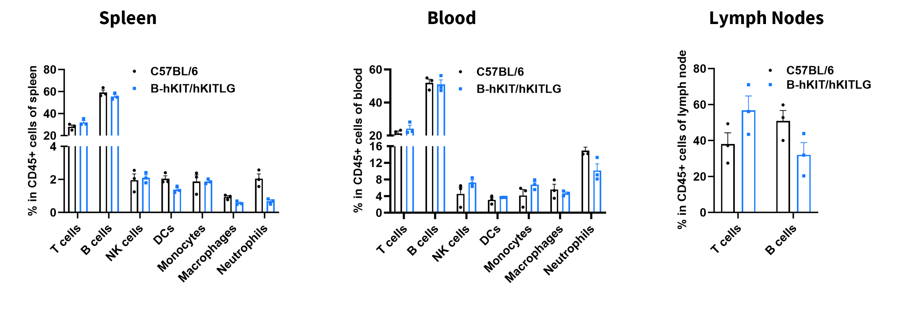
      • The percentages of T cells, B cells, NK cells, DCs, neutrophils, monocytes, and macrophages in homozygous B-hKIT/hKITLG mice were similar to those in C57BL/6 mice.
      • Humanization of KIT and KITLG does not affect normal immune cell development or splenic distribution.

      Analysis of leukocyte subpopulations by flow cytometry in immune organs and blood. Splenocytes, peripheral blood, and lymph nodes were isolated from female C57BL/6 and B-hKIT/hKITLG mice (female, 8-week-old, n = 3). Single live cells were gated on the CD45+ population and analyzed by flow cytometry as indicated. Values are expressed as mean ± SEM.

      Analysis of T Cell Subpopulations
      • The proportions of CD4⁺ T cells, CD8⁺ T cells, and Tregs in homozygous B-hKIT/hKITLG mice were comparable to those in C57BL/6 mice.
      • Humanization of KIT and KITLG does not affect normal T cell development, differentiation, or splenic distribution.

      Analysis of T-cell subpopulations by flow cytometry in immune organs and blood. Splenocytes, peripheral blood, and lymph nodes were isolated from female C57BL/6 and KIT and KITLG (female, 8-week-old, n = 3). Single live cells were gated on the CD3+ T-cell population and analyzed by flow cytometry as indicated. Values are expressed as mean ± SEM.

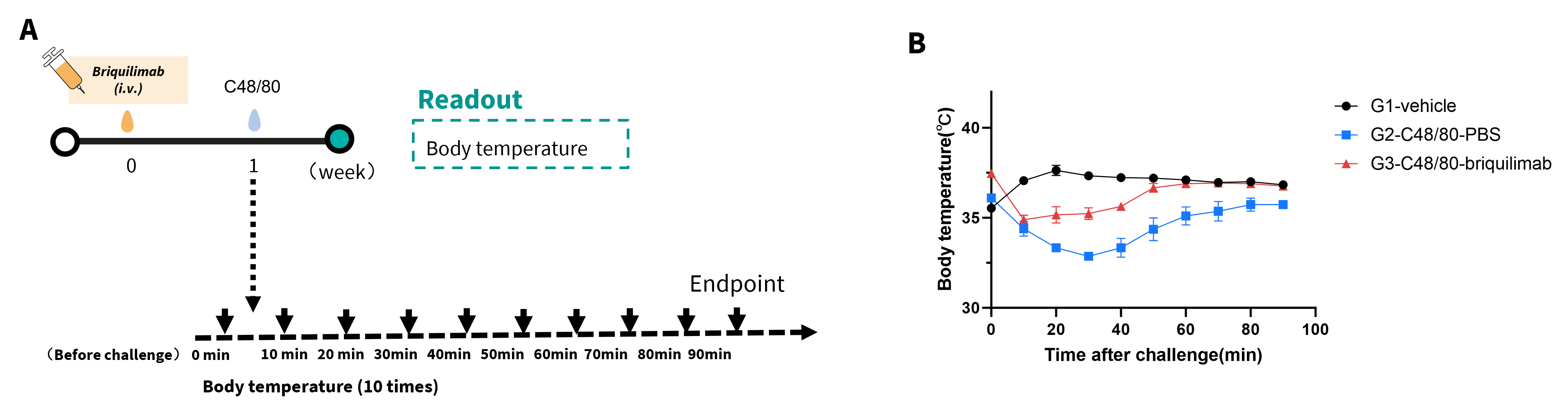
      Compound 48/80 (C48/80) Induced Systemic Anaphylaxis Reaction in B-hKIT Mice

      Effects of C48/80‑induced systemic anaphylaxis reaction. (A) Anti‑hKIT antibody briquilimab (25 mpk, commercially purchased) was injected before challenge at week 0. B‑hKIT mice (H/H) (n=3) were challenged with C48/80 at week 1. (B) Body temperature was recorded from 0 to 90 minutes after challenge with 2.5 mg/kg C48/80. briquilimab, by depleting mast cells, attenuated the decrease in body temperature induced by C48/80, demonstrating its inhibitory effect on the systemic anaphylaxis reaction in B-hKIT mice.
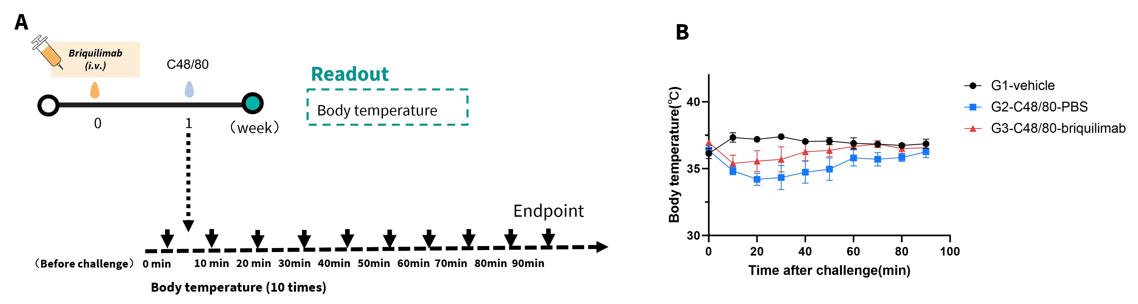
      Compound 48/80 (C48/80) Induced Systemic Anaphylaxis Reaction in B-hKIT/hKITLG Mice

      Effects of C48/80‑induced systemic anaphylaxis reaction. (A) Anti‑hKIT antibody briquilimab (25 mpk, commercially purchased) was injected before challenge at week 0. B-hKIT/hKITLG mice (H/H) (n=3) were challenged with C48/80 at week 1. (B) Body temperature was recorded from 0 to 90 minutes after challenge with 2.5 mg/kg C48/80. briquilimab, by depleting mast cells, attenuated the decrease in body temperature induced by C48/80, demonstrating its inhibitory effect on the systemic anaphylaxis reaction in B-hKIT/hKITLG mice.

      * When publishing results obtained using this animal model, please acknowledge the source as follows: The animal model [B-hKIT/hKITLG mice] (Cat# 112090) was purchased from Biocytogen.