Targeting Strategy
Gene targeting strategy for B-hPD-1/hLAG3 plus/hCD8 mice.
The exon 2 of mouse Pdcd1 gene encodes the IgV domain is replaced by human counterparts in B-hPD-1/hLAG3 plus/hCD8 mice. The promoter, 5’UTR and 3’UTR region of the mouse gene are also retained. The human PD-1 expression is driven by endogenous mouse Pdcd1 promoter, while mouse Pdcd1 gene transcription and translation will be disrupted.
The exons 2-7 of mouse Lag3 gene that encode extracellular domain are replaced by human counterparts in B-hPD-1/hLAG3 plus/hCD8 mice. The genomic region of mouse Lag3 gene that encodes transmembrane domain and cytoplasmic portion is retained. The promoter, 5’UTR and 3’UTR region of the mouse gene are also retained. The chimeric LAG3 expression is driven by endogenous mouse Lag3 promoter, while mouse Lag3 gene transcription and translation will be disrupted.
The exons 1-3 and partial exon 4 of mouse Cd8a gene that encode extracellular domain are replaced by human counterparts in B-hPD-1/hLAG3 plus/hCD8 mice. The genomic region of mouse Cd8a gene that encodes transmembrane domain and cytoplasmic portion is retained. The promoter, 5’UTR and 3’UTR region of the mouse gene are also retained. The chimeric CD8A expression is driven by endogenous mouse Cd8a promoter, while mouse Cd8a gene transcription and translation will be disrupted. The exons 1-3 and partial exon 4 of mouse Cd8b1 gene that encode extracellular domain are replaced by human counterparts in B-hPD-1/hLAG3 plus/hCD8 mice. The genomic region of mouse Cd8b gene that encodes transmembrane domain and cytoplasmic portion is retained. The promoter, 5’UTR and 3’UTR region of the mouse gene are also retained. The chimeric CD8B expression is driven by endogenous mouse Cd8b1 promoter, while mouse Cd8b1 gene transcription and translation will be disrupted.
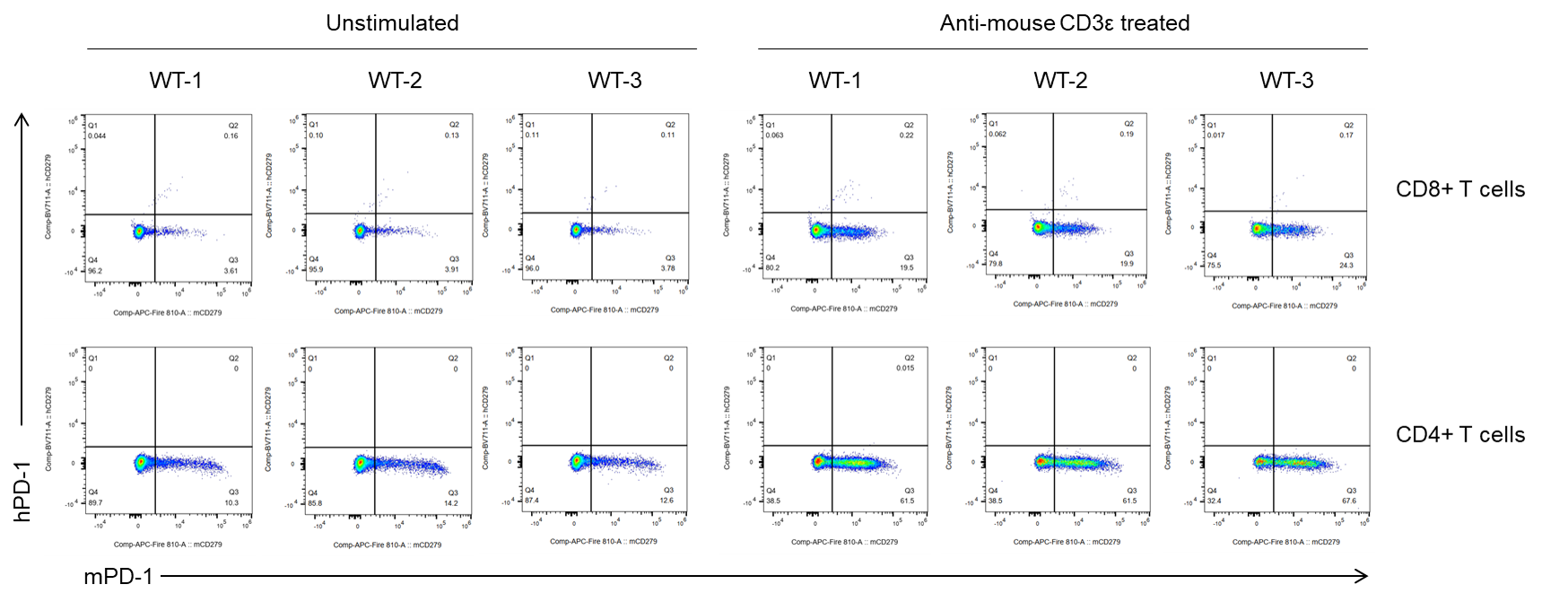
Protein Expression Analysis in Spleen of Wild-type (WT) Mice
Strain specific PD-1 expression analysis in wild-type mice by flow cytometry. Splenocytes were collected from wild-type C57BL/6JNifdc mice after stimulated with or without anti-mouse CD3ε antibody (7.5 μg, i.p.) in vivo for 24 hrs (female, 12-week-old, n=3). Protein expression was analyzed with anti-mouse PD-1 antibody (Biolegend, 135252), and anti-human PD-1 antibody (Biolegend, 329928) by flow cytometry. Mouse PD-1 was only detectable in wild-type C57BL/6JNifdc mice.
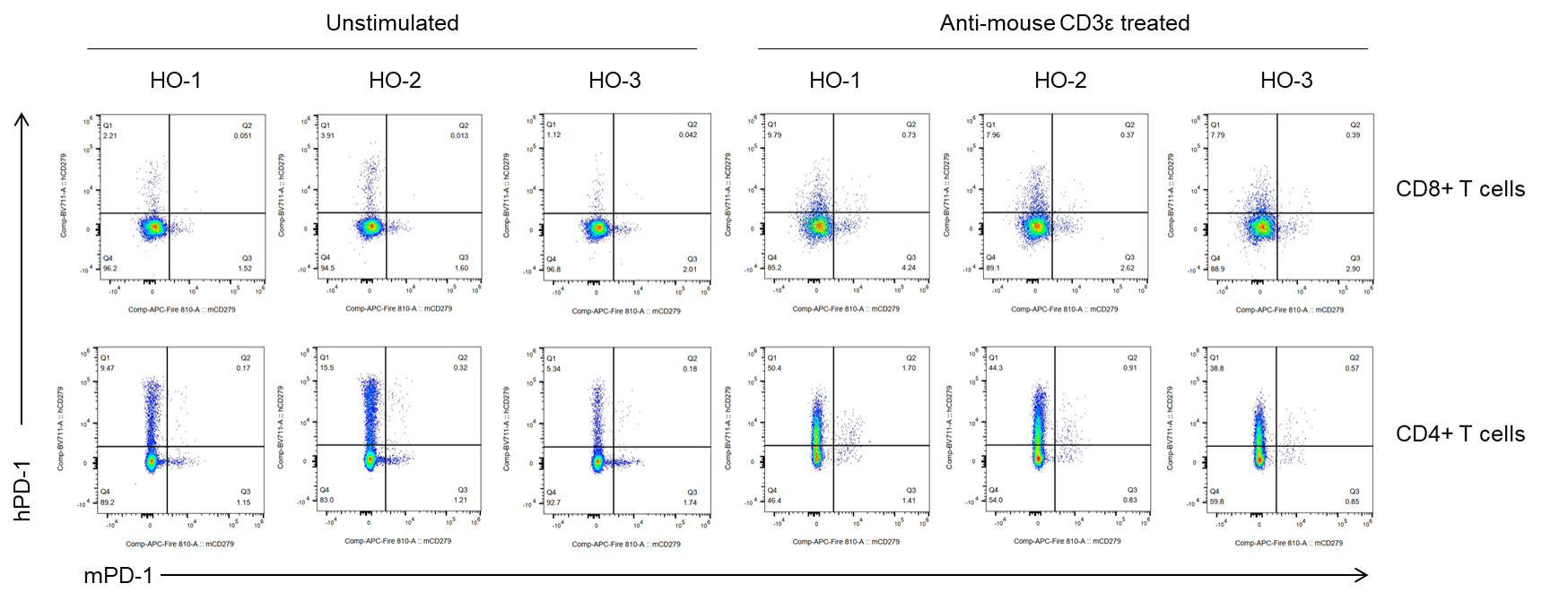
Protein Expression Analysis in Spleen of Homozygous (HO) Mice
Strain specific PD-1 expression analysis in homozygous B-hPD-1/hLAG3 plus/hCD8 mice by flow cytometry. Splenocytes were collected from homozygous B-hPD-1/hLAG3 plus/hCD8 mice after stimulated with or without anti-mouse CD3ε antibody (7.5 μg, i.p.) in vivo for 24 hrs (female, 12-week-old, n=3). Protein expression was analyzed with anti-mouse PD-1 antibody (Biolegend, 135252), and anti-human PD-1 antibody (Biolegend, 329928) by flow cytometry. Human PD-1 was only detectable in homozygous B-hPD-1/hLAG3 plus/hCD8 mice.
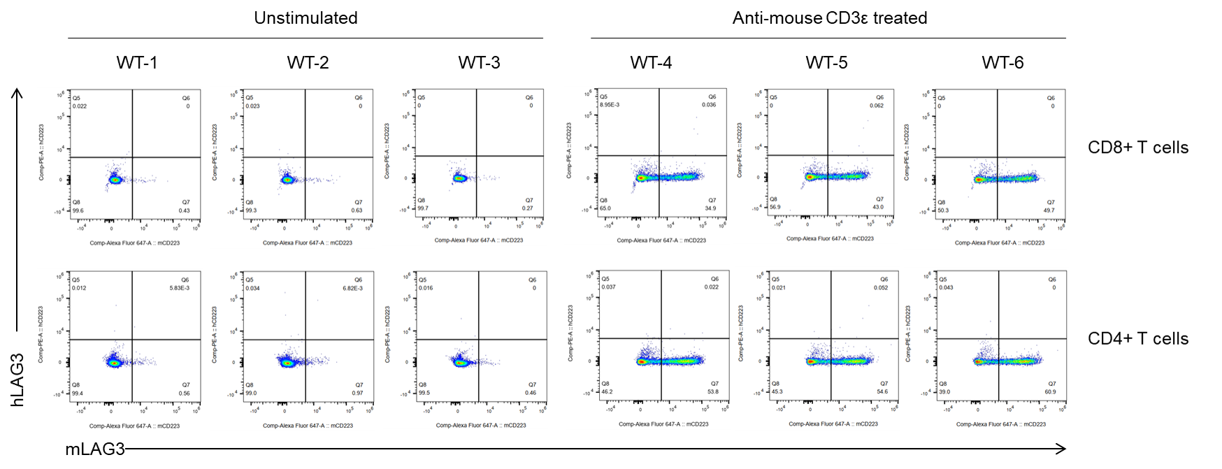
Protein Expression Analysis in Spleen of Wild-type (WT) Mice
Strain specific LAG3 expression analysis in wild-type mice by flow cytometry. Splenocytes were collected from wild-type C57BL/6JNifdc mice after stimulated with or without anti-mouse CD3ε antibody (7.5 μg, i.p.) in vivo for 24 hrs (female, 12-week-old, n=3). Protein expression was analyzed with anti-mouse LAG3 antibody (Biolegend, 125242), and anti-human LAG3 antibody (Biolegend, 369306) by flow cytometry. Mouse LAG3 was only detectable in wild-type C57BL/6JNifdc mice.
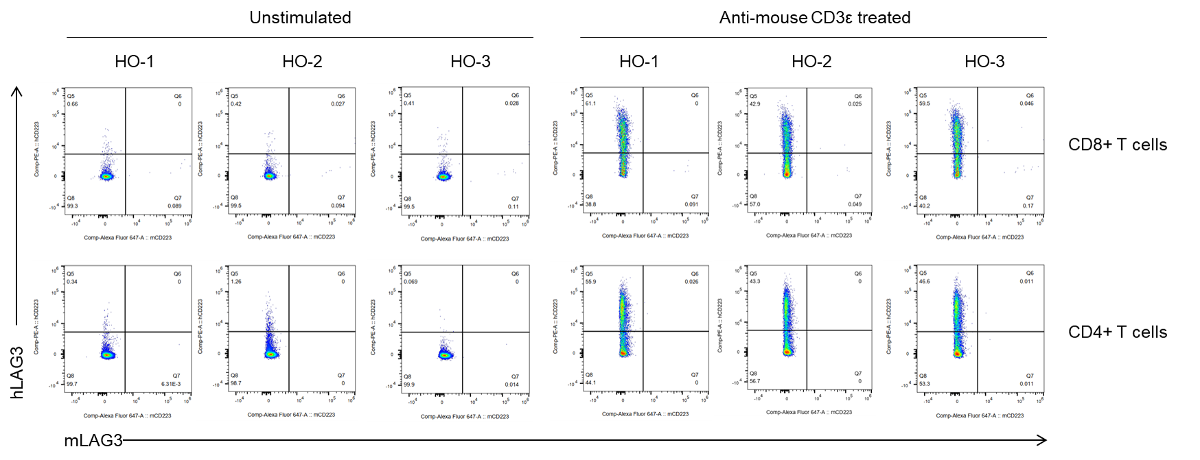
Protein Expression Analysis in Spleen of Homozygous (HO) Mice
Strain specific LAG3 expression analysis in homozygous B-hPD-1/hLAG3 plus/hCD8 mice by flow cytometry. Splenocytes were collected from homozygous B-hPD-1/hLAG3 plus/hCD8 mice after stimulated with or without anti-mouse CD3ε antibody (7.5 μg, i.p.) in vivo for 24 hrs (female, 12-week-old, n=3). Protein expression was analyzed with anti-mouse LAG3 antibody (Biolegend, 125242), and anti-human LAG3 antibody (Biolegend, 369306) by flow cytometry. Human LAG3 was only detectable in B-hPD-1/hLAG3 plus/hCD8 mice.
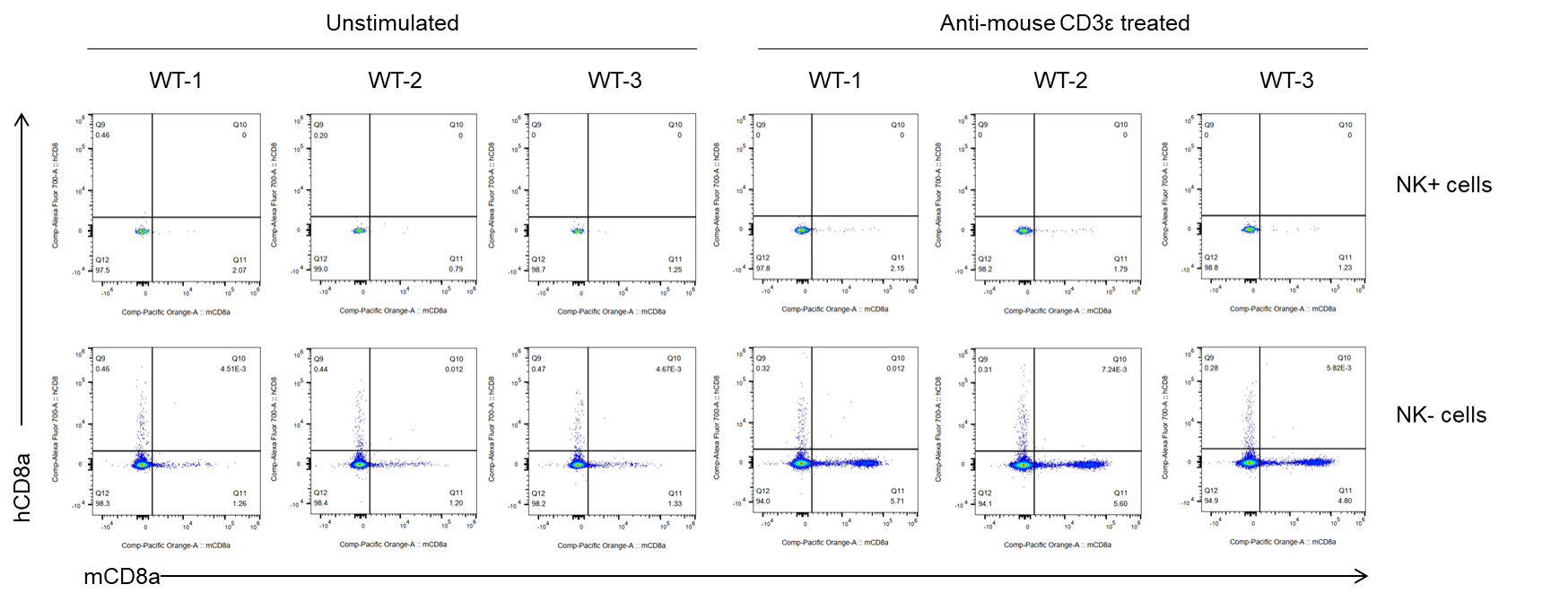
Protein Expression Analysis in Spleen of Wild-type (WT) Mice
Strain specific CD8 expression analysis in wild-type mice by flow cytometry. Splenocytes were collected from wild-type C57BL/6JNifdc mice after stimulated with or without anti-mouse CD3ε antibody (7.5 μg, i.p.) in vivo for 24 hrs (female, 12-week-old, n=3). Protein expression was analyzed with anti-mouse LAG3 antibody (Invitrogen, MCD0830), and anti-human CD8a antibody (Biolegend, 300920) by flow cytometry. Mouse CD8a was only detectable in wild-type C57BL/6JNifdc mice.
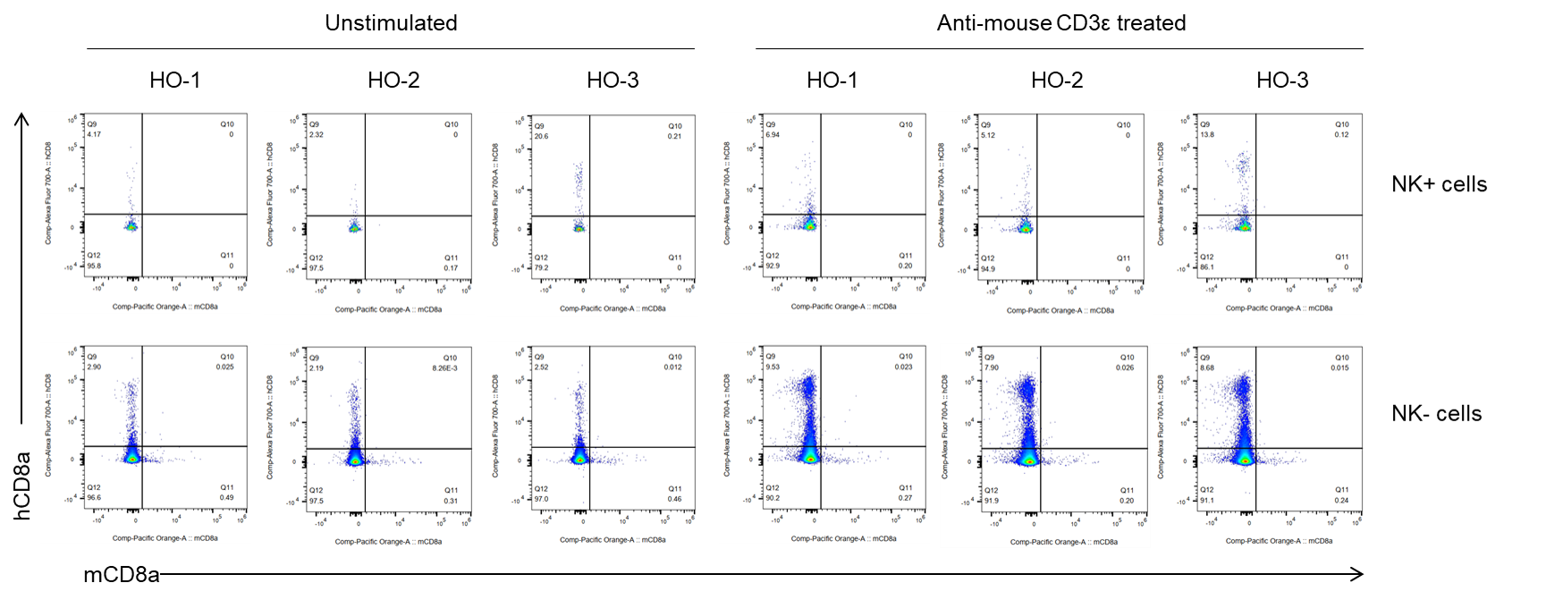
Protein Expression Analysis in Spleen of Homozygous (HO) Mice
Strain specific CD8 expression analysis in wild-type mice by flow cytometry. Splenocytes were collected from homozygous B-hPD-1/hLAG3 plus/hCD8 mice after stimulated with or without anti-mouse CD3ε antibody (7.5 μg, i.p.) in vivo for 24 hrs (female, 12-week-old, n=3). Protein expression was analyzed with anti-mouse LAG3 antibody (Invitrogen, MCD0830), and anti-human CD8a antibody (Biolegend, 300920) by flow cytometry. Human CD8a was only detectable in detectable in B-hPD-1/hLAG3 plus/hCD8 mice.
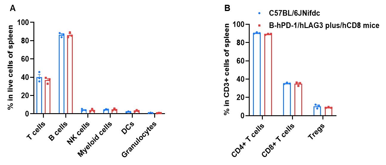
Frequency of Leukocyte Subpopulations in Spleen
Frequency of leukocyte subpopulations in spleen by flow cytometry. Splenocytes were isolated from wild-type C57BL/6JNifdc mice and homozygous in B-hPD-1/hLAG3 plus/hCD8 mice (female, 12-week-old, n=3). A. Flow cytometry analysis of the splenocytes was performed to assess the frequency of leukocyte subpopulations. B. Frequency of T cell subpopulations. Frequencies of T cells, B cells, NK cells, dendritic cells, myeloid cells, granulocytes, CD4+ T cells, CD8+ T cells, and Tregs in B-hPD-1/hLAG3 plus/hCD8 mice were similar to those in C57BL/6JNifdc mice, demonstrating that humanization of PD-1, LAG3, and CD8 does not change the frequency or distribution of these cell types in spleen. The frequency of leukocyte subpopulations in blood, bone marrow and thymus of B-hPD-1/hLAG3 plus/hCD8 mice were also comparable to wild-type C57BL/6 mice (Data not shown).
* When publishing results obtained using this animal model, please acknowledge the source as follows: The animal model [B-hPD-1/hLAG3 plus/hCD8 mice] (Cat# 114083) was purchased from Biocytogen.