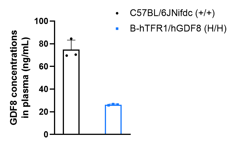
Protein expression analysis of GDF8
GDF8 protein expression analysis in homozygous B-hTFR1/hGDF8 mice by ELISA. Plasma were collected from wild-type C57BL/6JNifdc mice (female, n=3, 9-week-old) and homozygous B-hTFR1/hGDF8 mice (female, n=3, 9-week-old). Protein expression level of GDF8 was analyzed by ELISA with anti-GDF8 ELISA kit (R&D, DGDF80). GDF8 was detectable in both wild-type C57BL/6JNifdc mice and homozygous B-hTFR1/hGDF8 mice, as the antibody in the ELISA kit was cross-reactive between human and mouse.
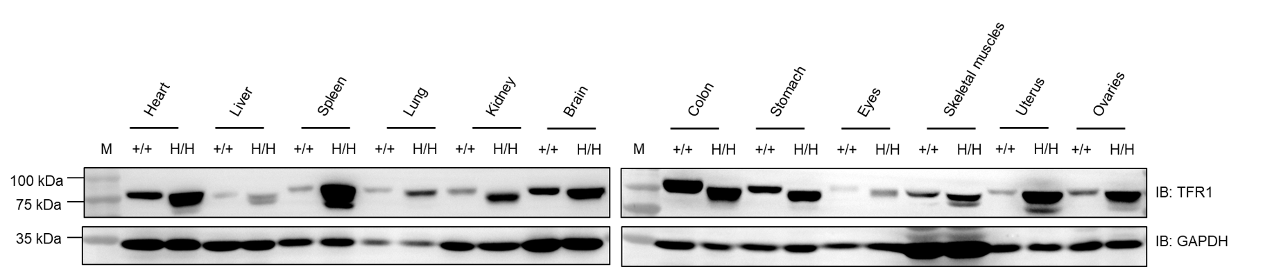
Protein expression analysis of TFR1 in multiple tissues
Western blot analysis of TFR1 protein expression in wild-type C57BL/6JNidc mice and homozygous B-hTFR1/hGDF8 mice by WB. Various tissues were collected from wild-type C57BL/6JNifdc mice (+/+) and homozygous B-hTFR1/hGDF8 mice (H/H), and then analyzed by western blot with anti-TFR1 antibody (abcam, ab214039). 40 μg total protein was loaded for western blotting analysis. GAPDH was detected as an internal control. TFR1 was detectable in heart, liver, spleen, lung, kidney, brain, stomach, colon, skeletal muscle, eyes, uterus and ovaries from both C57BL/6JNifdc and homozygous B-hTFR1/hGDF8 mice, as the antibody was cross-reactive between human and mouse. M, marker.
Frequency of leukocyte subpopulations in spleen
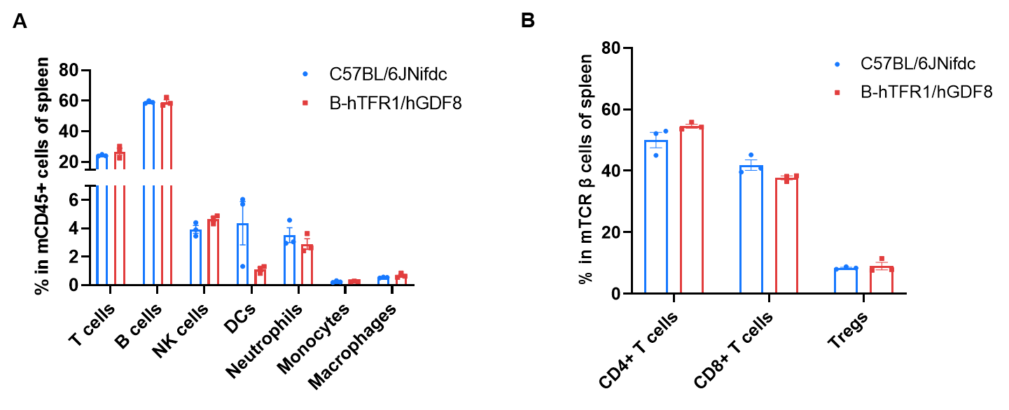
Frequency of leukocyte subpopulations in spleen by flow cytometry. Splenocytes were isolated from wild-type C57BL/6JNifdc mice (female, n=3, 9-week-old) and homozygous B-hTFR1/hGDF8 mice (female, n=3, 9-week-old). A. Flow cytometry analysis of the splenocytes was performed to assess the frequency of leukocyte subpopulations. B. Frequency of T cell subpopulations. Percentages of T cells, B cells, NK cells, DCs, neutrophils, monocytes, macrophages, CD4+T cells, CD8+T cells and Tregs in B-hTFR1/hGDF8 mice were similar to those in C57BL/6JNifdc mice. Humanization of TFR1 does not change the overall frequency or distribution of immune cell types in spleen, blood and lymph nodes. The frequency of leukocyte subpopulations in bone marrow, lymph nodes and blood of B-hTFR1/hGDF8 mice were also comparable to wild-type C57BL/6JNifdc mice (Data not shown). Values are expressed as mean ± SEM. Significance was determined by two-way ANOVA test. *p < 0.05, **p < 0.01, ***p < 0.001.
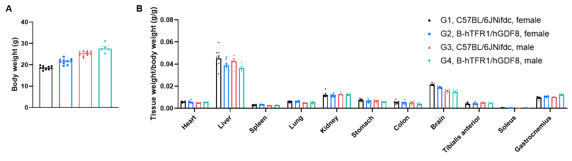
Body weights and tissue weights
Comparison of body weights and tissue weights between wild-type C57BL/6JNifdc and homozygous B-hTFR1/hGDF8 mice. A. The body weights of humanized homozygous B-hTFR1/hGDF8 mice (female, 9-week-old, n=10; male, 9-week-old, n=5) was comparable with wild-type C57BL/6JNifdc mice (female, 9-week-old, n=10; male, 9-week-old, n=10). B. There is no difference in tissue weight between the wild-type C57BL/6JNifdc mice and homozygous B-hTFR1/hGDF8 mice.
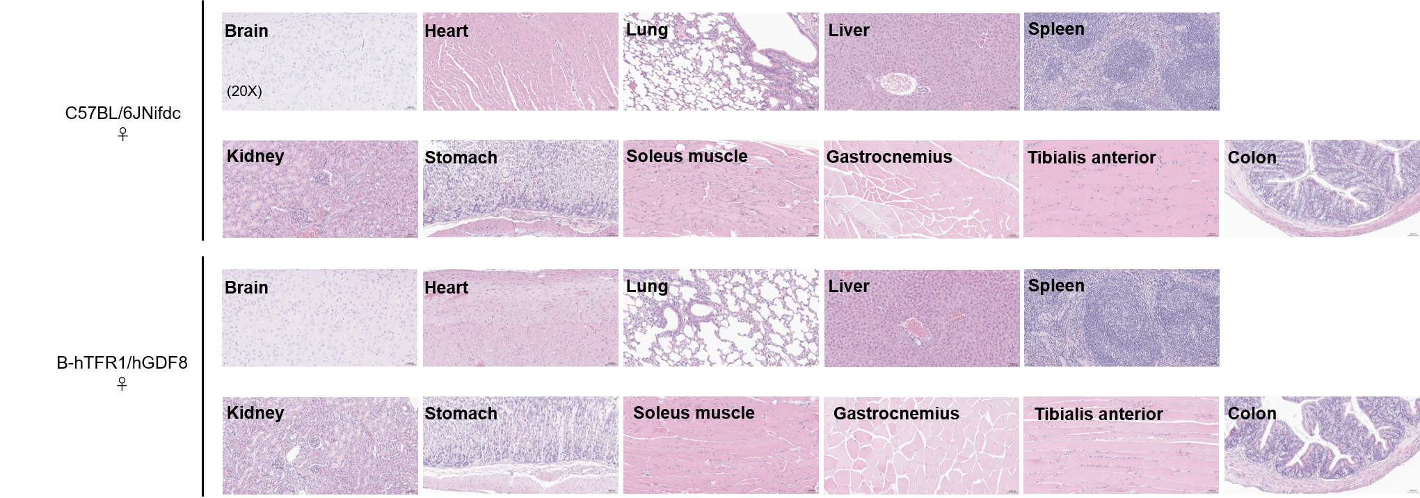
Histopathological Analysis
Histopathological analysis of organs in B-hTFR1/hGDF8 mice. The main organs of B-hTFR1/hGDF8 mice were isolated at 9 weeks of age and analyzed with H&E staining (female, n=3). Results showed that no obvious abnormalities were found in all of the organs (brain, heart, lung, liver, spleen, kidney, stomach, soleus muscle, gastrocnemius, tibialis anterior and colon).
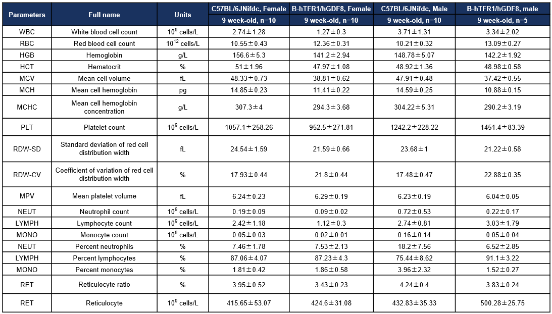
Hematology analysis
Complete blood count (CBC) of B-hTFR1/hGDF8 mice. Values are expressed as mean ± SD.
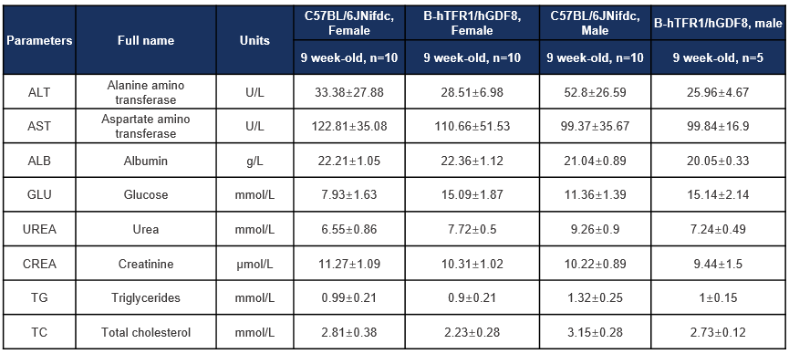
Biochemistry analysis
Blood chemistry tests of B-hTFR1/hGDF8 mice. Serum from male and female C57BL/6JNifdc and B-hTFR1/hGDF8 mice (female, n=10, 9 week-old; male, n=5, 9 week-old) were collected for biochemistry analysis. There were no differences in either measurement between C57BL/6JNifdc and B-hTFR1/hGDF8 mice, indicating that humanization of TFR1 and GDF8 does not change the biochemistry of blood. Values are expressed as mean ± SD.
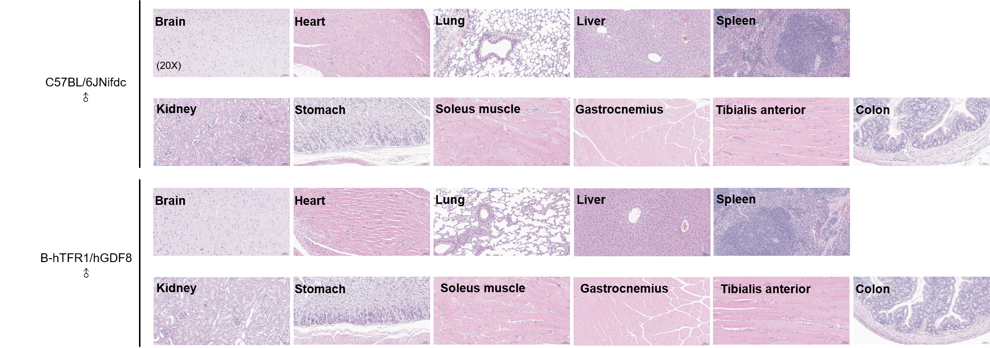
Histopathological Analysis
Histopathological analysis of organs in B-hTFR1/hGDF8 mice. The main organs of B-hTFR1/hGDF8 mice were isolated at 9 weeks of age and analyzed with H&E staining (male, n=3). Results showed that no obvious abnormalities were found in all of the organs (brain, heart, lung, liver, spleen, kidney, stomach, soleus muscle, gastrocnemius, tibialis anterior and colon). Scale bar: 100 μm.
* When publishing results obtained using this animal model, please acknowledge the source as follows: The animal model [B-hTFR1/hGDF8 mice] (Cat# 113849) was purchased from Biocytogen.