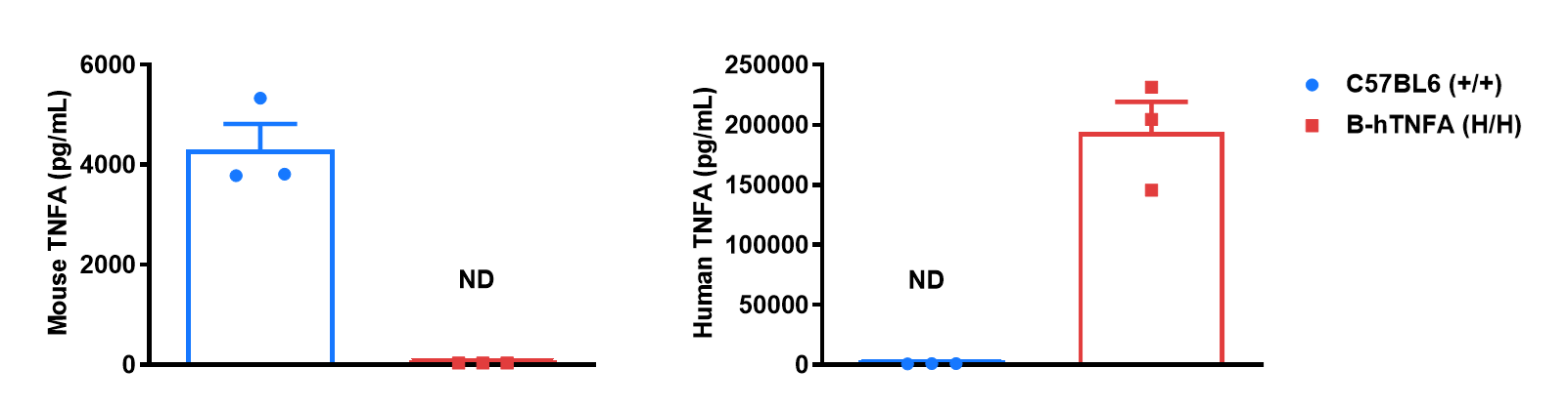
Protein expression analysis in serum
Strain specific TNFA expression analysis in wild-type C57BL/6 mice and homozygous humanized B-hTNFA mice by ELISA. Serum was collected from wild-type C57BL/6 mice (+/+) and homozygous B-hTNFA mice (H/H) stimulated with LPS (20 μg/mice) in vivo for 1 hour (female, 8-week-old, n=3). Expression level of TNFA were analyzed by ELISA. Mouse TNFA was only detectable in wild-type C57BL/6 mice. Human TNFA was exclusively detectable in homozygous B-hTNFA mice. Values are expressed as mean ± SEM. ND: not detectable.
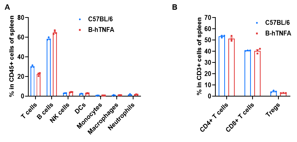
Frequency of leukocyte subpopulations in spleen
Frequency of leukocyte subpopulations in spleen by flow cytometry. Splenocytes were isolated from wild-type C57BL/6 mice and homozygous B-hTNFA mice (female, 8-week-old, n=3). A. Flow cytometry analysis of the splenocytes was performed to assess the frequency of leukocyte subpopulations. B. Frequencies of T cell subpopulations. Percentages of T cells, B cells, NK cells, DCs, monocytes, macrophages, neutrophils, CD4+ T cells, CD8+ T cells and Tregs in B-hTNFA mice were similar to those in C57BL/6 mice, demonstrating that humanization of TNFA does not change the frequency or distribution of these cell types in spleen. Values are expressed as mean ± SEM.
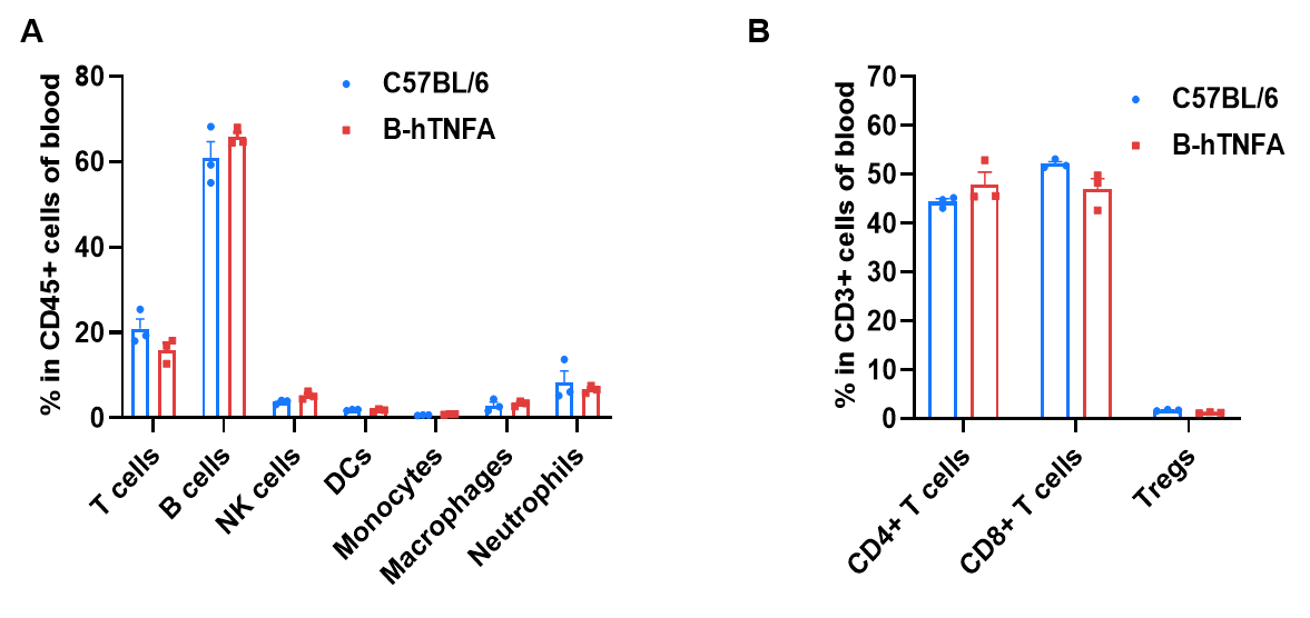
Frequency of leukocyte subpopulations in blood
Frequency of leukocyte subpopulations in blood by flow cytometry. Blood cells were isolated from wild-type C57BL/6 mice and homozygous B-hTNFA mice (female, 8-week-old, n=3). A. Flow cytometry analysis of the blood cells was performed to assess the frequency of leukocyte subpopulations. B. Frequencies of T cell subpopulations. Percentages of T cells, B cells, NK cells, DCs, monocytes, macrophages, neutrophils, CD4+ T cells, CD8+ T cells and Tregs in B-hTNFA mice were similar to those in C57BL/6 mice, demonstrating that humanization of TNFA does not change the frequency or distribution of these cell types in blood. Values are expressed as mean ± SEM.
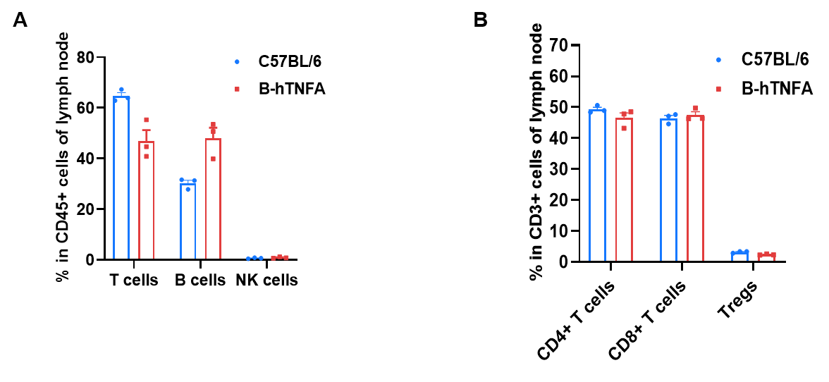
Frequency of leukocyte subpopulations in lymph nodes
Frequency of leukocyte subpopulations in lymph nodes by flow cytometry. Leukocytes were isolated from wild-type C57BL/6 mice and homozygous B-hTNFA mice (female, 8-week-old, n=3). A. Flow cytometry analysis of the leukocytes was performed to assess the frequency of leukocyte subpopulations. B. Frequencies of T cell subpopulations. Percentages of T cells, B cells, NK cells, CD4+ T cells, CD8+ T cells and Tregs in B-hTNFA mice were similar to those in C57BL/6 mice, demonstrating that humanization of TNFA does not change the frequency or distribution of these cell types in lymph nodes. Values are expressed as mean ± SEM.
In vivo efficacy of anti-human TNFA antibody in CAIA model

Efficacy of anti-human TNFA antibody in B-hTNFA mice with collagen antibody induced arthritis (CAIA) model. B-hTNFA mice (female, n=10, 9-10 weeks-old) were intravenously injected with collagen antibody (mAb) on Day 0, followed by intraperitoneally injection of LPS on day 3 (A). Body weight (B) and clinical score (C) were measured on Day 0 and daily from Day 3 to Day 20 of the study. The treatment group received either anti-human TNFA antibody Adalimumab analog (in house) or methotrexate (purchased from MCE). The model group (G2 group) exhibited an arthritis phenotype after LPS injection, and its clinical score stabilized after 8 days. This indicated that the CAIA model was successfully established in B-hTNFA mice.. The clinical score of Adalimumab analog (in house) treatment group and methotrexate (purchased from MCE) were significantly lower than those of the model group (G2 group). The results indicated B-hTNFA mice provide a powerful preclinical CAIA mouse model for in vivo evaluation of anti-human TNFA antibody.
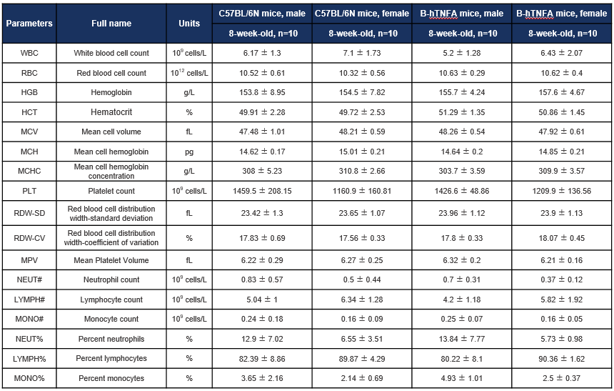
Hematology analysis
Complete blood count (CBC) of B-hTNFA mice. Values are expressed as mean ± SD.
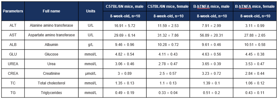
Biochemistry analysis
Biochemical test of B-hTNFA mice. Values are expressed as mean ± SD.
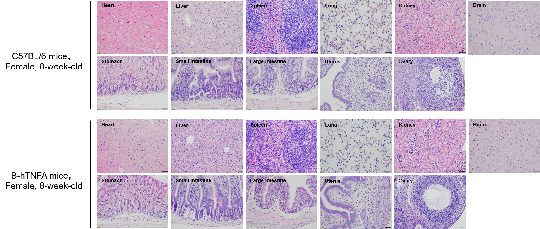
Histopathological analysis
Histopathological analysis of organs in B-hTNFA mice. The main organs of B-hTNFA mice and C57BL/6 mice (female, 8-week-old, n=10) were isolated and analyzed with H&E staining. Results showed that no obvious abnormalities were found in all of the organs (heart, liver, spleen, lung, kidney, brain, stomach, small intestine, large intestine, uterus and ovary). Scale bar: 100 μm.
* When publishing results obtained using this animal model, please acknowledge the source as follows: The animal model [B-hTNFA mice] (Cat# 110002) was purchased from Biocytogen.