Description
CD3/CD28/PSMA: A tri-targeted signaling axis for precise T-cell redirecting and sustained activation
- CD3 Molecule: As a vital component of the T-cell receptor (TCR) complex, CD3 is responsible for transducing antigen recognition signals into the cell interior. The engagement and ligation of CD3 can directly induce T-cell activation.
- Gene Information: CD3 is a primary T-cell co receptor complex transducing Signal 1; CD28 is a critical T-cell costimulatory receptor supplying Signal 2; PSMA (Prostate Specific Membrane Antigen) is a tumor associated cellsurface antigen highly overexpressed on prostate carcinoma cells and tumor vasculature.
- Protein Expression: Both CD3 and CD28 are constitutively and highly expressed on the surface of mature naive and memory T lymphocytes. This ubiquitous co expression profile makes them ideal foundational targets for universally engaging and activating T cells. PSMA is a type II transmembrane glycoprotein belonging to the zincdependent exopeptidase superfamily, possessing folate hydrolase and neurocarboxypeptidase activities. It is expressed at low levels in tissues such as the kidney, brain, salivary glands, liver, ganglia, and small intestine, among others, but is highly expressed across all stages of prostate cancer. Targeting PSMA recruits and anchors T-cells into spatial proximity with the tumor cells to direct specific lysis.
- T-Cell Engagement (TCE) & Dual Signaling: Simultaneous engagement of CD3 (Signal 1) and PSMA brings T-cells into close spatial proximity with tumor cells to initiate target specific lysis. Concurrent activation of CD28 (Signal 2) triggers downstream costimulatory cascades, preventing T-cell exhaustion and sustaining long term anti tumor immune responses.
- Therapeutic Engagement: By engaging CD3 for tumor targeting and CD28 for costimulation, the trispecific strategy delivers both activation signals, overcoming T cell anergy and enabling full activation to mediate potent and sustained lysis of PSMA positive malignancies.
Key Advantages
- Accurate Modeling of Human Biology Expresses and Function:This effectively overcomes the significant species-specific differences inherent to traditional mouse models.
- An intact immune system:The model provides stable and repeatable data, making it ideal model for large-scale, high-throughput molecular screening.
- Enhanced R&D Efficiency:It enables effective preclinical evaluation of antibodies, thereby improving candidate screening accuracy, reducing failure costs, and shortening the R&D cycle.
Key Applications
- Efficacy Evaluation:To evaluate the efficacy and safety of a CD3/CD28/PSMA-targeted trispecific antibody, disease models were established by engrafting gene-humanized cell lines.
- Safety and CRS Research:This model assesses the safety of T-cell engagers, particularly the risk of Cytokine Release Syndrome (CRS).
- Novel Therapeutic Screening:Support the discovery, screening, and optimization of new treatment strategies, leveraging a relevant humanized models for translational research.
Targeting Strategy
CD3EDG
- The chimeric human CD3EDG was expressed, while mouse Cd3edg were knocked out in B-hCD3EDG/hCD28/hPSMA mice.
CD28
- The exons 2-3 of mouse Cd28 gene that encode the extracellular domain were replaced by human CD28 exons 2-3 in B-hCD3EDG/hCD28/hPSMA mice.
PSMA
- A chimeric CDS that encodes human PSMA extracellular domain, mouse Psma transmembrane and cytoplasmic domain, followed by mouse 3’UTR-STOP is inserted into mouse Psma exon 2.
- The genomic region of mouse Psma gene that encodes cytoplasmic portion and transmembrane domain is retained. The promoter, 5’UTR and 3’UTR region of the mouse gene are also retained. The chimeric PSMA expression is driven by endogenous mouse Psma promoter, while mouse Psma gene transcription and translation will be disrupted.
CD3E Protein Expression
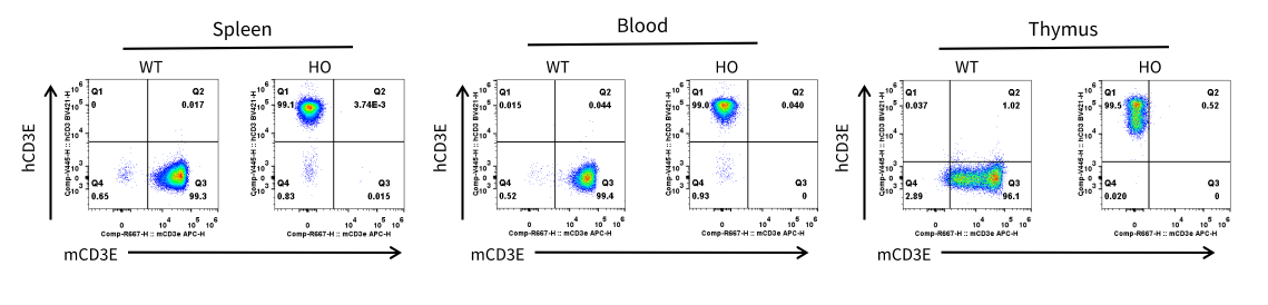
Strain specific CD3E expression analysis in homozygous B-hCD3EDG/hCD28/hPSMA mice by flow cytometry. Splenocytes, blood and thymus were collected from wild-type C57BL/6JNifdc mice (WT) and homozygous B-hCD3EDG/hCD28/hPSMA mice (HO), and analyzed by flow cytometry with species-specific anti-mouse CD3E antibody (Biolegend, 100312) and anti-human CD3E antibody (Biolegend, 317344).
CD28 Protein Expression
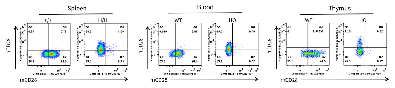
Strain specific CD28 expression analysis in homozygous B-hCD3EDG/hCD28/hPSMA mice by flow cytometry. Splenocytes, blood and thymus were collected from wild-type C57BL/6JNifdc mice (WT) and homozygous B-hCD3EDG/hCD28/hPSMA mice (HO), and analyzed by flow cytometry with species-specific anti-mouse CD28 antibody (Biolegend, 102105) and anti-human CD28 antibody (Biolegend, 302912).
PSMA Protein Expression
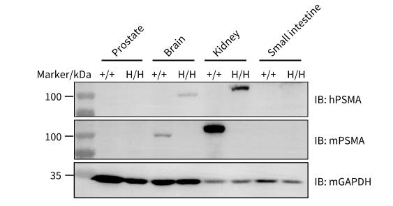
Western blot analysis of hPSMA protein expression in homozygous B-hCD3EDG/hCD28/hPSMA mice. Various tissue lysates were collected from wild-type C57BL/6JNifdc mice (+/+) and homozygous B-hCD3EDG/hCD28/hPSMA mice (H/H), and then analyzed by western blot with species-specific anti-human PSMA antibody (CST, 12703) and anti-mouse PSMA antibody (abcam, ab134142), 40 μg of total proteins were loaded for western blot analysis.
- Human PSMA was detected in brain and kidney of B-hCD3EDG/hCD28/hPSMA mice, as the antibody cross-reacted with mouse and humanPSMA.
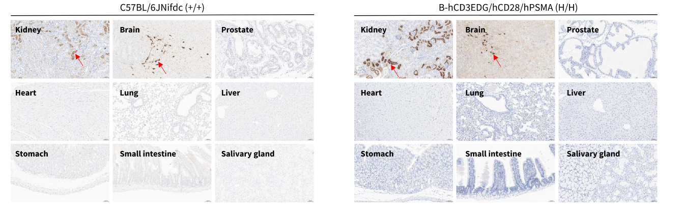
Immunohistochemical (IHC) analysis of PSMA protein expression in homozygous B-hCD3EDG/hCD28/hPSMA mice. Various tissue lysates were collected from wild-type C57BL/6JNifdc mice (+/+) and homozygous B-hCD3EDG/hCD28/hPSMA mice (H/H), and then analyzed by IHC with anti-PSMA antibody (abcam, ab133579).
Analysis of Leukocyte Subpopulations
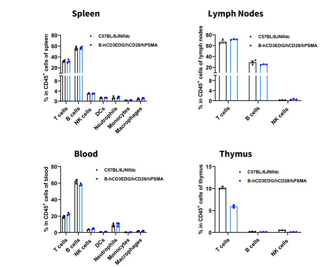
Analysis of leukocyte subpopulations by flow cytometry in immune organs and blood.Splenocytes, peripheral blood, lymph nodes, and thymus were isolated from C57BL/6JNifdc and B-hCD3EDG/hCD28/hPSMA mice (female, 8-week-old, n=3). Single live cells were gated on the CD45⁺ population and analyzed by flow cytometry as indicated. Values are expressed as mean ± SEM.
Analysis of T Cell Subpopulations
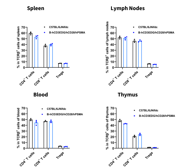
Analysis of T-cell subpopulations by flow cytometry in immune organs and blood.Splenocytes, peripheral blood, lymph nodes, and thymus were isolated from C57BL/6JNifdc and B-hCD3EDG/hCD28/hPSMA mice (female, 8-week-old, n=3). Single live cells were gated on the TCRβ⁺ T-cell population and analyzed by flow cytometry as indicated. Values are expressed as mean ± SEM.
* When publishing results obtained using this animal model, please acknowledge the source as follows: The animal model [B-hCD3EDG/hCD28/hPSMA mice] (Cat# 114939) was purchased from Biocytogen.