Description
- The mechanisms of retinal neovascularization are thought to be mediated by various growth factors including vascular endothelial growth factor (VEGF), one of the most potent angiogenic factors known, and has a pivotal role during normal retinal vasculature development.
- To generate a mouse model for retinal neovascularisation through vascular endothelial growth factor (VEGF) upregulation, transgenic mice were generated by containing the human VEGF coding sequence CDS and 3’UTR, using the human rhodopsin promoter to drive photoreceptor-specific expression of human VEGF in transgenic mice.
- B-Tg(hVEGFA) mice are valuable research tools to study excess VEGF-related molecular and cellular changes and provide additional opportunities to test anti-angiogenic therapies.
Targeting strategy
Gene targeting strategy for B-Tg(hVEGFA) mice. The exogenous human rhodopsin promoter and human VEGFA coding sequence CDS and 3’UTR were inserted into the mouse genome randomly.
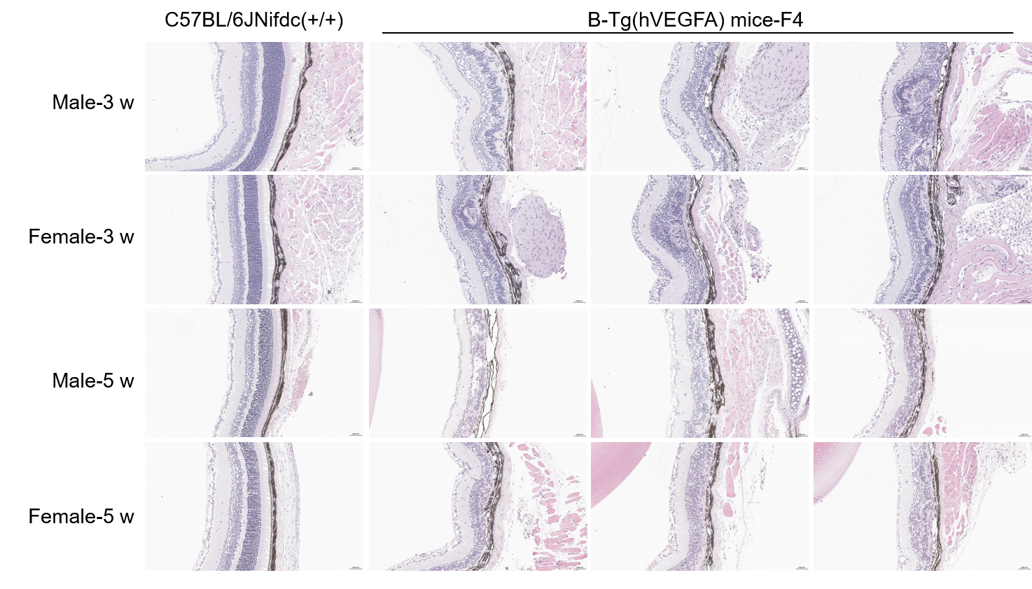
H&E staining of B-Tg(hVEGFA) mice
Representative images of HE staining of wild-type C57BL/6JNifdc mice and B-Tg(hVEGFA) mice. Retina tissues of wild-type C57BL/6JNifdc mice (+/+) and B-Tg(hVEGFA) mice (3 weeks old and 5 weeks old, n=3) were collected and analyzed with H&E staining. Compared with wild-type mice, B-Tg(hVEGFA) mice at 3 weeks of age displayed disorganized retinal layers (ganglion cell, inner nuclear, outer plexiform, outer nuclear) and an indistinct photoreceptor layer. By 5 weeks, the pathology worsened, with the outer nuclear layer becoming indistinct. Scale bar, 50 μm.
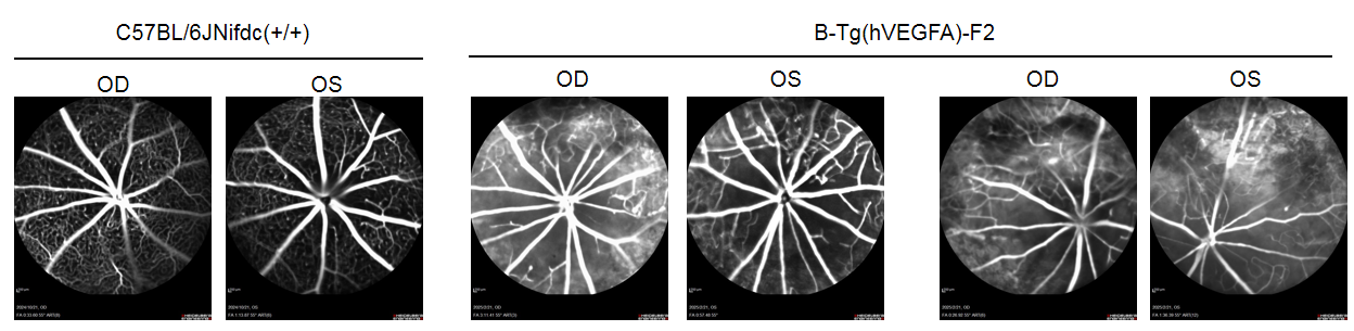
Fundus Fluorescein Angiography (FFA) of B-Tg(hVEGFA) mice
Fundus Fluorescein Angiography (FFA) of wild-type C57BL/6JNifdc mice and B-Tg(hVEGFA) mice. The FFA images of wild-type C57BL/6JNifdc mice (+/+) and B-Tg(hVEGFA) mice (12 weeks old, male). The results showed that there were uneven vascular fluorescence with patchy areas of intense fluorescence suggests vascular permeability leakage or localized pathology in B-Tg(hVEGFA) mice compared to the C57BL/6JNifdc mice.
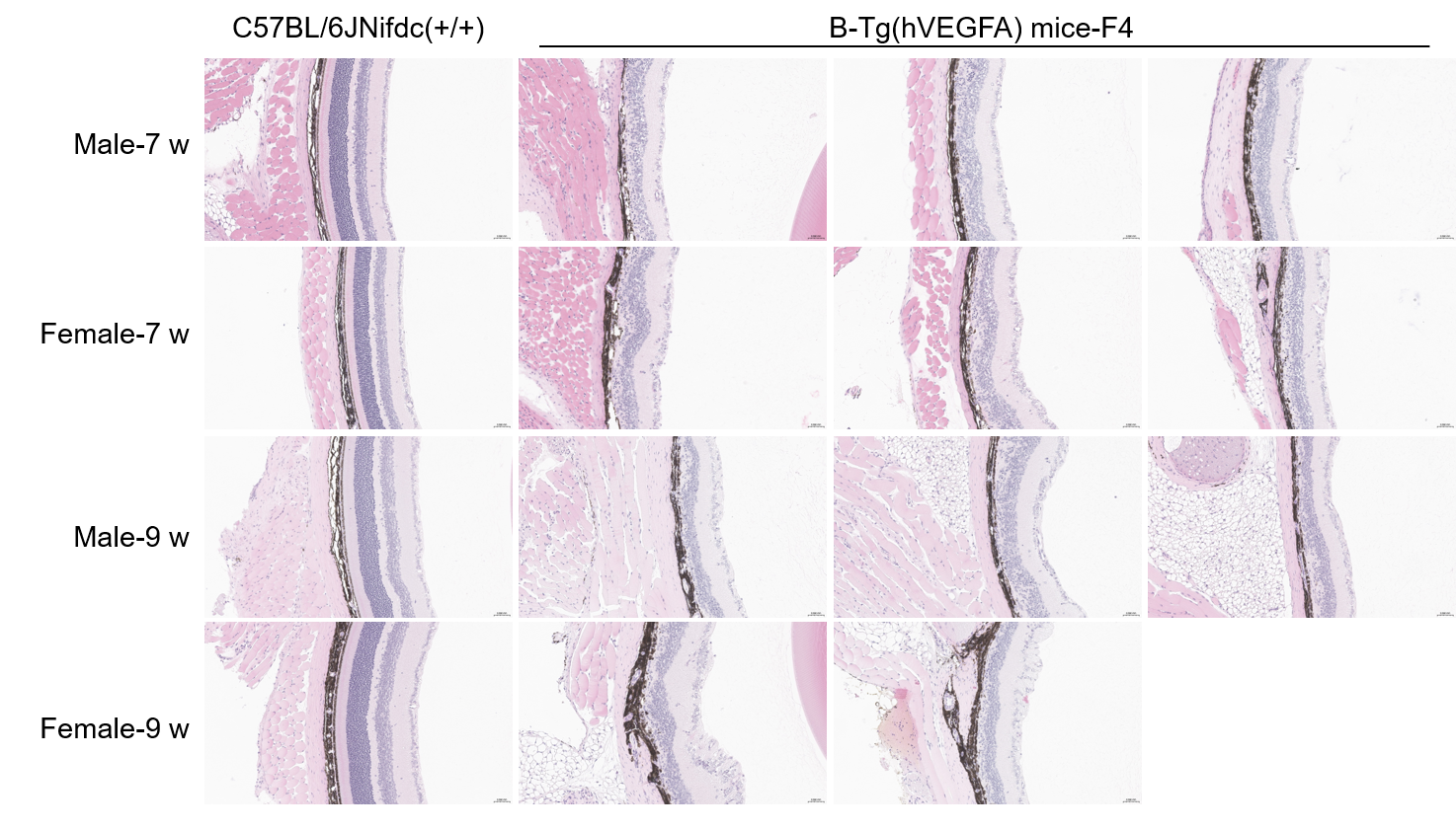
H&E staining of B-Tg(hVEGFA) mice
Representative images of HE staining of wild-type C57BL/6JNifdc mice and B-Tg(hVEGFA) mice. Retina tissues of wild-type C57BL/6JNifdc mice (+/+) and B-Tg(hVEGFA) mice (7 weeks old, n=3; 9 weeks old, male n=3 and female n=2) were collected and analyzed with H&E staining. The results showed that the ganglion cell layer, inner nuclear layer, outer plexiform layer, outer nuclear layer were disorganized, with the photoreceptor and outer nuclear layers being indistinct in B-Tg(hVEGFA) mice compared to the C57BL/6JNifdc mice. Scale bar, 50 μm.
* When publishing results obtained using this animal model, please acknowledge the source as follows: The animal model [B-Tg(hVEGFA) mice] (Cat# 113126) was purchased from Biocytogen.