
• 321917
| Product name | B-hPD-L1 plus/hHER2 MC38 |
|---|---|
| Catalog number | 321917 |
| Strain background | C57BL/6 |
| NCBI gene ID | 60533,13866 (Human) |
| Aliases | B7h1; Pdl1; Pdcd1l1; Pdcd1lg1; A530045L16Rik; Neu; HER2; HER-2; c-neu; Erbb-2; c-erbB2; l11Jus8; mKIAA3023 |
| Tissue | Colon |
| Disease | Colon carcinoma |
| Species | Mouse |
| Application | B-hPD-L1 plus/hHER2 MC38 |
このページで
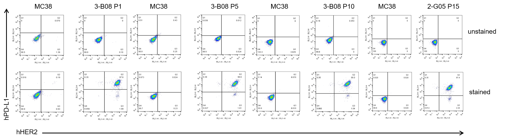
Passage stability analysis in B-hPD-L1 plus/hHER2 MC38 cells by flow cytometry. Single cell suspensions from wild-type MC38 and B-hPD-L1 plus/hHER2 MC38 cultures were stained with species-specific anti-hHER2 antibody and anti-PD-L1 antibody. Human HER2 and human PD-L1 were specifically detected on the surface of B-hPD-L1 plus/hHER2 MC38 cells and remained stable across multiple passages, with no significant change in expression levels observed. No expression was detected in wild-type MC38 cells.
The passage number for this cell line is calculated starting from the first subculture after revival from the master cell bank, which is designated as P1.
The exogenous promoter and human PD-L1 coding sequence was inserted to replace part of murine exon 3. The insertion disrupts the endogenous murine Pdl1 gene, resulting in a non-functional transcript.
The exogenous CAG promoter and the coding sequence composed of human ERBB2 extracellular domain, mouse Erbb2 transmembrane domain and mouse Erbb2 intracellular domain were inserted to replace part of murine exon 2 and all of exons 3-7. The insertion disrupts the endogenous murine Erbb2 gene, resulting in a non-functional transcript.
Gene targeting strategy for B-hPD-L1 plus/hHER2 MC38 cells.
The exogenous promoter and human PD-L1 coding sequence was inserted to replace part of murine exon 3. The insertion disrupts the endogenous murine Pdl1 gene, resulting in a non-functional transcript.
The exogenous CAG promoter and the coding sequence composed of human ERBB2 extracellular domain, mouse Erbb2 transmembrane domain and mouse Erbb2 intracellular domain were inserted to replace part of murine exon 2 and all of exons 3-7. The insertion disrupts the endogenous murine Erbb2 gene, resulting in a non-functional transcript.
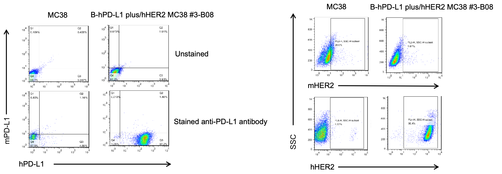
PD-L1 and HER2 expression analysis in B-hPD-L1 plus/hHER2 MC38 cells by flow cytometry. Single cell suspensions from wild-type MC38 and B-hPD-L1 plus/hHER2 MC38 cultures were stained with species-specific anti-PD-L1 and anti-HER2 antibody. Human PD-L1 and HER2 were detected on the surface of B-hPD-L1 plus/hHER2 MC38 cells but not wild-type MC38 cells. The 3-B08 clone of B-hPD-L1 plus/hHER2 MC38 cells was used for in vivo tumor growth assays.
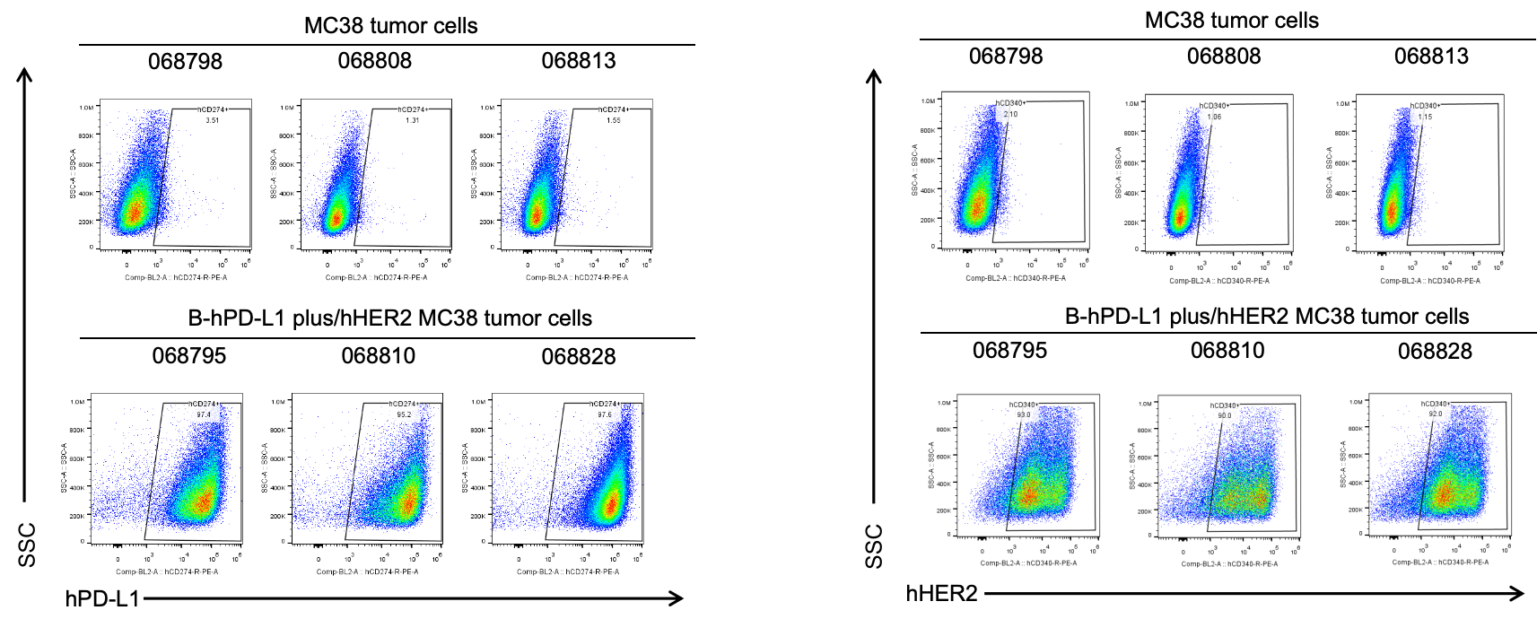
B-hPD-L1 plus/hHER2 MC38 cells were subcutaneously transplanted into C57BL/6 mice (n=6), and on 39 days post inoculation, tumor cells were harvested and assessed for human PD-L1 and HER2 expression by flow cytometry. As shown, human PD-L1 and HER2 were highly expressed on the surface of tumor cells. Therefore, B-hPD-L1 plus/hHER2 MC38 cells can be used for in vivo efficacy studies of PD-L1 and HER2 therapeutics.
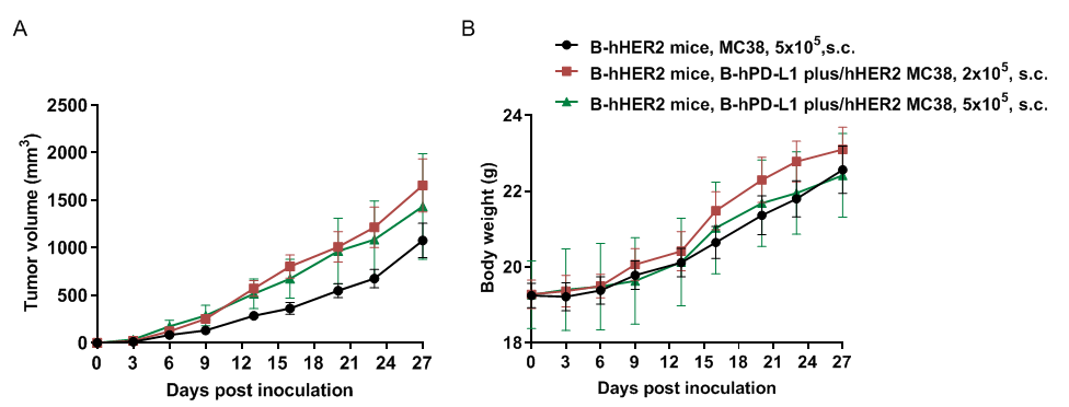
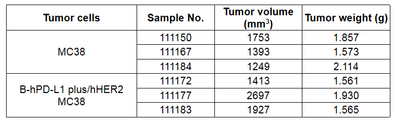
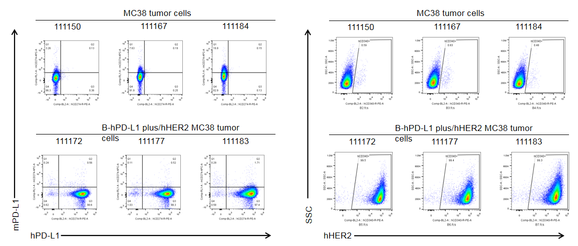
B-hPD-L1 plus/hHER2 MC38 cells were subcutaneously transplanted into homozygous B-hHER2 mice (n=6), and on 30 days post inoculation, tumor cells were harvested and assessed for human PD-L1 and HER2 expression by flow cytometry. As shown, human PD-L1 and HER2 were highly expressed on the surface of tumor cells. Therefore, B-hPD-L1 plus/hHER2 MC38 cells can be used for in vivo efficacy studies of PD-L1 and HER2 therapeutics.