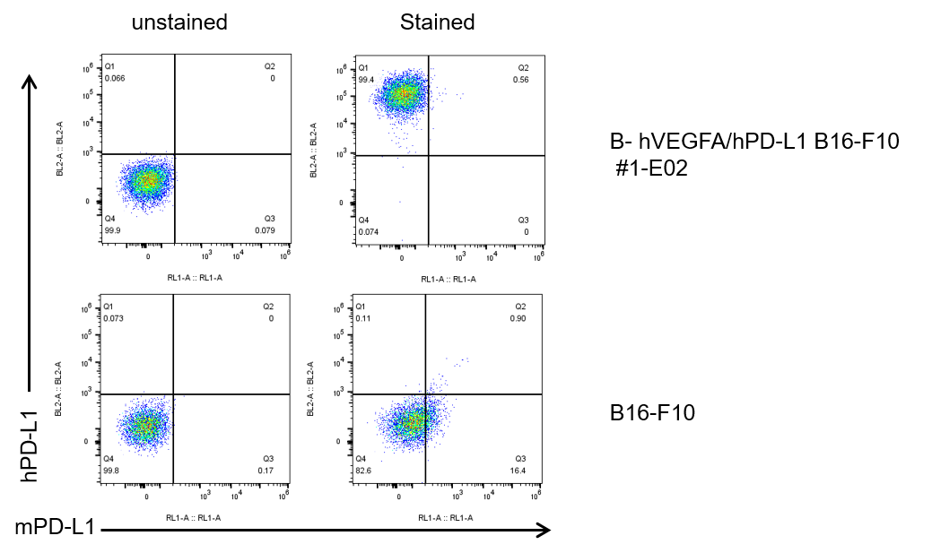
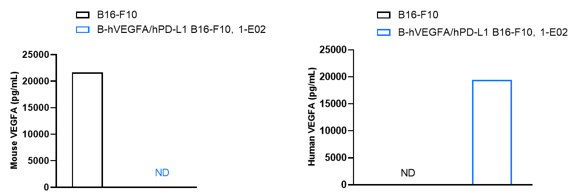
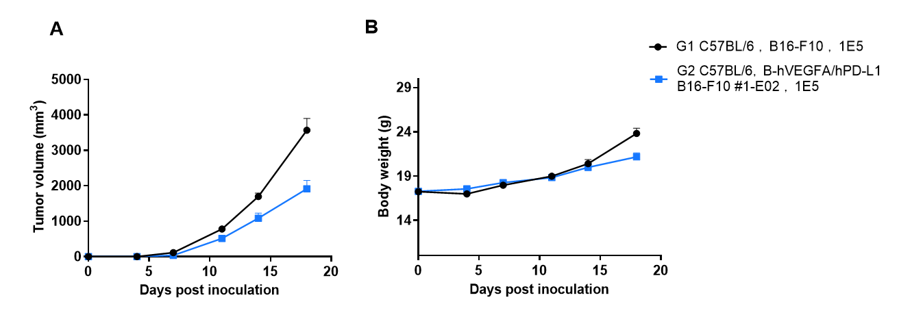
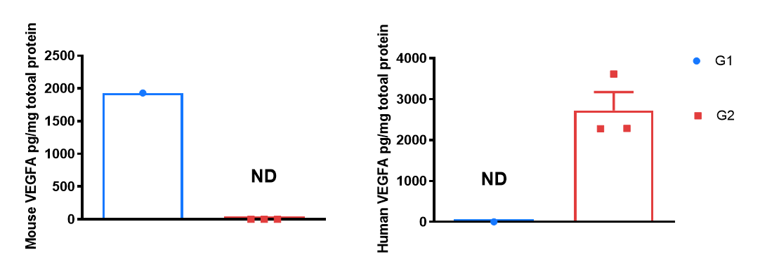
B-hVEGFA/hPD-L1 B16-F10
Catalog Number: 322475
Strain Name: NA
Strain Background: C57BL/6
NCBI gene ID: 60533,22339 (Human)
Aliases: B7h1; Pdl1; Pdcd1l1; Pdcd1lg1; A530045L16Rik; Vpf; Vegf; L-VEGF
---
ライセンスオプション提供可能