Targeting strategy
Gene targeting strategy for B-hAPLNR mice. The exon 1 of mouse Aplnr gene that encodes the full-length protein was replaced by human APLNR exon 1 in B-hAPLNR mice.
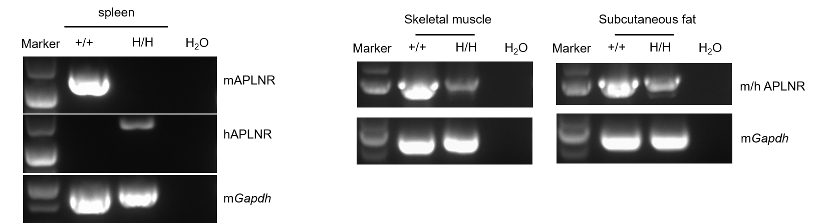
mRNA expression analysis
Strain specific analysis of APLNR mRNA expression in wild-type C57BL/6N mice and B-hAPLNR mice by RT-PCR. Spleen, Skeletal muscle, Subcutaneous fat RNA were isolated from wild-type C57BL/6N mice (+/+) and homozygous B-hAPLNR mice (H/H), then cDNA libraries were synthesized by reverse transcription, followed by PCR with mouse or human APLNR primers. Mouse Aplnr mRNA was detectable only in wild-type C57BL/6N mice. Human APLNR mRNA was detectable only in homozygous B-hAPLNR mice but not in wild-type mice.
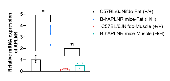
Strain specific analysis of APLNR gene expression in wild-type C57BL/6N mice and B-hAPLNR mice by RT-qPCR. Inguinal fat and hindlimb calf muscle RNA were isolated from wild-type C57BL/6N mice (+/+) (male, 6w, n=3) and homozygous B-hAPLNR mice (H/H) (male, 6w, n=3). The mRNA expression of human APLNR in homozygous B-hAPLNR mice was higher compared with mouse Aplnr in wild-type C57BL/6N mice. Values are expressed as mean ± SEM. *P < 0.05, **P < 0.01, ***P < 0.001. ns, no significant.
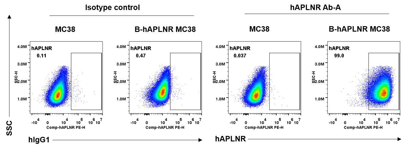
Human APLNR antibody binding Assay
Anti-human APLNR antibody binding assessment in B-hAPLNR MC38. B-hAPLNR MC38 and MC38 were analyzed by flow cytometry. Anti-hAPLNR antibody hAPLNR Ab-A (provided by a client) can bind B-hAPLNR MC38 but not bind MC38.
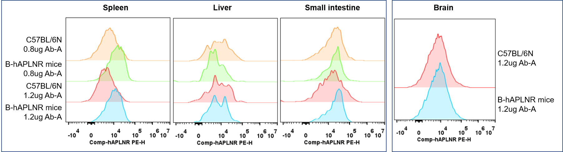
Protein expression analysis in tissue macrophages
Strain specific APLNR expression analysis with macrophages from homozygous B-hAPLNR mice by flow cytometry. Macrophages in Spleen,Liver, Small intestine and Brain were collected from wild-type C57BL/6N mice (+/+) (male, 6-week-old) and homozygous B-hAPLNR mice (H/H) (male, 6-week-old) , and analyzed by flow cytometry with anti-human APLNR antibody Ab-A (provided by a client) . Human APLNR was exclusively detectable in spleen of homozygous B-hAPLNR mice.
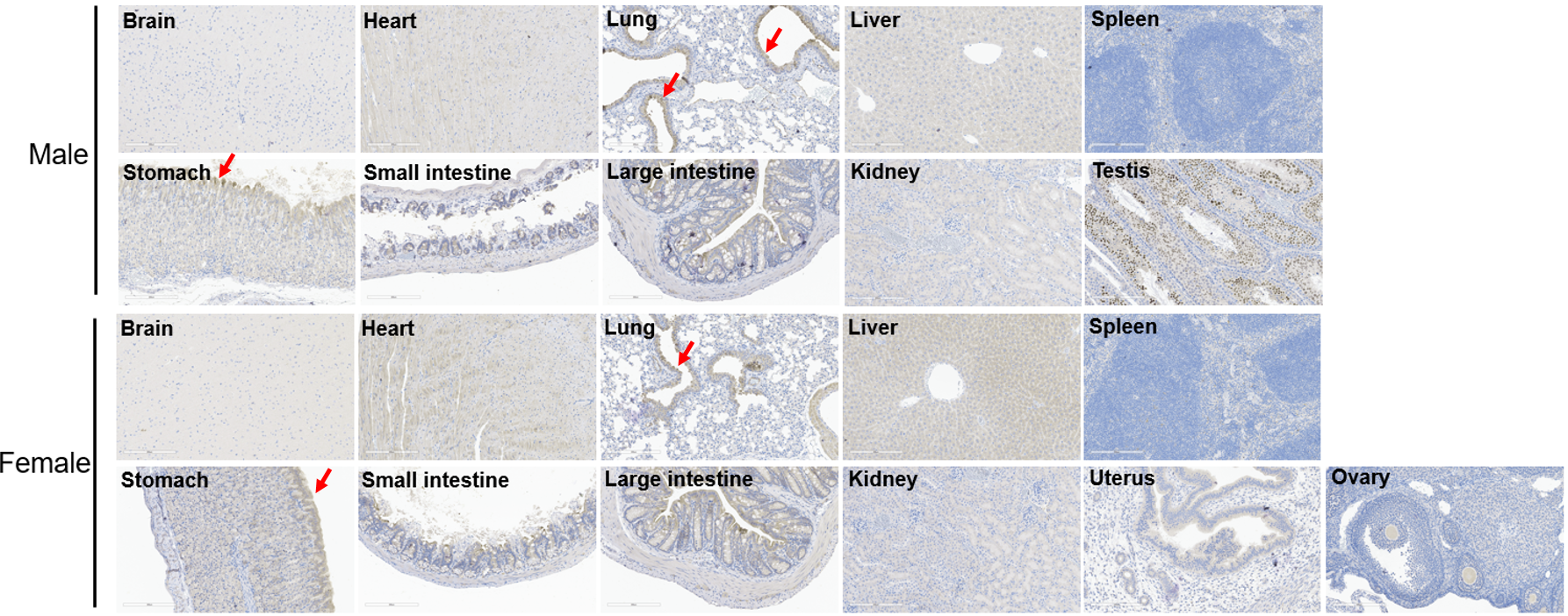
Protein expression analysis by Immunohistochemical analysis(IHC)
Immunohistochemical analysis of organs in B-hAPLNR mice . The main organs of B-hAPLNR mice(H/H) were isolated from female(6-week-old, n=1) and male(9-week-old,n=1). Human APLNR Antibody(LS-A64) used for IHC analysis. Considering that APLNR is a membrane protein, typical positive signals are observed in the mucosal epithelium of lung and stomach tissues. Staining in other tissues may represent nonspecific binding signals of the antibody. Scale bar: 200 μm.
* When publishing results obtained using this animal model, please acknowledge the source as follows: The animal model [B-hAPLNR mice] (Cat# 111408) was purchased from Biocytogen.