


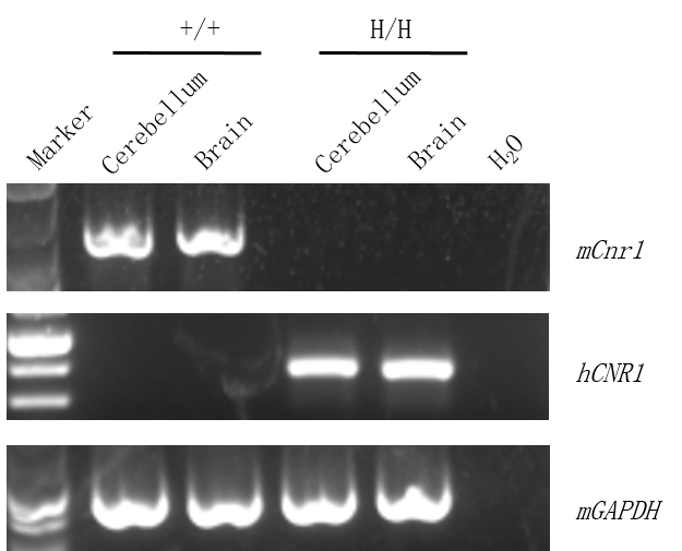
Strain specific analysis of CNR1 mRNA expression in wild-type C57BL/6JNifdc mice and homozygous CB1 humanized mice by RT-PCR. Cerebellum and brain tissue were collected from wild-type C57BL/6JNifdc mice (+/+) and homozygous B-hCB1 mice (H/H). Mouse Cnr1 mRNA was detectable only in wild-type C57BL/6JNifdc mice. Human CNR1 mRNA was detectable only in homozygous B-hCB1 mice, but not in wild-type C57BL/6JNifdc mice.
Mouse and human CB1 were detectable in wild-type C57BL/6JNifdc mice and homozygous CB1 humanized mice.

Strain specific CB1 expression analysis in wild-type C57BL/6JNifdc mice and homozygous CB1 humanized mice by western blot. Cerebellum and brain tissue were collected from wild-type C57BL/6 mice (+/+) and homozygous B-hCB1 mice (H/H) and analyzed by western blot with anti-CB1 antibody (Thermo Fisher, PA5-18427) which was cross reactive between mouse and human. Mouse and human CB1 were detectable in wild-type mice and homozygous B-hCB1 mice.

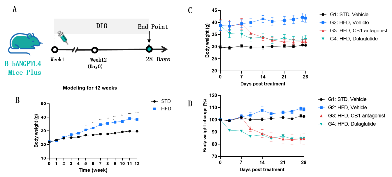
Body weight in DIO CB1 humanized mice. Homozygous B-hCB1 mice (male, n=30) were randomly assigned to two groups based on body weight: Group A (n = 8), fed a standard diet (STD), and Group B (n = 22), fed a high-fat diet (HFD). After 12 weeks of HFD feeding to induce DIO, the mice in Group B were randomly divided into three groups (G2, G3, and G4) and administered vehicle, an antagonist, or dulaglutide, respectively (n = 7–8 per group). (A) The schematic diagram of experimental processing. (B) Body weight after 12 weeks of DIO. (C-D) Body weight and weight changes in each group after treatment. As shown in Figure B, body weight was significantly higher in the HFD group than in the STD group, indicating that HFD feeding successfully induced obesity in B-hCB1 mice. As shown in Figures C and D, treatment with CB1 antagonists or dulaglutide significantly reduced body weight compared with the HFD vehicle group. Values are expressed as mean ± SEM.
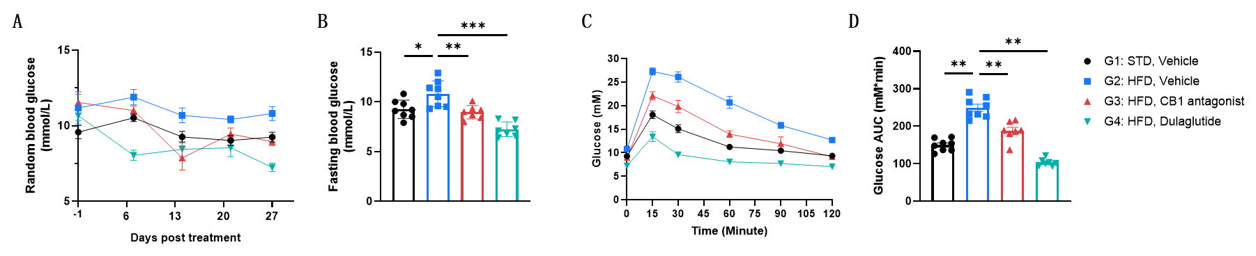
The CB1 antagonist and dulaglutide improved glucose homeostasis in DIO CB1 humanized mice. (A) Random blood glucose during treatment. (B) Fasting blood glucose on Day 27 post-treatment. (C-D) Glucose tolerance test. As shown in the figure, the CB1 antagonist and dulaglutide can reduce random blood glucose and fasting blood glucose and improve glucose tolerance in DIO B-hCB1 mice. Values are expressed as mean ± SEM.
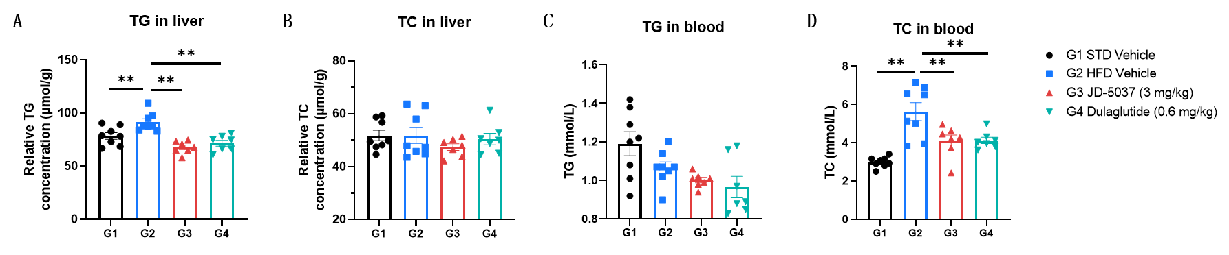
The CB1 antagonist and dulaglutide improved lipid metabolism in DIO CB1 humanized mice. (A) TG in liver. (B) TC in liver. (C) TG in blood. (D) TC in blood. As shown in the figure, the CB1 antagonist and dulaglutide can reduce the level of TG in liver and TC in blood. Values are expressed as mean ± SEM.

The CB1 antagonist and dulaglutide reduced liver damage in DIO CB1 humanized mice.(A) Representative H&E-stained liver images (representative dataset from n = 7-8 per group). (B) Statistical analysis of total histological score for hepatocellular steatosis, lobular inflammation and ballooning degeneration. Values are expressed as mean ± SD.