Key Advantages
Validation
Applications

CD5A

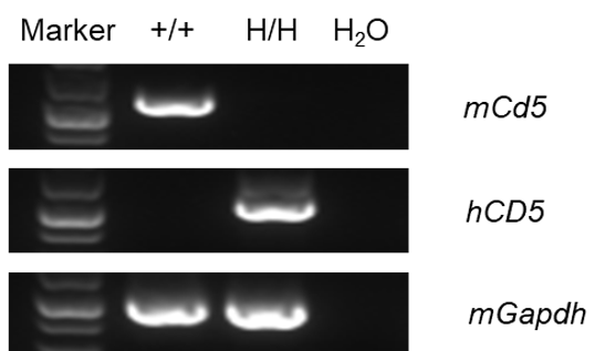
Strain-specific analysis of mouse and human CD5 mRNA expression in wild-type C57BL/6N mice and CD5 humanized mice by RT-PCR. Total RNA was isolated from thymus tissues of wild-type C57BL/6N mice (+/+) and homozygous CD5 humanized mice (H/H). cDNA was synthesized by reverse transcription, followed by PCR amplification using mouse- or human-specific CD5 primers.
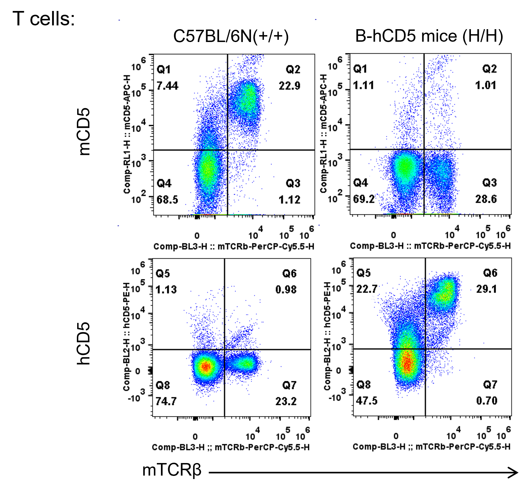
Strain-specific analysis of mouse and human CD5 protein expression in wild-type C57BL/6N mice and CD5 humanized mice by flow cytometry. Splenocytes were collected from wild-type C57BL/6N mice (+/+) and homozygous CD5 humanized mice (H/H). Mouse and human CD5 protein expression was analyzed by flow cytometry using species-specific anti-mouse CD5 (BioLegend, 100625) and anti-human CD5 (BioLegend, 300607) antibodies.
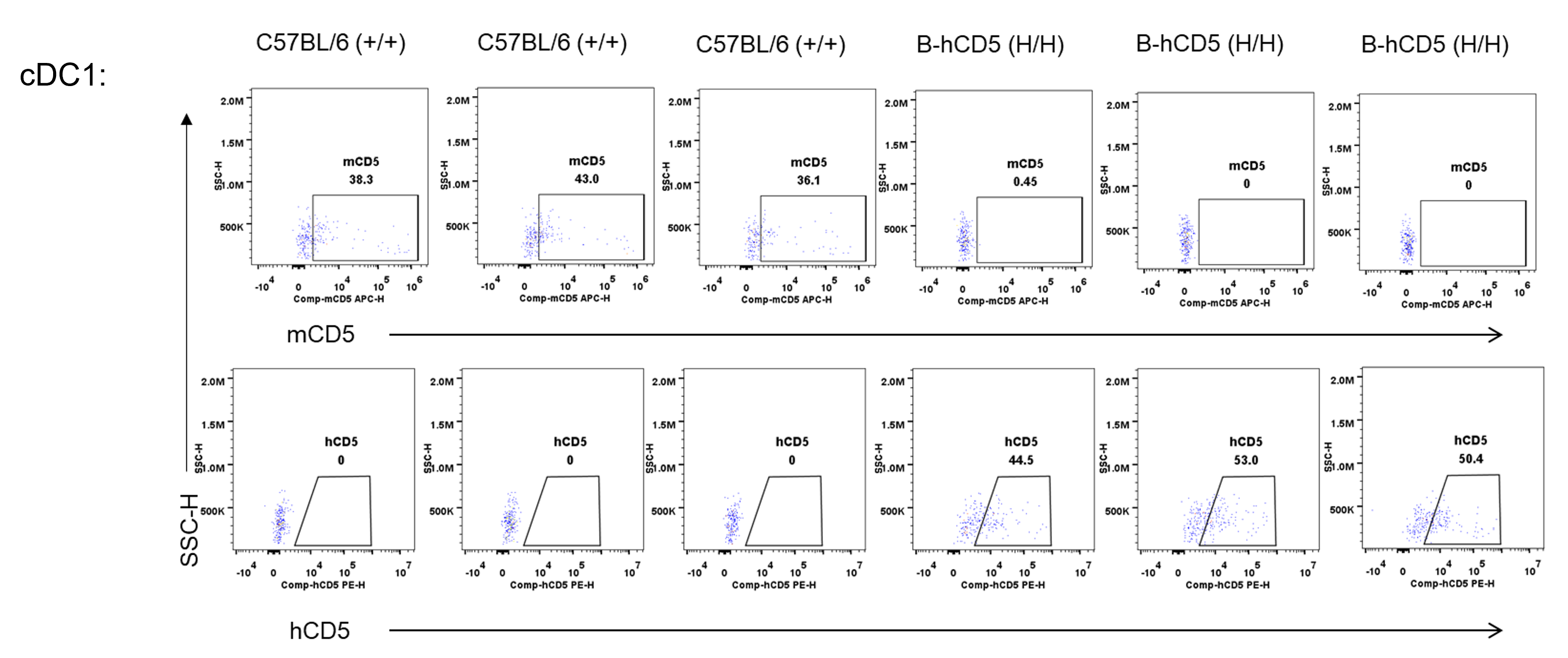
Strain-specific analysis of mouse and human CD5 protein expression in wild-type C57BL/6 mice and CD5 humanized mice by flow cytometry. Spleens were collected from wild-type C57BL/6 mice (+/+) and homozygous CD5 humanized mice (H/H). Splenocytes were analyzed by flow cytometry using species-specific anti-CD5 antibodies. Mouse CD5 was detected in wild-type C57BL/6 mice, whereas human CD5 was detected exclusively in homozygous CD5 humanized mice.
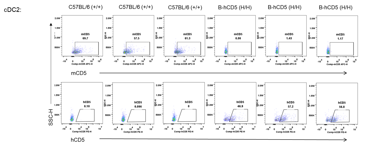
Strain-specific analysis of mouse and human CD5 protein expression in wild-type C57BL/6 mice and CD5 humanized mice by flow cytometry. Spleens were collected from wild-type C57BL/6 mice (+/+) and homozygous CD5 humanized mice (H/H). Splenocytes were analyzed by flow cytometry using species-specific anti-CD5 antibodies.
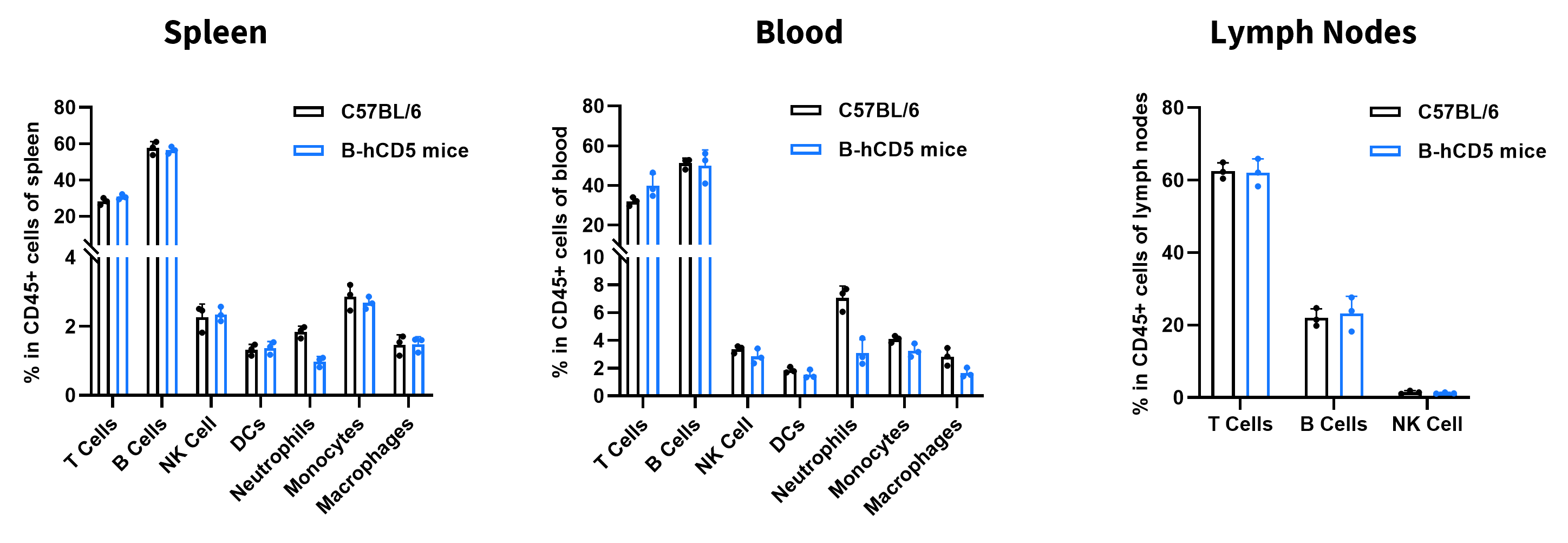
Flow cytometry analysis of leukocyte subpopulations in immune organs and peripheral blood of CD5 humanized mice. Splenocytes, peripheral blood, and lymph nodes were isolated from female wild-type C57BL/6 mice and CD5 humanized mice (8 weeks old, n = 3). Single live cells were gated on the CD45⁺ population and analyzed by flow cytometry as indicated. Values are expressed as mean ± SEM.
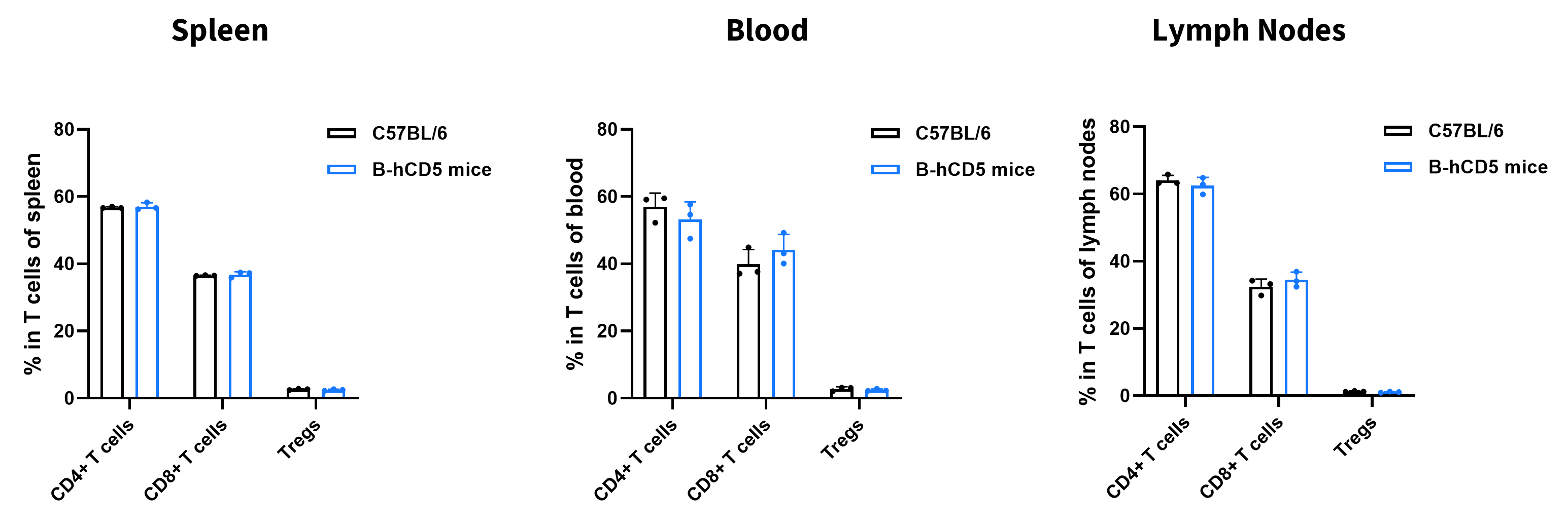
Flow cytometry analysis of T-cell subpopulations in immune organs and peripheral blood of CD5 humanized mice. Splenocytes, peripheral blood, and lymph nodes were isolated from female wild-type C57BL/6 mice and CD5 humanized mice (8 weeks old, n = 3). Single live cells were gated on the T-cell population and analyzed by flow cytometry as indicated. Values are expressed as mean ± SEM.
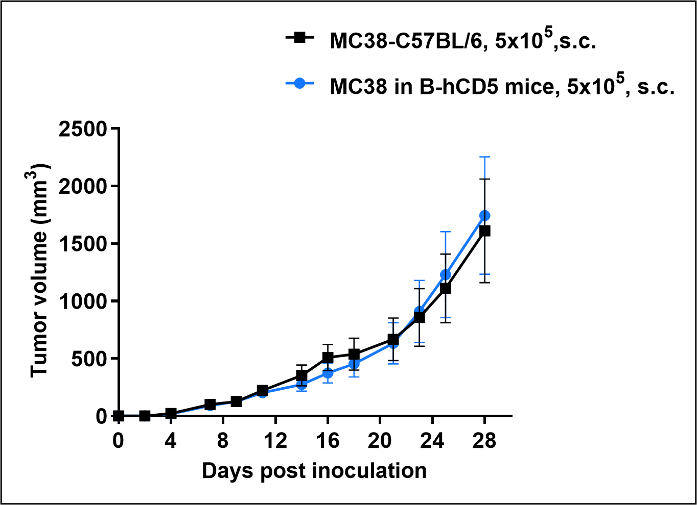
Subcutaneous MC38 homograft tumor growth in CD5 humanized mice. MC38 mouse colon carcinoma cells (5 × 10⁵) were subcutaneously implanted into wild-type C57BL/6 mice and CD5 humanized mice (female, 11 weeks old, n = 6). Tumor volumes were measured twice weekly and calculated using the formula: V = 0.5 × long diameter × (short diameter)². Tumor volumes are expressed in mm³, and data are presented as mean ± SEM. MC38 cells successfully established subcutaneous tumors in CD5 humanized mice, demonstrating the suitability of this model for in vivo antitumor efficacy studies.