Description
CD79A: CD79A Transduces B-Cell Receptor Signals for B-Lymphocyte Activation.
- Gene Information: CD79A (CD79A) encodes immunoglobulin alpha (Igα), a type I transmembrane glycoprotein located on human chromosome 19q13.2 that forms a critical heterodimer with CD79B to constitute the B-cell receptor complex.
- Protein Expression: Expressed selectively throughout B-cell lineage development—from early pre-B cells to mature B cells—CD79A expression is downregulated during terminal differentiation into plasma cells, making it a highly specific lineage marker.
- Signaling Pathway: Upon antigen binding, Src-family kinases phosphorylate the immunoreceptor tyrosine-based activation motif (ITAM) on CD79A, recruiting and activating Syk kinase to trigger downstream PI3K/Akt, and NF-κB signaling cascades.
- Therapeutic Inhibition: Targeting CD79A with monoclonal antibodies or antibody-drug conjugates (ADCs) induces BCR internalization and B-cell depletion, providing an effective therapeutic strategy for B-cell malignancies and autoimmune diseases.
Human CD79A Protein Expression Analysis in Spleen of CD79A Humanized Mice
- Mouse CD79A was detectable in wild-type C57BL/6N mice and homozygous CD79A humanized mice due to the cross-reactivity of anti-mouse CD79A antibody .
- Human CD79A was detectable in homozygous CD79A humanized mice, but not in wild-type C57BL/6N mice.
Strain specific CD79A expression analysis in wild-type C57BL/6N mice and homozygous CD79A humanized mice by flow cytometry. Splenocytes were collected from wild-type C57BL/6N mice and homozygous B-hCD79A mice. Protein expression was analyzed with anti-mouse CD79A antibody (Biolegend, 133105) and anti-human CD79A antibody (RD, FAB69201G) by flow cytometry.
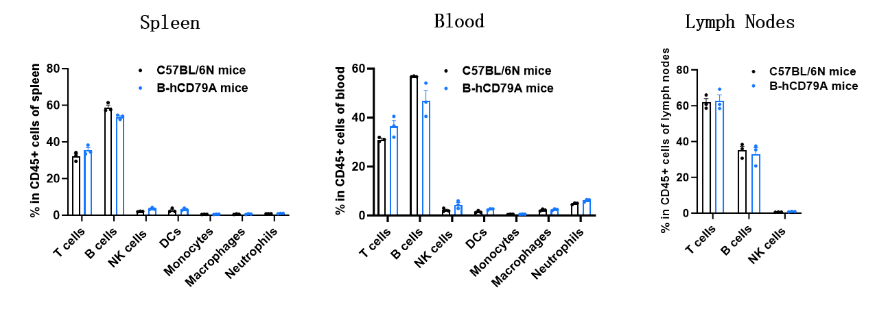
Animal State Evaluation-Leukocyte Profiling in CD79 Humanized Mice
- The percentages of T cells, B cells, NK cells, DCs, neutrophils, monocytes, and macrophages in homozygous CD79A humanized mice were similar to those in C57BL/6N mice.
- Humanization of CD79A does not affect normal immune cell development or distribution.
Analysis of leukocyte subpopulations by flow cytometry in immune organs and blood.Splenocytes, peripheral blood, and lymph nodes were isolated from C57BL/6N mice and homozygous B-hCD79A mice (female, 8-week-old, n = 3). Single live cells were gated on the CD45⁺ population and analyzed by flow cytometry as indicated. Values are expressed as mean ± SEM.
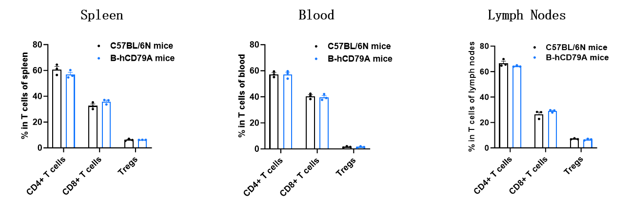
Animal State Evaluation-T Cell Profiling in CD79 Humanized Mice
- The proportions of CD4⁺ T cells, CD8⁺ T cells, and Tregs in homozygous CD79A humanized mice were comparable to those in C57BL/6N mice.
- Humanization of CD79A does not affect normal T cell development, differentiation, or distribution.
Analysis of T-cell subpopulations by flow cytometry in immune organs and blood.Splenocytes, peripheral blood, and lymph nodes were isolated from C57BL/6N mice and homozygous B-hCD79A mice (female, 8-week-old, n = 3). Single live cells were gated on the CD3⁺ T-cell population and analyzed by flow cytometry as indicated. Values are expressed as mean ± SEM.
* When publishing results obtained using this animal model, please acknowledge the source as follows: The animal model [B-hCD79A mice] (Cat# 111095) was purchased from Biocytogen.