Description
EPCAM: A key adhesion molecule in tumor malignancy and its therapeutic intervention
- Gene Information: EPCAM is a transmembrane glycoprotein featuring both extracellular (EpEx) and intracellular (EpICD) domains. Cleavage of EPCAM releases EpICD, which translocates to the nucleus as a transcriptional co-factor.
- Protein Expression: EPCAM is expressed in various human epithelial tissues, cancerous tissues, progenitor cells, and stem cells. It is widely overexpressed in a variety of solid tumors and tumor-initiating cells (TICs), playing a vital role in fueling cancer progression, particularly in colorectal cancer (CRC).
- Signaling Pathway: EpICD interacts with Wnt receptor promoters (FZD6 and LRP5/6) to upregulate their transcription and activate Wnt signaling. Additionally, activation of Wnt kinases (GSK3β and CK1) in the b-catenin destruction complex induces γ-secretase activity to enhance EpICD shedding, forming a positive feedback loop.
- Therapeutic Inhibition: By blocking EPCAM‑mediated Wnt receptor upregulation and signaling, thereby inhibiting tumor growth and prolonging survival in CRC models.
Targeting strategy
Gene targeting strategy for B-hEPCAM mice. The exons 3-8 of mouse Epcam genes that encode the extracellular protein were replaced by human EPCAM exons 2-7 in B-hEPCAM mice.
Protein Expression Analysis
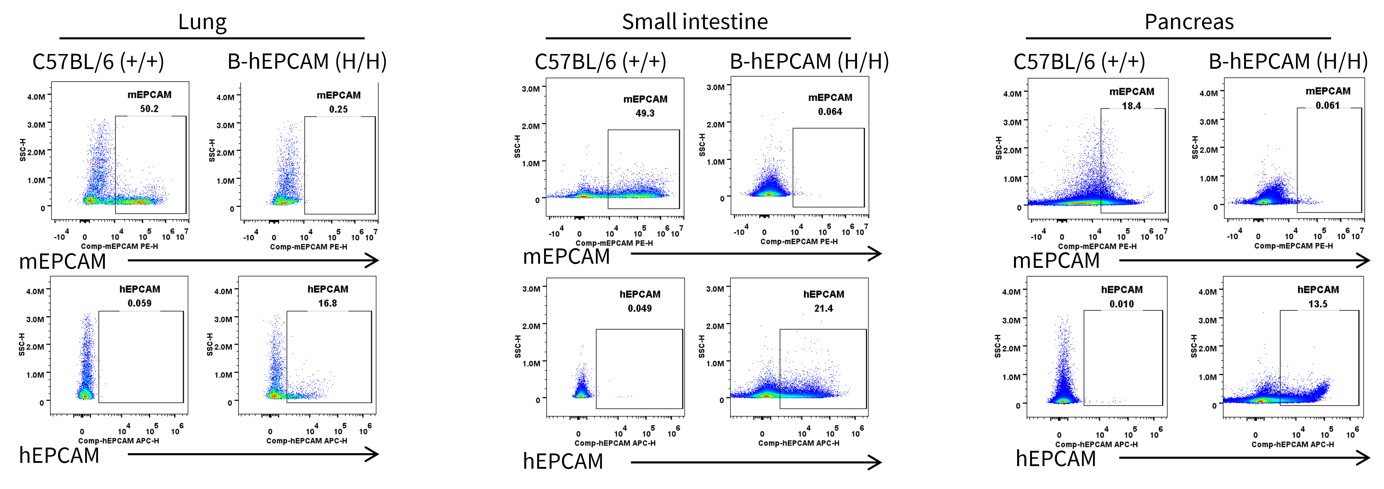
- Mouse EPCAM was detectable in wild-type C57BL/6 mice, but not in homozygous B-hEPCAM mice.
- Human EPCAM was exclusively detectable in homozygous B-hEPCAM mice.
Strain specific EPCAM expression analysis in homozygous B-hEPCAM mice by flow cytometry. Lung, small intestine and pancreas were collected from wild-type C57BL/6 mice (+/+) and homozygous B-hEPCAM mice (H/H), and CD45- cells were analyzed by flow cytometry with species-specific anti-mouse EPCAM antibody and anti-human EPCAM antibody.
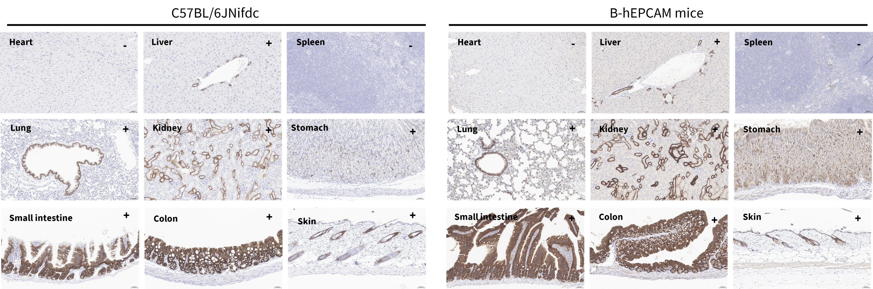
Immunohistochemical (IHC) analysis of EPCAM expression in B-hEPCAM mice. The heart, liver, spleen, lung, kidney, stomach, small intestine, colon and skin were collected from wild-type C57BL/6JNifdc mice and B-hEPCAM mice (male, 7-week-old), analyzed by IHC with anti-EPCAM antibody. “+” indicates that the tissue is positive, and “-” indicates that the tissue is negative.
* When publishing results obtained using this animal model, please acknowledge the source as follows: The animal model [B-hEPCAM mice] (Cat# 110826) was purchased from Biocytogen.