Description
IL2RB/hIL15: IL2RB and IL15 Coordinate to Drive Lymphocyte Activation and Survival.
- Gene Information: IL2RB and IL15 encode key cytokine-receptor components (IL2RB on human chromosome 22q12.3 and IL15 on chromosome 4q31.21) that cooperatively regulate cytotoxic lymphocyte signaling and adaptive immune homeostasis.
- Protein Expression: IL15 is expressed and secreted by antigen-presenting cells, monocytes, and stromal cells, while IL2RB (CD122) is constitutively expressed on NK cells, memory CD8+ T cells, and regulatory T cells.
- Signaling Pathway: IL15 binds to its complex with IL2RB and IL2RG, triggering receptor dimerization that activates JAK1/JAK3 kinase complexes, driving downstream STAT3/STAT5, PI3K/Akt, and MAPK/ERK pathways to induce cell proliferation.
- Therapeutic Inhibition: Targeting the IL2RB/IL15 axis suppresses hyperactive NK and T-cell signaling, providing promising therapeutic strategies for autoimmune diseases, graft-versus-host disease, chronic inflammation, and specific T-cell or NK-cell malignancies.
Human IL15 Protein Expression Analysis in Serum of IL2RB/IL15 Humanized Mice
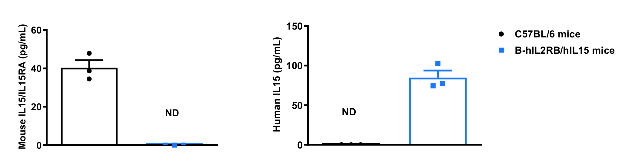
- Mouse IL15 was detected in wild-type C57BL/6 mice.
- Human IL15 was detected in homozygous IL2RB/IL15 humanized mice, but not in wild-type C57BL/6 mice.
Strain specific IL15 expression analysis in wild-type C57BL/6 mice and homozygous IL2RB/IL15 humanized mice by ELISA. Serum was collected from wild-type C57BL/6 mice and homozygous B-hIL2RB/hIL15 mice. Expression level of mouse and human IL15 were analyzed by ELISA (anti-mouse IL15 ELISA Kit: RD, D1500; anti-human IL15 ELISA Kit: Invitrogen, BMS6023). Values are expressed as mean ± SEM. ND: not detectable.
Human IL2RB Protein Expression Analysis in Spleen of IL2RB/IL15 Humanized Mice
- Mouse IL2RB was detectable in T cells and NK cells from spleen of wild-type C57BL/6 mice. Human IL2RB was detectable in T cells and NK cells from spleen of homozygous IL2RB/IL15 humanized mice, but not in wild-type C57BL/6 mice.
Strain specific IL2RB expression analysis in wild-type C57BL/6 mice and homozygous IL2RB/IL15 humanized mice by flow cytometry. Splenocytes were collected from wild-type C57BL/6 mice and homozygous B-hIL2RB/hIL15 mice stimulated with anti-mouse CD3ε antibody (7.5 μg, i.p.) in vivo for 24 hours. Protein expression was analyzed with anti-mouse IL2RB antibody (Biolegend, 105911) and anti-human IL2RB antibody (Biolegend, 339005) by flow cytometry.
- Mouse IL2RB was detectable in CD4+ T cells and CD8+ T cells from spleen of wild-type C57BL/6 mice.
- Human IL2RB was detectable in CD4+ T cells and CD8+ T cells from spleen of homozygous IL2RB/IL15 humanized mice, but not in wild-type C57BL/6 mice.
Strain specific IL2RB expression analysis in wild-type C57BL/6 mice and homozygous IL2RB/IL15 humanized mice by flow cytometry. Splenocytes were collected from wild-type C57BL/6 mice and homozygous B-hIL2RB/hIL15 mice stimulated with anti-mouse CD3ε antibody (7.5 μg, i.p.) in vivo for 24 hours. Protein expression was analyzed with anti-mouse IL2RB antibody (Biolegend, 105911) and anti-human IL2RB antibody (Biolegend, 339005) by flow cytometry.
Animal State Evaluation-Leukocyte Profiling in IL2RB/IL15 Humanized Mice
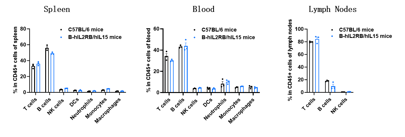
- The percentages of T cells, B cells, NK cells, DCs, neutrophils, monocytes, and macrophages in homozygous IL2RB/IL15 humanized mice were similar to those in C57BL/6mice.
- Humanization of IL2RB and IL15 does not affect normal immune cell development or distribution.
Analysis of leukocyte subpopulations by flow cytometry in immune organs and blood.Splenocytes, peripheral blood, and lymph nodes were isolated from C57BL/6 mice and homozygous B-hIL2RB/hIL15 mice (female, 8-week-old, n = 3). Single live cells were gated on the CD45⁺ population and analyzed by flow cytometry as indicated. Values are expressed as mean ± SEM.
Animal State Evaluation-T Cell Profiling in IL2RB/IL15 Humanized Mice
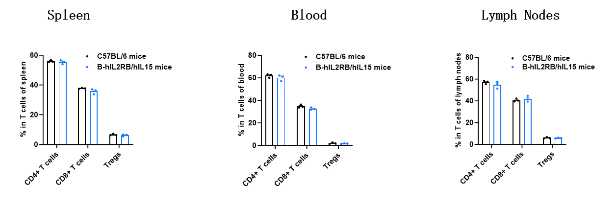
- The proportions of CD4⁺ T cells, CD8⁺ T cells, and Tregs in homozygous IL2RB/IL15 humanized mice were comparable to those in C57BL/6 mice.
- Humanization of IL2RB and IL15 does not affect normal T cell development, differentiation, or distribution.
Analysis of T-cell subpopulations by flow cytometry in immune organs and blood.Splenocytes, peripheral blood, and lymph nodes were isolated from C57BL/6 mice and homozygous B-hIL2RB/hIL15 mice (female, 8-week-old, n = 3). Single live cells were gated on the CD3⁺ T-cell population and analyzed by flow cytometry as indicated. Values are expressed as mean ± SEM.
* When publishing results obtained using this animal model, please acknowledge the source as follows: The animal model [B-hIL2RB/hIL15 mice] (Cat# 111945) was purchased from Biocytogen.