Description
CD3 and 4-1BB: A New Trends of T cell engager therapeutic
- Gene Information: CD3: Encoded by CD3E, D, G; part of the Ig superfamily. It forms the essential signaling backbone of the T-cell receptor (TCR) complex. 4-1BB: Encoded by TNFRSF9; a member of the TNF receptor superfamily. It acts as a potent inducible costimulatory gene.
- Protein Expression: CD3: Constitutive and universal marker for all mature T cells (CD4+, CD8+). Always present on the cell surface. 4-1BB: Inducible; absent on resting cells. It is expressed only after T-cell activation, marking tumor-reactive or “effector” lymphocytes.
- Signaling Pathway: CD3 (Signal 1): Operates via ITAM phosphorylation and ZAP-70. It triggers the initial "on" switch, Ca2+ flux, and immediate cytotoxicity. 4-1BB (Signal 2): Operates via TRAF1/2 and NF-κB. It drives metabolic fitness, prevents apoptosis (Bcl-xL), and sustains long-term T-cell memory.
- Therapeutic Inhibition: Uses bispecifics (BiTEs) to force T-cell killing of tumor cells regardless of MHC restriction; Cytokine Release Syndrome (CRS) is the primary safety concern. Focuses on boosting T-cell persistence; modern multi-specific formats (CD3×41BB ×TAA) localize activation to the tumor microenvironment.
CDH17: A key adhesion molecule in tumor malignancy and its therapeutic intervention
- Gene Information: CDH17 is a member of the cadherin superfamily, encoding a calcium‑dependent membrane‑associated glycoprotein. The protein has a cadherin‑like structure: it possesses an extracellular region with seven cadherin domains and a transmembrane segment, but it lacks the conserved cytoplasmic domain.
- Protein Expression: In normal tissue, CDH17 restricted expression epithelium of small intestine and colon. CDH17 was overexpressed in colorectal adenocarcinoma, gastric adenocarcinoma and pancreatic adenocarcinoma. It can also be detected in liver metastases originated from colorectal adenocarcinoma.
- Signaling Pathway: It drives cancer progression—specifically cell adhesion, proliferation, migration, and invasion—by activating the Wnt/β-catenin, Ras/Raf/MEK/ERK, and α2/β1 integrin pathways (via its RGD motif), making its knockdown a promising therapeutic strategy to inhibit tumor malignancy.
- Therapeutic Inhibition: By blocking CDH17‑mediated adhesion and signaling, a CDH17‑targeted agent inhibits tumor progression and improves clinical outcomes, including reduced tumour growth, decreased metastasis, and prolonged survival.
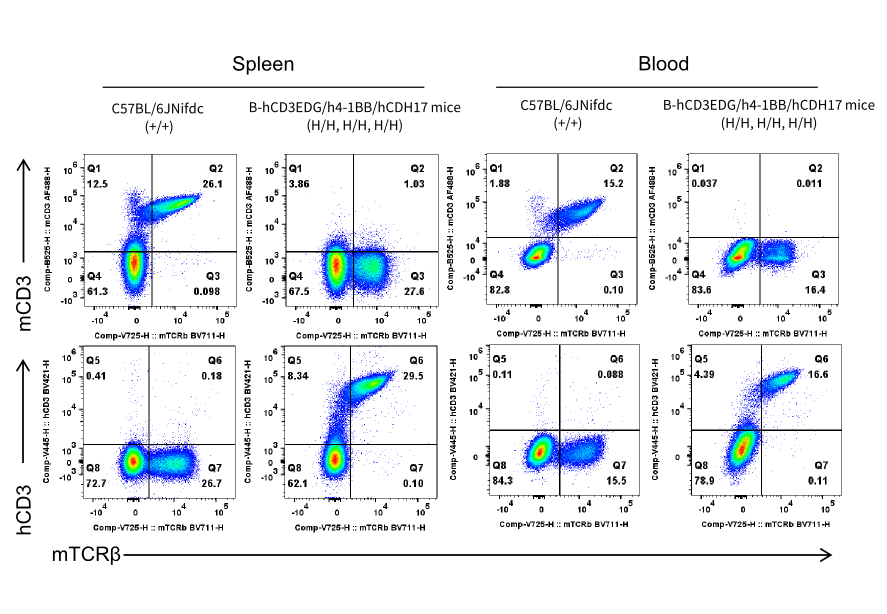
CD3E Protein Expression
CD3E expression analysis in wild-type C57BL/6JNifdc mice and homozygous humanized B-hCD3EDG/h4-1BB/hCDH17 mice by flow cytometry. Splenocytes and blood were collected from wild-type C57BL/6JNifdc mice (+/+) and homozygous B-hCD3EDG/h4-1BB/hCDH17 mice (H/H, H/H, H/H) (female, 7-week-old). Protein expression was analyzed with anti-mouse CD3E antibody (Biolegend, 100210) and anti-human CD3E antibody (Biolegend, 317344) by flow cytometry.
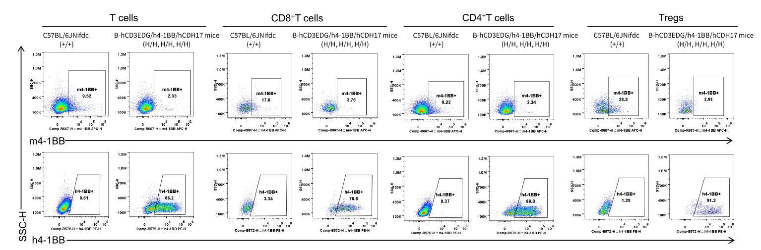
4-1BB Protein Expression
Mouse and human 4-1BB expression analysis in splenocytes. Splenocytes were collected from wild-type C57BL/6JNifdc mice and B-hCD3EDG/h4-1BB/hCDH17 mice that treated with anti-m/hCD3E antibody (7.5 μg/mice, i.p., 24 hours). 4-1BB expression was analyzed by flow cytometry using anti-mouse 4-1BB antibody (Biolegend, 106110) and anti-human 4-1BB antibody (Biolegend, 309804).
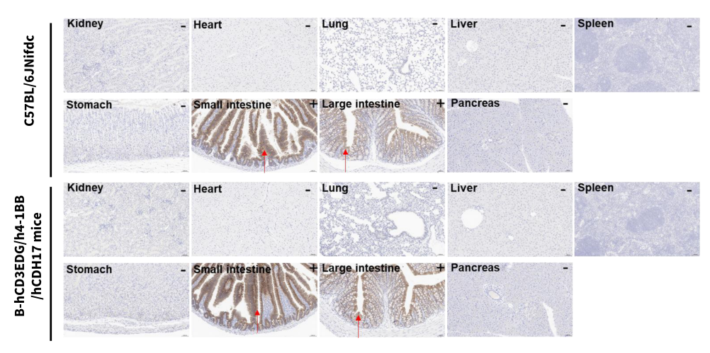
CDH17 Protein Expression
Immunohistochemical (IHC) analysis of CDH17 expression in C57BL/6JNifdc mice and B-hCD3EDG/h4-1BB/hCDH17 mice. Various tissues were collected from wild-type C57BL/6JNifdc mice (WT) and homozygous B-hCD3EDG/h4-1BB/hCDH17 mice (HO) and analyzed by IHC with anti-CDH17 antibody (abcam, ab109190).
* When publishing results obtained using this animal model, please acknowledge the source as follows: The animal model [B-hCD3EDG/h4-1BB/hCDH17 mice] (Cat# 115953) was purchased from Biocytogen.