B-hCD3EDG/hCD8/hTRAC/hTRBC1/hTRBC2 mice
C57BL/6-Cd3etm1(CD3E)Bcgen Cd3dtm1(CD3D)Bcgen Cd3gtm1(CD3G)Bcgen Cd8atm1(CD8A)Bcgen Cd8btm1(CD8B)Bcgen Tractm1(TRAC)Bcgen Trbc1tm1(TRBC1)Bcgen Trbc2tm1(TRBC2)Bcgen/Bcgen • 114950

B-hCD3EDG/hCD8/hTRAC/hTRBC1/hTRBC2 mice

Catalog Number
114950
Strain Name
C57BL/6-Cd3etm1(CD3E)Bcgen Cd3dtm1(CD3D)Bcgen Cd3gtm1(CD3G)Bcgen Cd8atm1(CD8A)Bcgen Cd8btm1(CD8B)Bcgen Tractm1(TRAC)Bcgen Trbc1tm1(TRBC1)Bcgen Trbc2tm1(TRBC2)Bcgen/Bcgen
Strain Background
C57BL/6
NCBI gene ID
Aliases
CD3epsilon, IMD18, T3E, TCRE; CD3-DELTA, CD3DELTA, IMD19, T3D; CD3-GAMMA, CD3GAMMA, IMD17, T3G; IMD7, TCRA, TRCA; BV05S1J2.2, TCRB, TCRBC1; TCRBC2; CD8, CD8alpha, IMD116, Leu2, p32; CD8B1, CD8beta, LEU2, LY3, LYT3, Ly-3, P37

このページで

  • Description
  • Targeting strategy
  • Phenotypic analysis

ポスター

すべて表示

    出版物

      Description

      CD3EDG: Signaling in Tregs for Mechanism and Therapeutic Potential

      •  Gene Information: The CD3E, CD3D, and CD3G genes, located in a cluster on chromosome 11q23, encode the three invariant polypeptide chains that assemble into the CD3εδ and CD3εγ heterodimers of the T-cell receptor (TCR) complex.
      •  Protein Expression: These proteins are exclusively and constitutively expressed on the surface of mature T lymphocytes and thymocytes, serving as definitive lineage markers for the T-cell population.
      • Signaling Pathway: Upon TCR engagement by an antigen-MHC complex, the CD3 chains transduce activation signals through their cytoplasmic Immunoreceptor Tyrosine-based Activation Motifs (ITAMs), which recruit ZAP-70 to trigger downstream MAPK, NF-κB, and calcium signaling pathways.
      • Therapeutic Inhibition: Therapeutic strategies utilize monoclonal antibodies or bispecific T-cell engagers (TCEs) to either modulate TCR signaling for immunosuppression in transplantation or to bypass MHC restriction by directly linking T cells to tumor cells for targeted cytotoxicity.

      CD8: A pivotal co-receptor target for engaging and activating cytotoxic T cells.

      •  Gene Information: CD8 is a transmembrane glycoprotein composed of alpha and beta chains. It functions as a critical co-receptor for the T cell receptor, essential for MHC class I-restricted antigen recognition and activation.
      • Protein Expression: CD8 is predominantly and stably expressed on the surface of cytotoxic T lymphocytes. This highly specific expression makes it an ideal cellular marker and therapeutic anchor for directing immune responses.
      •  Signaling Pathway: Upon interacting with MHC class I molecules, CD8 recruits the tyrosine kinase Lck to the TCR complex. This initiates essential downstream phosphorylation cascades, ultimately driving robust T cell activation, clonal expansion, cytokine production, and potent anti-tumor cytotoxicity.
      •  Therapeutic Targeting: CD8 is widely used in T-cell engagers to redirect cytotoxicity. Crucially, it has emerged as a premier target for cutting-edge in vivo CAR-T therapies, where CD8-targeted vectors (like LNPs or viral vectors) are utilized for direct in vivo delivery of CAR genes to specifically engineer patient T cells internally.

      Targeting the TRAC/TRBC1/TRBC2: Biological Roles and Therapeutic Strategies

      •  Gene Information: TRAC, TRBC1, and TRBC2 encode the constant regions of the T-cell receptor (TCR) alpha and beta chains, where mutually exclusive TRBC1 or TRBC2 selection establishes a distinct genetic marker for clonal T-cell populations.
      •  Protein Expression: These proteins form the structural backbone of the surface αβTCR-CD3 complex, which is universally present on healthy T cells but exhibits strict monotypic restriction (either TRBC1 or TRBC2) on malignant T-cell clones.
      •  Signaling Pathway: Antigen engagement shifts the TCR constant domain to trigger downstream CD3 phosphorylation cascades, activating essential transcription factors for T-cell proliferation and survival.
      •  Therapeutic Inhibition: Targeting the specific TRBC subunit expressed by the lymphoma allows CAR-T cells or antibodies to selectively eradicate the malignancy while preserving the reciprocal, healthy T-cell compartment to maintain immunity.
      Targeting strategy

      CD3EDG

      • The chimeric human CD3EDG was expressed, while mouse Cd3edg were knocked out in B-hCD3EDG/hCD8/hTRAC/hTRBC1/hTRBC2 mice.

      CD8

      • The exons 1-3 and partial exon 4 of mouse Cd8a gene that encode signal peptide and extracellular domain are replaced by human counterparts in B-hCD3EDG/hCD8/hTRAC/hTRBC1/hTRBC2 mice. The genomic region of mouse Cd8a gene that encodes transmembrane domain and cytoplasmic portion is retained. The promoter, 5′ UTR and 3′ UTR region of the mouse gene are also retained. The chimeric CD8A expression is driven by endogenous mouse Cd8a promoter, while mouse Cd8a gene transcription and translation will be disrupted.
      •  The exons 1-3 and partial exon 4 of mouse Cd8b1 gene that encode signal peptide and extracellular domain are replaced by human counterparts in B-hCD3EDG/hCD8/hTRAC/hTRBC1/hTRBC2 mice. The genomic region of mouse Cd8b1 gene that encodes transmembrane domain and cytoplasmic portion is retained. The promoter, 5′ UTR and 3′ UTR region of the mouse gene are also retained. The chimeric CD8B1 expression is driven by endogenous mouse Cd8b1 promoter, while mouse Cd8b1 gene transcription and translation will be disrupted.

      TRAC

      •  The partial exon 1, exon 2, partial exon 3 and introns 1-2 of mouse Trac were replaced by partial exon 1, exon 2, partial exon 3 and introns 1-2 of human TRAC in the B-hCD3EDG/hCD8/hTRAC/hTRBC1/hTRBC2 mice.

      TRBC1/TRBC2

      • The exons 1-2, partial exon 3 and introns 1-2 of mouse Trbc1 and Trbc2 were replaced by human exons 1-2, partial exon 3 and introns 1-2 of human TRBC1 and TRBC2 in B-hCD3EDG/hCD8/hTRAC/hTRBC1/hTRBC2 mice.
      CD3E Protein Expression Analysis

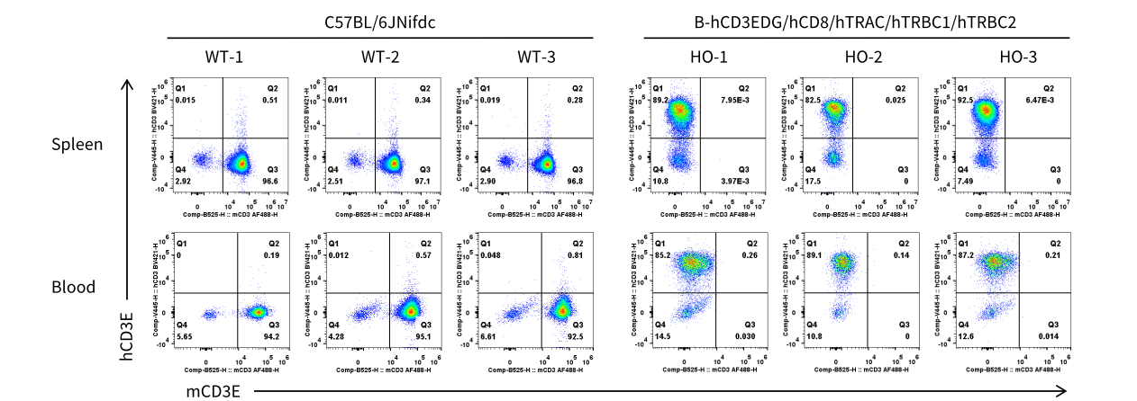
      Strain specific CD3E expression analysis in homozygous B-hCD3EDG/hCD8/hTRAC/hTRBC1/hTRBC2 mice by flow cytometry. Splenocytes and blood cells were collected from wild-type (WT) C57BL/6JNifdc mice and homozygous (HO) B-hCD3EDG/hCD8/hTRAC/hTRBC1/hTRBC2 mice (male, 9-week-old, n=3). Protein expression was analyzed with anti-mouse CD3ε antibody (Biolegend, 100312) and anti-humanCD3 antibody (BD, 556612) by flow cytometry.
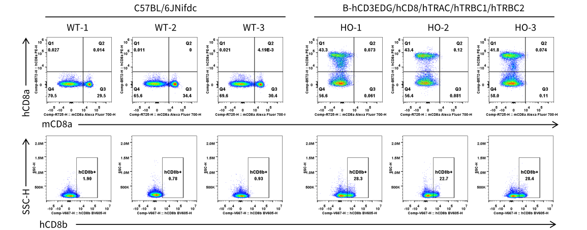
      CD8 Expression in Spleen T Cells

      Strain specific CD8 expression analysis in homozygous B-hCD3EDG/hCD8/hTRAC/hTRBC1/hTRBC2 mice by flow cytometry. Splenocytes were collected from wild-type (WT) C57BL/6JNifdc mice and homozygous (HO) B-hCD3EDG/hCD8/hTRAC/hTRBC1/hTRBC2 mice. CD8 expression was analyzed by flow cytometry using anti-mouse CD8 antibody (Biolegend, 100730), anti-human CD8a antibody (Biolegend, 300908) and anti-human CD8b antibody (BD,742392).
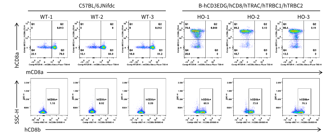
      CD8 Expression in Spleen CD4- T Cells

      Strain specific CD8 expression analysis in homozygous B-hCD3EDG/hCD8/hTRAC/hTRBC1/hTRBC2 mice by flow cytometry. Splenocytes were collected from wild-type (WT) C57BL/6JNifdc mice and homozygous (HO) B-hCD3EDG/hCD8/hTRAC/hTRBC1/hTRBC2 mice. CD8 expression was analyzed by flow cytometry using anti-mouse CD8 antibody (Biolegend, 100730), anti-human CD8a antibody (Biolegend, 300908) and anti-human CD8b antibody (BD,742392).
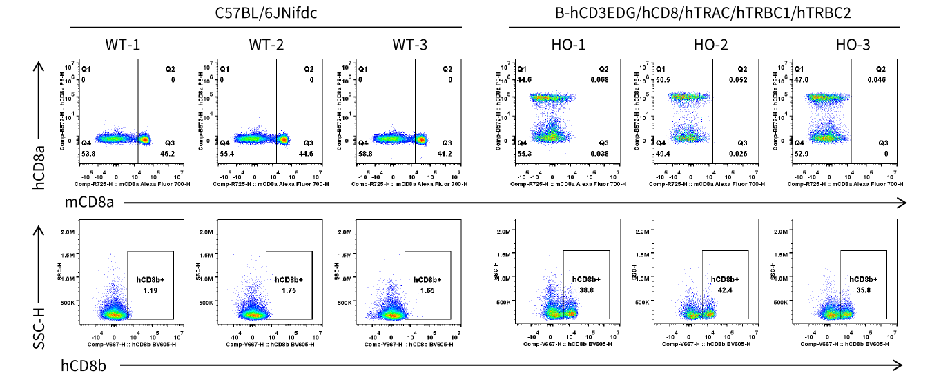
      CD8 Expression in Blood T Cells

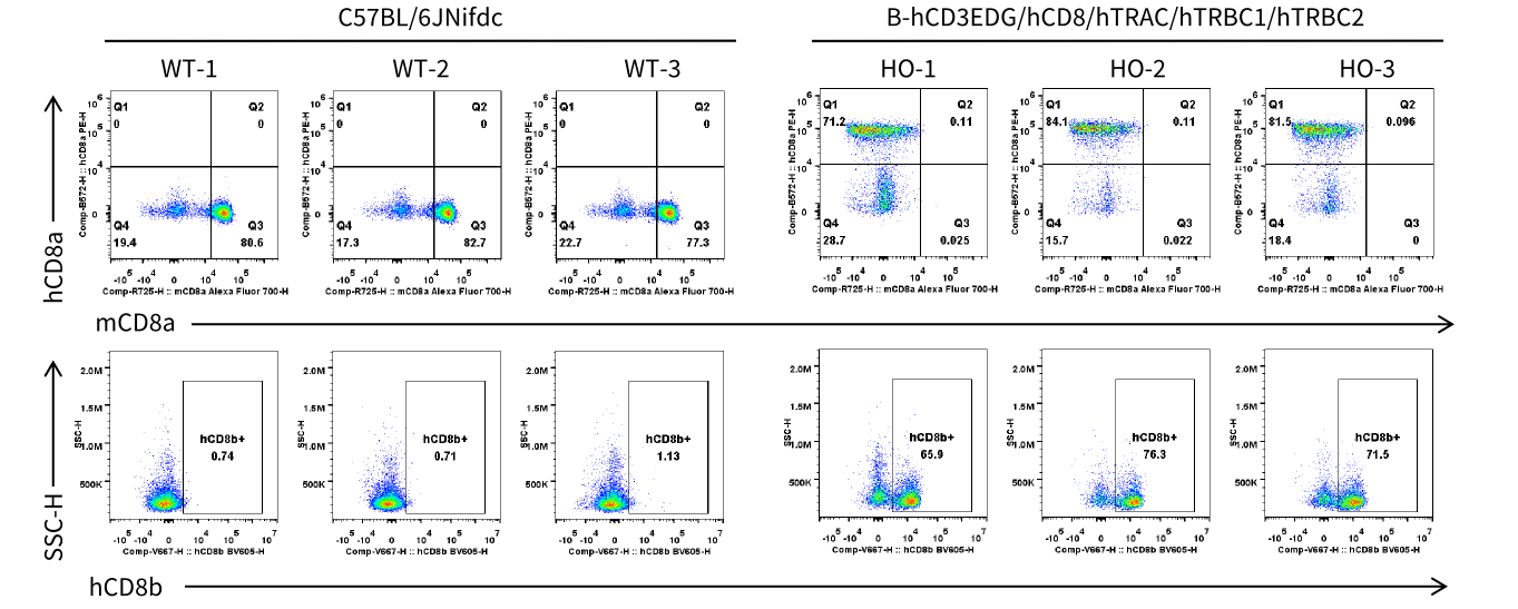
      Strain specific CD8 expression analysis in homozygous B-hCD3EDG/hCD8/hTRAC/hTRBC1/hTRBC2 mice by flow cytometry. Blood cells were collected from wild-type (WT) C57BL/6JNifdc mice and homozygous (HO) B-hCD3EDG/hCD8/hTRAC/hTRBC1/hTRBC2 mice. CD8 expression was analyzed by flow cytometry using anti-mouse CD8 antibody (Biolegend, 100730), anti-human CD8a antibody (Biolegend, 300908) and anti-human CD8b antibody (BD, 742392).
      CD8 Expression in Blood CD4- T Cells

      Strain specific CD8 expression analysis in homozygous B-hCD3EDG/hCD8/hTRAC/hTRBC1/hTRBC2 mice by flow cytometry. Blood cells were collected from wild-type (WT) C57BL/6JNifdc mice and homozygous (HO) B-hCD3EDG/hCD8/hTRAC/hTRBC1/hTRBC2 mice. CD8 expression was analyzed by flow cytometry using anti-mouse CD8 antibody (Biolegend, 100730), anti-human CD8a antibody (Biolegend, 300908) and anti-human CD8b antibody (BD, 742392).
      TRAC mRNA Expression Analysis

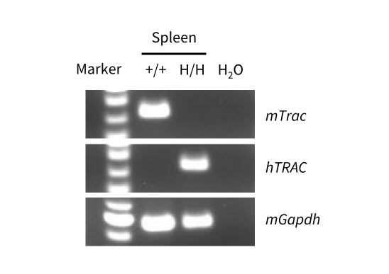
      Strain specific analysis of TRAC mRNA expression in wild-type C57BL/6JNifdc mice and B-hCD3EDG/hCD8/hTRAC/hTRBC1/hTRBC2 mice by RT-PCR. Spleen RNA was isolated from wild-type C57BL/6JNifdc mice (+/+) and homozygous B-hCD3EDG/hCD8/hTRAC/hTRBC1/hTRBC2 mice (H/H), and then cDNA libraries were synthesized by reverse transcription, followed by PCR with mouse or human TRAC primers.
      TRAC Protein Expression Analysis

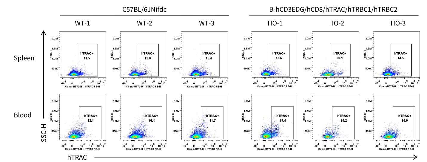
      Strain specific TRAC expression analysis in homozygous B-hCD3EDG/hCD8/hTRAC/hTRBC1/hTRBC2 mice by flow cytometry. Splenocytes and blood cells were collected from wild-type (WT) C57BL/6JNifdc mice and homozygous (HO) B-hCD3EDG/hCD8/hTRAC/hTRBC1/hTRBC2 mice (male, 9-week-old, n=3). TRAC expression was analyzed by flow cytometry using anti- TRAC antibody (Invitrogen, PA5-95587).
      TRBC1 Protein Expression Analysis

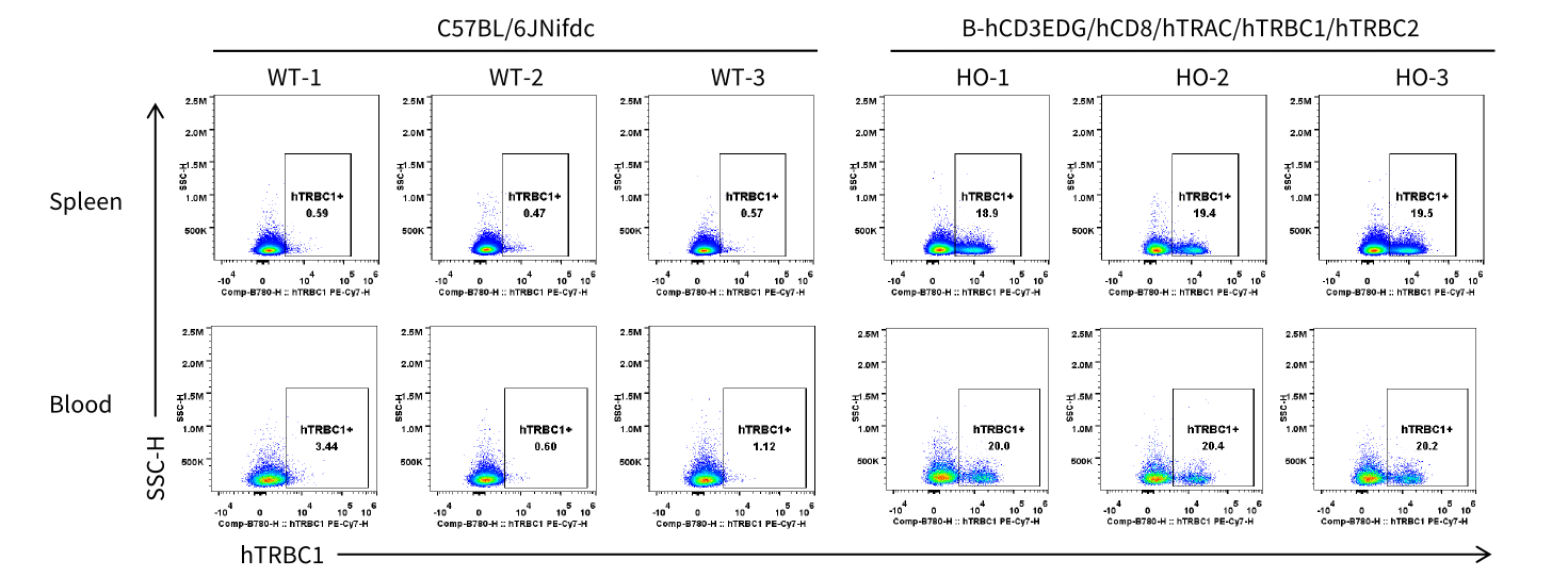
      Strain specific TRBC1 expression analysis in homozygous B-hCD3EDG/hCD8/hTRAC/hTRBC1/hTRBC2 mice by flow cytometry. Splenocytes and blood cells were collected from wild-type (WT) C57BL/6JNifdc mice and homozygous (HO) B-hCD3EDG/hCD8/hTRAC/hTRBC1/hTRBC2 mice (male, 9-week-old, n=3). TRBC1 expression was analyzed by flow cytometry using anti-human TRBC1 antibody (Biolegend, 383515).
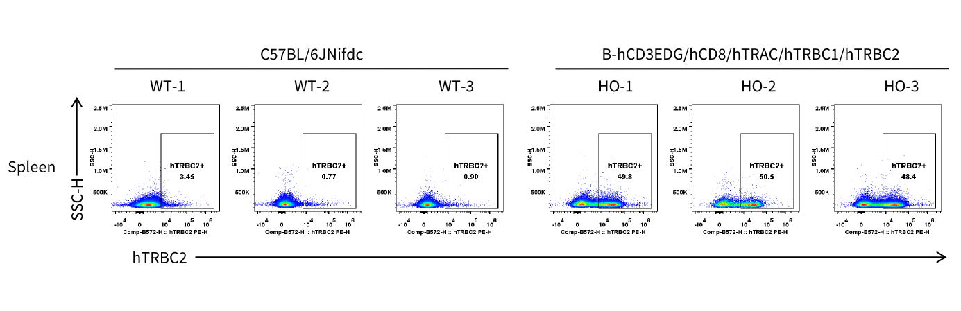
      TRBC2 Protein Expression Analysis

      Strain specific TRBC2 expression analysis in homozygous B-hCD3EDG/hCD8/hTRAC/hTRBC1/hTRBC2 mice by flow cytometry. Splenocytes were collected from wild-type (WT) C57BL/6JNifdc mice and homozygous (HO) B-hCD3EDG/hCD8/hTRAC/hTRBC1/hTRBC2 mice (male, 9-week-old, n=3).TRBC2 expression was analyzed by flow cytometry using anti-human TRBC2 antibody (BD, 571191).
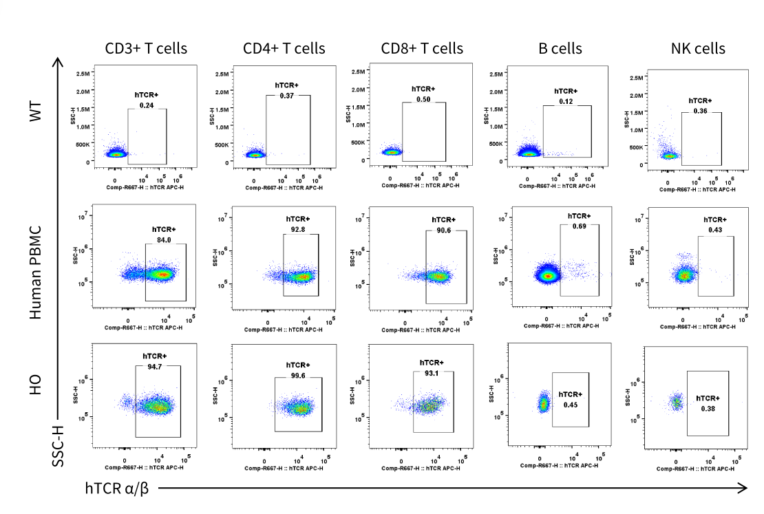
      TCR α/β Protein Expression Analysis

      Strain specific TCR α/β expression analysis inhomozygous
      B-hCD3EDG/hCD8/hTRAC/hTRBC1/hTRBC2 mice by flow cytometry.
      Splenocytes were collected from wild-type (WT) C57BL/6JNifdc mice and homozygous (HO) B-hCD3EDG/hCD8/hTRAC/hTRBC1/hTRBC2 mice (male, 9-week-old, n=1). TCR α/β expression was analyzed by flow cytometry using anti-human TCR α/β antibody (Biolegend, 306717).
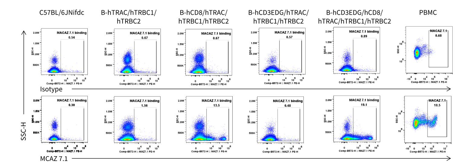
      The Binding of MCAZ 7.1 for Different TRAC/TRBC1/TRBC2-related Models

      FACS analysis of MACAZ7.1 binding to splenocytes from different humanized mouse strains. Splenocytes were collected from wild-type C57BL/6JNifdc mice and the indicated homozygous humanized mouse strains. The MCAZ7.1 was made in-house. Cells were incubated with MACAZ7.1 or a human IgG1 isotype control, followed by staining with a fluorescently labeled anti-human IgG1 secondary antibody. Antibody binding was analyzed by flow cytometry.
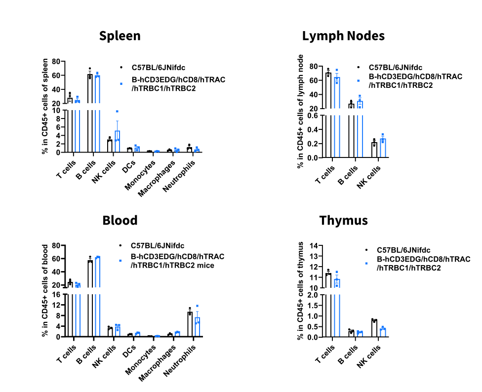
      Analysis of Leukocyte Subpopulations

      Analysis of leukocyte subpopulations by flow cytometry in immune organs and blood.Splenocytes, peripheral blood, lymph nodes and thymus were isolated from male C57BL/6JNifdc and B-hCD3EDG/hCD8/hTRAC/hTRBC1/hTRBC2 mice (male, n=3, 9-week-old). Single live cells were gated on the CD45⁺ population and analyzed by flow cytometry as indicated. Values are expressed as mean ± SEM.
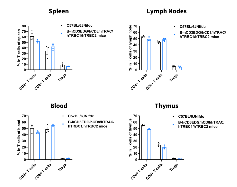
      Analysis of T Cell Subpopulations

      Analysis of T-cell subpopulations by flow cytometry in immune organs and blood. Splenocytes, peripheral blood, lymph nodes and thymus were isolated from male C57BL/6JNifdc and B-hCD3EDG/hCD8/hTRAC/hTRBC1/hTRBC2 mice (male, n = 3, 9-week-old). Single live cells were gated on the CD3⁺ T-cell population and analyzed by flow cytometry as indicated. Values are expressed as mean ± SEM.
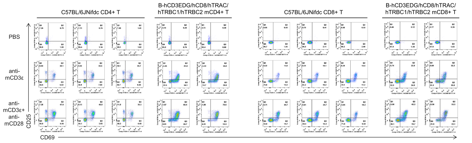
      Analysis of T Cell Activation

      In vitro T cell activation by anti-mCD3ε antibody with or without anti-mCD28 antibody in wild-type C57BL/6JNifdc mice and homozygous humanized B-hCD3EDG/hCD8/hTRAC/hTRBC1/hTRBC2 mice (24h). T cells were isolated from splenocytes of C57BL/6JNifdc (male, 9-week-old, n = 3) and B-hCD3EDG/hCD8/hTRAC/hTRBC1/hTRBC2 mice (male, 9-week-old, n = 2), and incubated in the presence of anti-mCD3ε antibody (2 ug/ml, BioXcell, BE0001-2), with or witnout anti-mCD28 antibody (5 ug/ml, BioXcell, BE0015-1) for 24h.
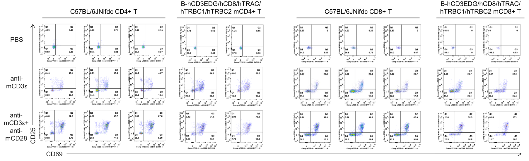
      In vitro T cell activation by anti-mCD3ε antibody with or without anti-mCD28 antibody in wild-type C57BL/6JNifdc mice and homozygous humanized B-hCD3EDG/hCD8/hTRAC/hTRBC1/hTRBC2 mice (48h). T cells were isolated from splenocytes of C57BL/6JNifdc (male, 9-week-old, n = 3) and B-hCD3EDG/hCD8/hTRAC/hTRBC1/hTRBC2 mice (male, 9-week-old, n = 2), and incubated in the presence of anti-mCD3ε antibody (2 ug/ml, BioXcell, BE0001-2), with or witnout anti-mCD28 antibody (5 ug/ml, BioXcell, BE0015-1) for 48h.

      * When publishing results obtained using this animal model, please acknowledge the source as follows: The animal model [B-hCD3EDG/hCD8/hTRAC/hTRBC1/hTRBC2 mice] (Cat# 114950) was purchased from Biocytogen.