


EPCAM: A key adhesion molecule in tumor malignancy and its therapeutic intervention

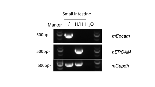
Human EPCAM was detectable in EPCAM Humanized Mice by RT-PCR and sequencing. Small intestine tissues were isolated from wild-type BALB/cCrSlcNifdc mice (+/+) and homozygous B-hEPCAM mice(C) (H/H). Primers were designed to detect mouse and human EPCAM. Sequencing of the PCR products confirmed that the amplified sequences were consistent with database reference sequences.
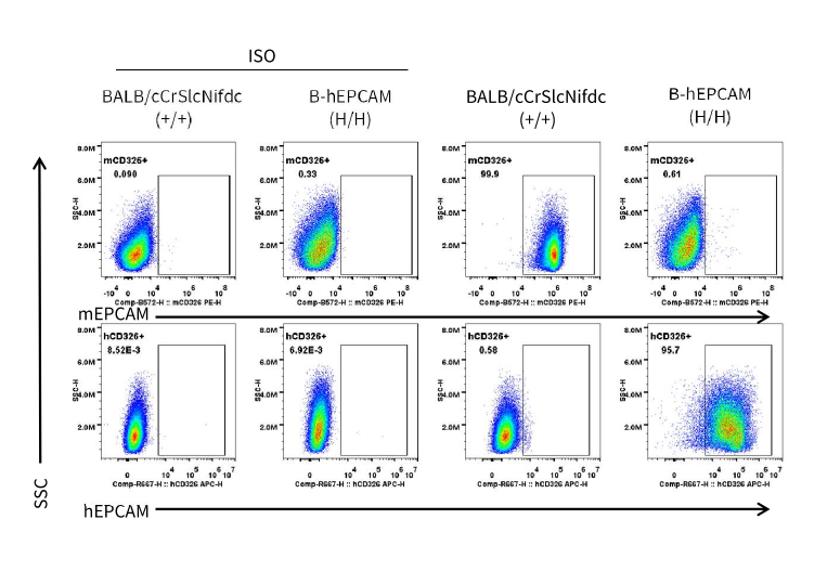
Strain-specific EPCAM expression analysis in wild-type BALB/cCrSlcNifdc mice and homozygous EPCAM humanized mice. Small intestine cells were collected from wild-type BALB/cCrSlcNifdc mice and homozygous B-hEPCAM mice (C). EPCAM expression was analyzed by flow cytometry using anti-human EPCAM antibody (Biolegend, 369809) and anti-mouse EPCAM antibody (Biolegend, 118205).
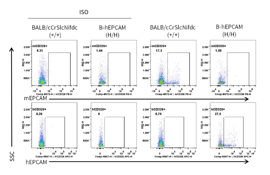
Strain specific EPCAM expression analysis in wild-type BALB/cCrSlcNifdc and homozygous EPCAM humanized mice by flow cytometry. Lung cells were collected from wild-type BALB/cCrSlcNifdc mice and homozygous B-hEPCAM mice(C). EPCAM expression was analyzed by flow cytometry using anti-human EPCAM antibody (Biolegend, 369809) and anti-mouse EPCAM antibody (Biolegend, 118205).
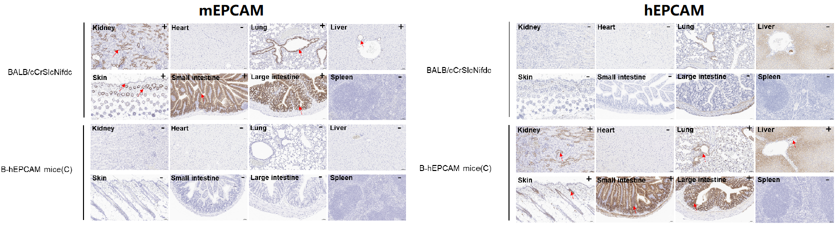
Immunohistochemical (IHC) analysis of EPCAM expression in EPCAM humanized mice. The kidney, heart, lung, liver, skin, small intestine, large intestine and spleen were collected from wild-type BALB/cCrSlcNifdc mice and B-hEPCAM mice(C) (female, 6-week-old), analyzed by IHC with anti-mEPCAM antibody (abcam, ab213501) and anti-hEPCAM antibody (abcam, ab223582). The arrow indicates tissue cells with positive EPCAM staining (brown). “+” indicates that the tissue is positive, and “-” indicates that the tissue is negative.