


TL1A: A key inflammation cytokine in chronic intestinal inflammation and fibrosis-related diseases

TL1A
The exons 1-4 of mouse Tl1a gene that encode extracellular domain were replaced by human counterparts in B-hTL1A/hIL23A/hIL12B/hα4β7, Rag2 KO mice.
The genomic region of mouse Tl1a gene that encodes transmembrane domain and cytoplasmic portion was retained. The promoter, 5’ UTR and 3’ UTR region of the mouse gene were also retained. The TL1A expression was driven by endogenous mouse Tl1a promoter, while mouse Tl1a gene transcription and translation will be disrupted.
IL23A
The exons 1-4 of mouse Il23a gene that encode the whole molecule (ATG to STOP codon) were replaced by human counterparts in B-hTL1A/hIL23A/hIL12B/hα4β7, Rag2 KO mice.
The promoter, 5’ UTR and 3’ UTR region of the mouse gene were retained. The human IL23A expression was driven by endogenous mouse Il23a promoter, while mouse Il23a gene transcription and translation will be disrupted.
IL12B
The exons 2-8 of mouse Il12b gene that encode the whole molecule (ATG to STOP codon), including 3’ UTR were replaced by human counterparts in B-hTL1A/hIL23A/hIL12B/hα4β7, Rag2 KO mice.
The promoter and 5’ UTR region of the mouse gene were retained. The human IL12B expression was driven by endogenous mouse Il12b promoter, while mouse Il12b gene transcription and translation will be disrupted.
ITGA4
The exons 1-27 of mouse Itga4 gene that encode the extracellular domain were replaced by human counterparts in B-hTL1A/hIL23A/hIL12B/hα4β7, Rag2 KO mice.
The promoter, 5’ UTR and 3’ UTR region of the mouse gene were retained. The human ITGA4 expression was driven by endogenous mouse Itga4 promoter, while mouse Itga4 gene transcription and translation will be disrupted.
ITGB7
The exons 2-14 of mouse Itgb7 gene that encode the extracellular domain were replaced by human counterparts in B-hTL1A/hIL23A/hIL12B/hα4β7, Rag2 KO mice.
The promoter, 5’ UTR and 3’ UTR region of the mouse gene were retained. The human ITGB7 expression was driven by endogenous mouse Itgb7 promoter, while mouse Itgb7 gene transcription and translation will be disrupted.
Rag2
The exon 3 and 3’ UTR region of mouse Rag2 were knocked out in B-hTL1A/hIL23A/hIL12B/hα4β7, Rag2 KO mice, resulting in a disruption of the Rag2 gene. B-hTL1A/hIL23A/hIL12B/hα4β7, Rag2 KO mice was obtained by deleting Rag2 on the background of B-hTL1A/hIL23A/hIL12B/hα4β7 mice (113826).

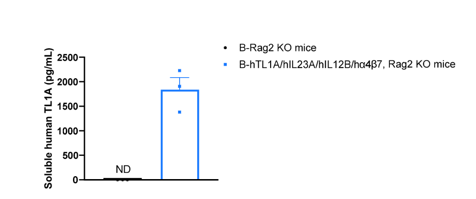
Soluble TL1A expression analysis in B-hTL1A/hIL23A/hIL12B/hα4β7, Rag2 KO mice by ELISA. Bone marrow derived dendritic cells (BMDCs) were produced by culturing the bone marrow from wild-type B-Rag2 KO mice (-/-) and homozygous B-hTL1A/hIL23A/hIL12B/hα4β7, Rag2 KO mice (H/H;H/H;H/H;H/H;H/H;-/-), which were stimulated with LPS in vitro. After stimulation, the supernatants were collected and the levels of soluble TL1A were measured using the species-specific human TL1A ELISA kit. Soluble human TL1A was exclusively detectable in homozygous B-hTL1A/hIL23A/hIL12B/hα4β7, Rag2 KO mice but not wild-type B-Rag2 KO mice. Values are expressed as mean ± SEM. ND: not detectable.
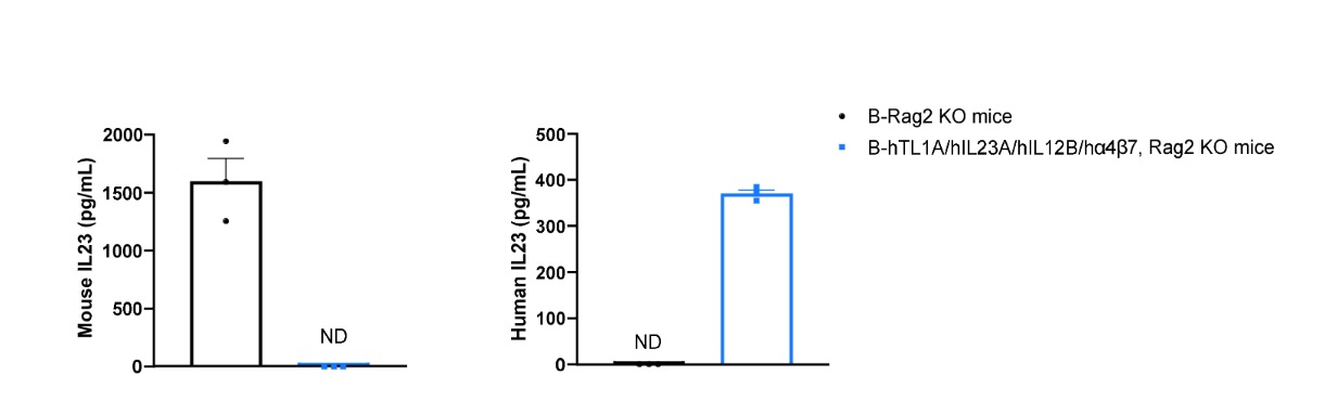
Mouse IL-23 and human IL-23 expression analysis in B-hTL1A/hIL23A/hIL12B/hα4β7, Rag2 KO mice by ELISA. Bone marrow derived dendritic cells (BMDCs) were produced by culturing the bone marrow from wild-type B-Rag2 KO mice (-/-) and homozygous B-hTL1A/hIL23A/hIL12B/hα4β7, Rag2 KO mice (H/H;H/H;H/H;H/H;H/H;-/-), which were stimulated with LPS in vitro. After stimulation, the supernatants were collected and the levels of mouse and human IL23 were analyzed by ELISA (R&D, M2300; R&D, D2300B). Mouse IL23 was only detectable in wild-type B-Rag2 KO mice. Human IL23 was exclusively detectable in homozygous B-hTL1A/hIL23A/hIL12B/hα4β7, Rag2 KO mice. Values are expressed as mean ± SEM. ND: not detectable.
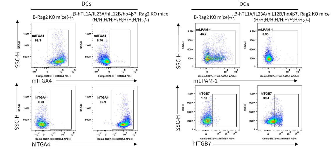
Strain specific ITGA4 and ITGB7 expression analysis in wild-type B-Rag2 KO mice and homozygous B-hTL1A/hIL23A/hIL12B/hα4β7, Rag2 KO mice by flow cytometry. Splenocytes were collected from wild-type B-Rag2 KO mice (-/-) and homozygous B-hTL1A/hIL23A/hIL12B/hα4β7, Rag2 KO mice (H/H;H/H;H/H;H/H;H/H;-/-). Protein expression was analyzed with anti-mouse ITGA4 antibody (Biolegend, 103705), anti-mouse LPAM-1 (Integrin α4β7) antibody (Biolegend, 120607), anti-human ITGA4 antibody (Biolegend, 304307) anti-human ITGB7 antibody (Invitrogen, MA5-23541) by flow cytometry. Mouse ITGA4 and ITGB7 were only detectable on DCs of wild-type B-Rag2 KO mice. Human ITGA4 and ITGB7 were exclusively detectable on DCs of homozygous B-hTL1A/hIL23A/hIL12B/hα4β7, Rag2 KO mice.
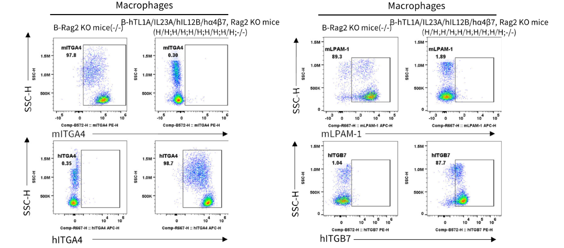
Strain specific ITGA4 and ITGB7 expression analysis in wild-type B-Rag2 KO mice and homozygous B-hTL1A/hIL23A/hIL12B/hα4β7, Rag2 KO mice by flow cytometry. Splenocytes were collected from wild-type B-Rag2 KO mice (-/-) and homozygous B-hTL1A/hIL23A/hIL12B/hα4β7, Rag2 KO mice (H/H;H/H;H/H;H/H;H/H;-/-). Protein expression was analyzed with anti-mouse ITGA4 antibody (Biolegend, 103705), anti-mouse LPAM-1 (Integrin α4β7) antibody (Biolegend, 120607), anti-human ITGA4 antibody (Biolegend, 304307) anti-human ITGB7 antibody (Invitrogen, MA5-23541) by flow cytometry. Mouse ITGA4 and ITGB7 were only detectable on macrophages of wild-type B-Rag2 KO mice. Human ITGA4 and ITGB7 were exclusively detectable on macrophages of homozygous B-hTL1A/hIL23A/hIL12B/hα4β7, Rag2 KO mice.
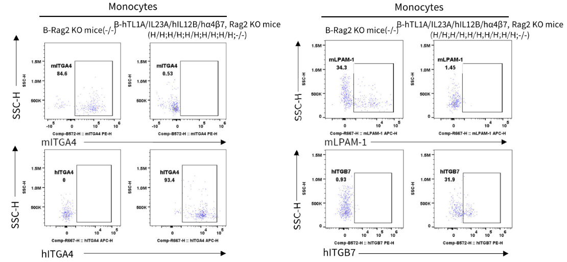
Strain specific ITGA4 and ITGB7 expression analysis in wild-type B-Rag2 KO mice and homozygous B-hTL1A/hIL23A/hIL12B/hα4β7, Rag2 KO mice by flow cytometry. Splenocytes were collected from wild-type B-Rag2 KO mice (-/-) and homozygous B-hTL1A/hIL23A/hIL12B/hα4β7, Rag2 KO mice (H/H;H/H;H/H;H/H;H/H;-/-). Protein expression was analyzed with anti-mouse ITGA4 antibody (Biolegend, 103705), anti-mouse LPAM-1 (Integrin α4β7) antibody (Biolegend, 120607), anti-human ITGA4 antibody (Biolegend, 304307) anti-human ITGB7 antibody (Invitrogen, MA5-23541) by flow cytometry. Mouse ITGA4 and ITGB7 were only detectable on monocytes of wild-type B-Rag2 KO mice. Human ITGA4 and ITGB7 were exclusively detectable on monocytes of homozygous B-hTL1A/hIL23A/hIL12B/hα4β7, Rag2 KO mice.
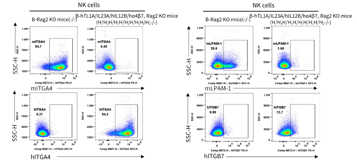
Strain specific ITGA4 and ITGB7 expression analysis in wild-type B-Rag2 KO mice and homozygous B-hTL1A/hIL23A/hIL12B/hα4β7, Rag2 KO mice by flow cytometry. Splenocytes were collected from wild-type B-Rag2 KO mice (-/-) and homozygous B-hTL1A/hIL23A/hIL12B/hα4β7, Rag2 KO mice (H/H;H/H;H/H;H/H;H/H;-/-). Protein expression was analyzed with anti-mouse ITGA4 antibody (Biolegend, 103705), anti-mouse LPAM-1 (Integrin α4β7) antibody (Biolegend, 120607), anti-human ITGA4 antibody (Biolegend, 304307) anti-human ITGB7 antibody (Invitrogen, MA5-23541) by flow cytometry. Mouse ITGA4 and ITGB7 were only detectable on NK cells of wild-type B-Rag2 KO mice. Human ITGA4 and ITGB7 were exclusively detectable on NK cells of homozygous B-hTL1A/hIL23A/hIL12B/hα4β7, Rag2 KO mice.
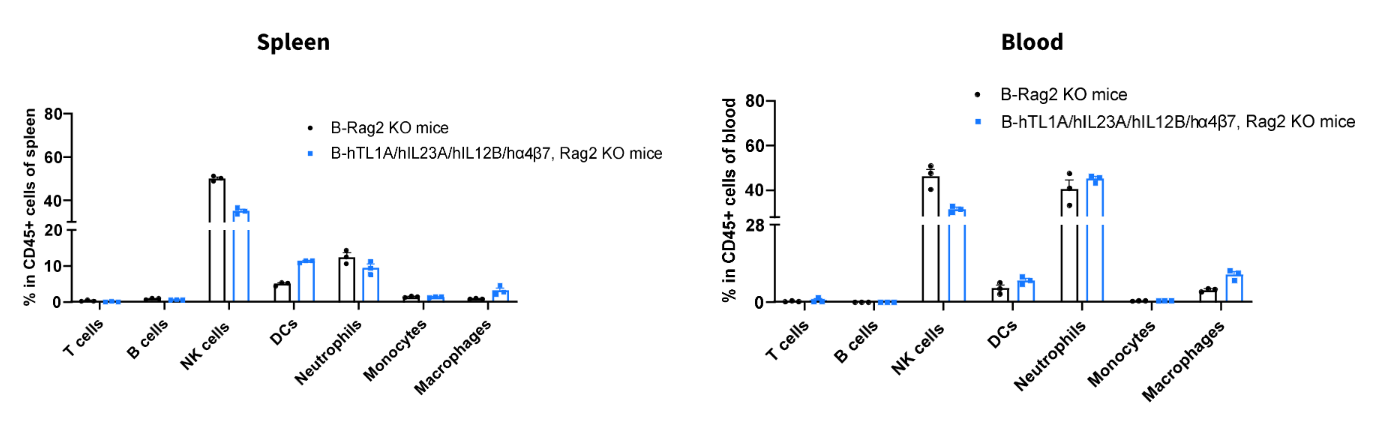
Analysis of leukocyte subpopulations by flow cytometry in immune organs and blood. Splenocytes and peripheral blood were isolated from wild-type B-Rag2 KO mice and B-hTL1A/hIL23A/hIL12B/hα4β7, Rag2 KO mice (female, n=3, 7-week-old). Single live cells were gated on the CD45⁺ population and analyzed by flow cytometry as indicated. Values are expressed as mean ± SEM.