- CDH3, also known as P-Cadherin, is a calcium-dependent cell-cell adhesion glycoprotein belonging to the classical type I cadherin family. Its structure is characterized by five extracellular cadherin repeats (EC1-EC5) that mediate homophilic binding (CDH3-to-CDH3 interaction between adjacent cells), a single-pass transmembrane domain, and a highly conserved cytoplasmic tail. This cytoplasmic domain interacts with catenin proteins (p120-catenin and β-catenin), which bridge the complex to the actin cytoskeleton, enabling strong adhesion and intracellular signaling.
- While its expression in normal adult tissues is largely restricted to basal layers (e.g., in the skin and mammary glands), CDH3 is frequently overexpressed in a variety of aggressive epithelial carcinomas, including breast (particularly triple-negative), gastric, ovarian, and pancreatic cancers. This aberrant overexpression is strongly correlated with poor prognosis, promoting tumorigenesis and metastasis through several key mechanisms:
1. Enhanced Invasion and Metastasis: Contrary to its typical role as a suppressor of invasion, CDH3 overexpression in tumors can disrupt normal cellular organization. It promotes an aggressive, motile phenotype, often by inducing Epithelial-to-Mesenchymal Transition (EMT), a process crucial for cancer cell dissemination.
2. Activation of Pro-Survival Signaling: The cytoplasmic engagement of β-catenin can lead to the stabilization and nuclear translocation of β-catenin, resulting in the transcriptional activation of oncogenic genes within the Wnt signaling pathway, thereby driving proliferation and inhibiting apoptosis.
3. Maintenance of Cancer Stem Cell (CSC) Populations: CDH3 is a key marker and functional driver in subpopulations of CSCs. It helps maintain stemness, self-renewal, and resistance to therapy, contributing to tumor initiation, recurrence, and metastasis.
4. Modulation of the Tumor Microenvironment: Through its adhesive functions, CDH3 facilitates interactions between tumor cells and the surrounding stroma, influencing pathways that support tumor growth, angiogenesis, and immune evasion.
- Due to its selective overexpression on cancer cells and its central role in driving malignancy, CDH3 has emerged as a promising therapeutic target for antibody-based therapies, including Antibody-Drug Conjugates (ADCs) and Bispecific Antibodies.
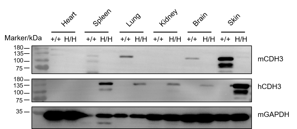
- In B-NDG hCDH3 mice, the exons 4-13 of mouse Cdh3 gene that encode the extracellular domain were replaced by human counterparts. A small amount of human CDH3 expression can be detected in the spleen, lung, kidney and brain of B-NDG hCDH3 mice, but a high level of human CDH3 expression can be detected in the skin of B-NDG hCDH3 mice.
- Application: This mouse model empowers the in vivo evaluation of CDH3-targeting therapeutic agents, such as antibodies, ADCs, and bispecifics, for efficacy and safety against human CDH3-expressing tumors.