Description
Background:
- The PDE6B gene, located on human chromosome 4p16.3, spans approximately 45 kb and comprises 22 exons. It encodes a member of the phosphodiesterase 6 (PDE6) subfamily, which specifically hydrolyzes cyclic guanosine monophosphate (cGMP). In rod photoreceptor cells, cGMP serves as a critical ligand for cyclic nucleotide-gated (CNG) ion channels in the outer segment membrane and plays a pivotal role in phototransduction.
- Loss-of-function mutations in PDE6B lead to pathological cGMP accumulation in photoreceptor cells, resulting in persistent CNG channel opening and subsequent cation influx. This disrupts cellular ion homeostasis, ultimately inducing photoreceptor toxicity and degeneration. As retinal photoreceptors progressively deteriorate, the light-to-electrical signal conversion capacity of the retina is severely impaired.
- PDE6B mutations are a major cause of autosomal recessive retinitis pigmentosa (RP). Bioinformatic analyses suggest that mutations at any site within the PDE6B gene can alter the three-dimensional conformation of PDE6β, leading to structural destabilization and functional impairment. Beyond recessive RP, aberrant PDE6B expression or function has also been implicated in other inherited retinal disorders, including: Autosomal dominant retinitis pigmentosa and Congenital stationary night blindness (CSNB).
Targeting strategy:
- The exons 1-22 of mouse Pde6b gene were knocked out in B-Pde6b KO mice.
Verification:
- Mouse PDE6B protein was only detected in the eye of C57BL/6JNifdc mice, but not in homozygous B-Pde6b KO mice.
- Mouse Pde6b mRNA was only detected in wild-type mice, but not in homozygous B-Pde6b KO mice.
Application:
- This product can be utilized to assess the pharmacodynamic effects of therapies targeting autosomal recessive retinitis pigmentosa (RP) and Leber congenital amaurosis (LCA).
Targeting strategy
Gene targeting strategy for B-Pde6b KO mice. The exons 1-22 of mouse Pde6b gene were knocked out in B-Pde6b KO mice. As a result, mouse PDE6B protein not expressed anymore.
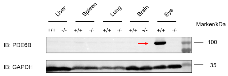
Protein expression analysis
Western blot analysis of PDE6B protein expression in homozygous B-Pde6b KO mice. Various tissue lysates were collected from wild-type C57BL/6JNifdc mice (+/+) and homozygous B-Pde6b KO mice (-/-), and then analyzed by western blot with anti-mouse PDE6B antibody (Proteintech, 22063-1-AP). 40 μg total proteins were loaded for western blotting analysis. Mouse PDE6B was only detected in the eye of C57BL/6JNifdc mice but not in homozygous B-Pde6b KO mice.
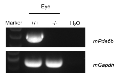
mRNA expression analysis
Strain specific analysis of mPde6b mRNA expression in wild-type C57BL/6JNifdc mice and B-Pde6b KO mice by RT-PCR. Eye RNA were isolated from wild-type C57BL/6JNifdc mice (+/+) and homozygous B-Pde6b KO mice (-/-), then cDNA libraries were synthesized by reverse transcription, followed by PCR with mouse Pde6b primers. Mouse Pde6b mRNA was only detectable in wild-type mice but not in homozygous B-Pde6b KO mice.
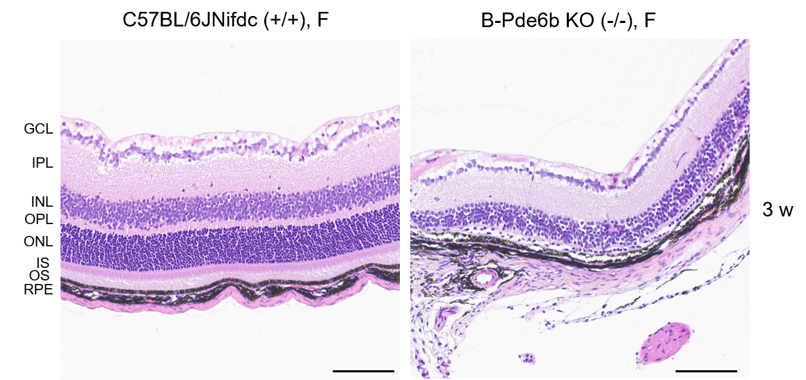
H&E staining of B-Pde6b KO mice
Representative images of HE staining of wild-type C57BL/6JNifdc mice and B-Pde6b KO mice. Retina tissues of wild-type C57BL/6JNifdc mice (+/+) and B-Pde6b KO mice (-/-) (3 weeks old, female) were collected and analyzed with H&E staining. The results showed that the retinal layers were disordered, and multiple layers such as the inner nuclear layer, the outer reticular layer and the cone-rod layer disappeared in B-Pde6b KO mice compared to the C57BL/6JNifdc mice. Meanwhile, the cells in the outer nuclear layer are reduced in B-Pde6b KO mice. Scale bar, 100 μm.
RPE: Retinal pigment epithelium, OS: Outer segment, IS: Inner segment, ONL: Outer nuclear layer, OPL: Outer plexiform layer, INL: Inner nuclear layer, IPL: Inner plexiform layer,
GCL: Ganglion cell layer.
* When publishing results obtained using this animal model, please acknowledge the source as follows: The animal model [B-Pde6b KO mice] (Cat# 113759) was purchased from Biocytogen.