The gastric inhibitory polypeptide receptor (GIPR) belongs to the G protein-coupled receptor family and is expressed in many tissues including the pancreas, stomach, brain, liver, etc. It plays a crucial role in regulating insulin secretion, glucose, and lipid metabolism. Mutations within this gene are associated with health conditions including obesity and diabetes.
In GIPR humanized mice (B-hGIPR), exons 2-14 of the mouse Gipr gene (encoding the entire protein from ATG to STOP codon, including the 3'UTR) were replaced by the human GIPR CDS and 3’ UTR. The human GIPR expression is driven by the retained endogenous mouse promoter and 5‘ UTR, while mouse gene transcription and translation are disrupted.
Validation confirms that human GIPR mRNA is exclusively detectable in GIPR humanized mice (B-hGIPR) across key metabolic tissues such as brain, epididymal white adipose tissue (eWAT), and inguinal white adipose tissue (ingWAT). Functional studies demonstrate that recombinant human GIP effectively promotes insulin secretion during intraperitoneal glucose tolerance tests (IPGTT) in this model. Furthermore, in high-fat diet (HFD)-induced obese GIPR humanized mice (B-hGIPR), anti-human GIPR antibodies (e.g., Amgen 2G10 analog), alone or in combination with Semaglutide, significantly reduce body weight, decrease food intake, and improve insulin resistance, validating the model's utility for metabolic drug evaluation.
Key Advantages
Validation
Applications
In vivo efficacy, pharmacodynamics, and safety evaluation of GIPR-targeting agonists, antagonists, and related combination therapies for metabolic diseases.

GIPR

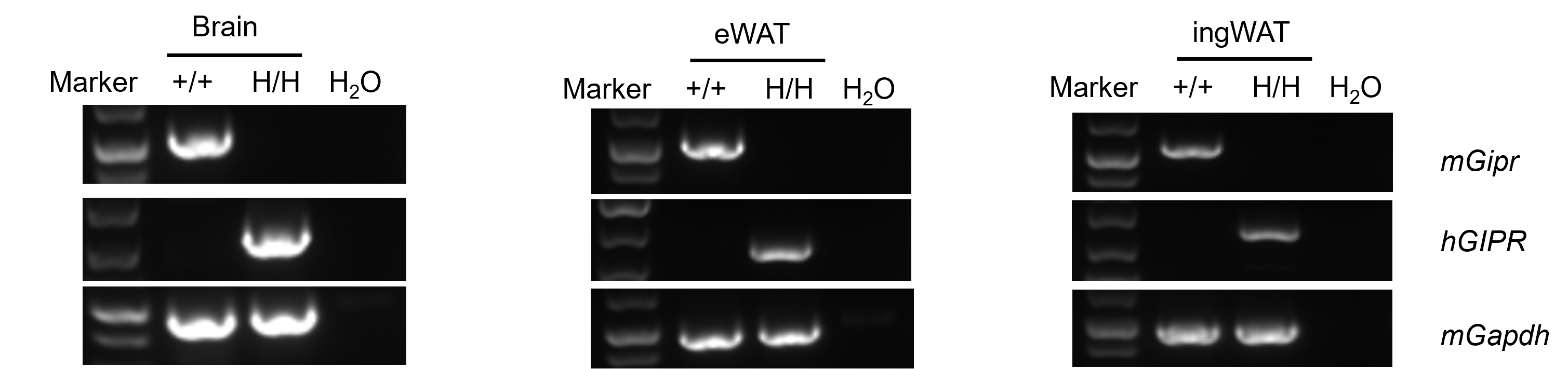
Strain specific analysis of GIPR mRNA expression in wild-type C57BL/6JNifdc mice and GIPR humanized mice by RT-PCR. Brain, eWAT, and ingWAT RNA were isolated from wild-type C57BL/6JNifdc mice (+/+) and homozygous GIPR humanized mice (H/H), then cDNA libraries were synthesized by reverse transcription, followed by PCR with mouse or human GIPR primers. Mouse Gipr mRNA was detectable only in wild-type C57BL/6JNifdc mice. Human GIPR mRNA was detectable only in homozygous GIPR humanized mice but not in wild-type mice. eWAT, epididymal white adipose tissue; ingWAT, inguinal white adipose tissue.
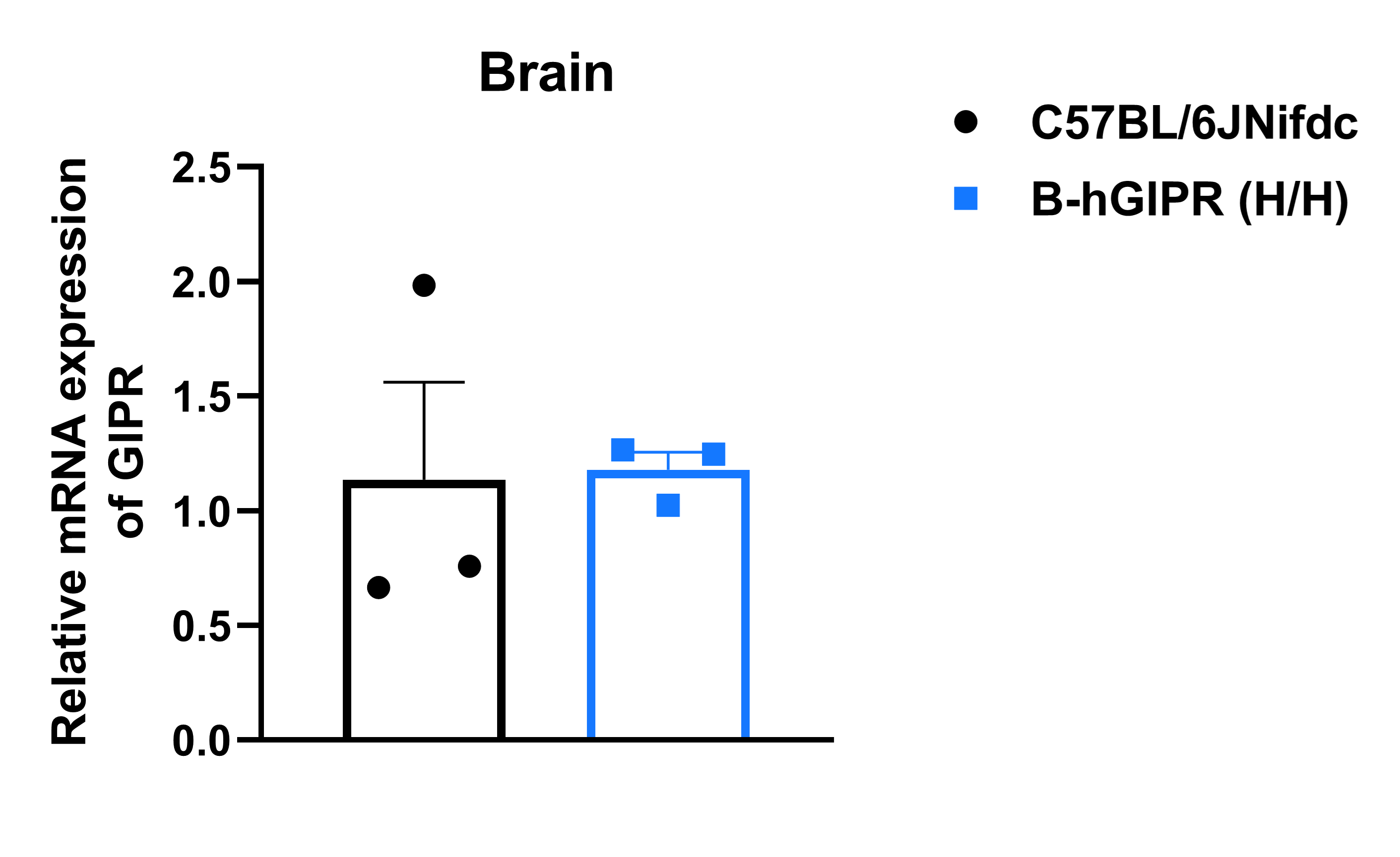
Analysis of GIPR mRNA expression in wild-type C57BL/6JNifdc and GIPR humanized mice by RT-qPCR. RNA was isolated from wild-type C57BL/6JNifdc (+/+) and homozygous GIPR humanized mice (H/H), then cDNA libraries were synthesized by reverse transcription, followed by PCR with GIPR primers. Values are expressed as mean ± SEM. (The primer pair was designed in the 5’UTR to amplify both mouse and human GIPR mRNA.)
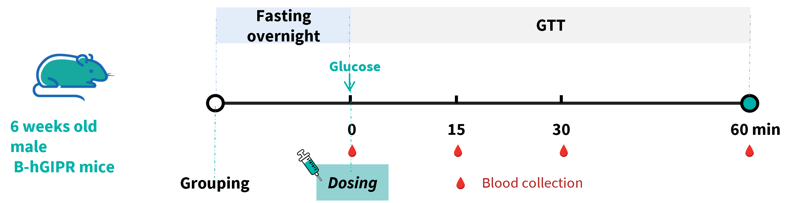
In vivo function of m/hGIP during IPGTT. Wild-type C57BL/6JNifdc mice and GIPR humanized mice (male, 6-week-old, n=6) were treated i.p. with mouse GIP (Cat. HY-P77948, MCE) or human GIP (Cat. HY-P0276, MCE) at 0.25 mg/kg. Then the Intraperitoneal Glucose Tolerance Test (IPGTT) was performed for 2 g/kg D-glucose (40% solution, 5 μL/g).
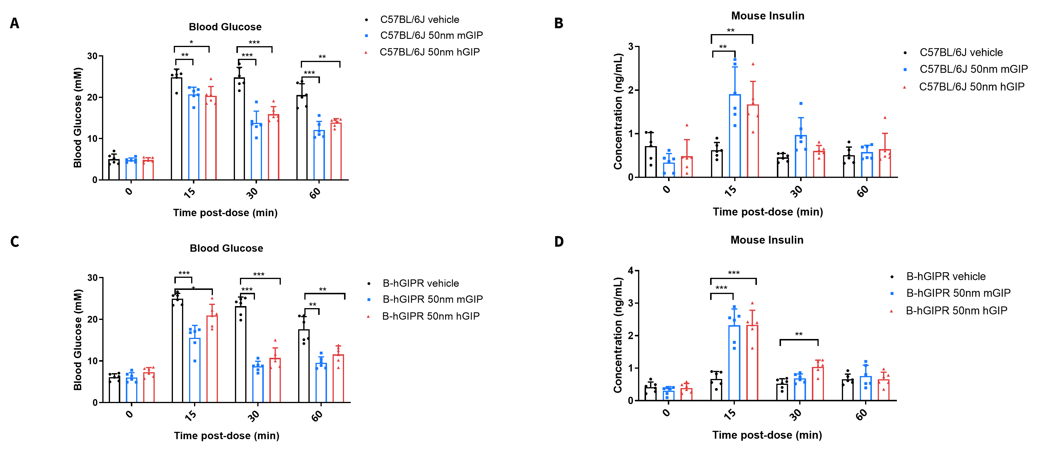
In vivo function of m/hGIP during IPGTT. Wild-type C57BL/6JNifdc mice and GIPR humanized mice (male, 6-week-old, n=6) were treated i.p. with mouse GIP (Cat. HY-P77948, MCE) or human GIP (Cat. HY-P0276, MCE) at 0.25 mg/kg. Then the IPGTT was performed for 2 g/kg D-glucose (40% solution, 5 μL/g). (A, C) Plasma glucose during IPGTT. (B, D) Mouse insulin during IPGTT. Data represent means ± SEM. Analyzed by 2way-ANOVA,*P<0.05, **P<0.01, ***P<0.001.

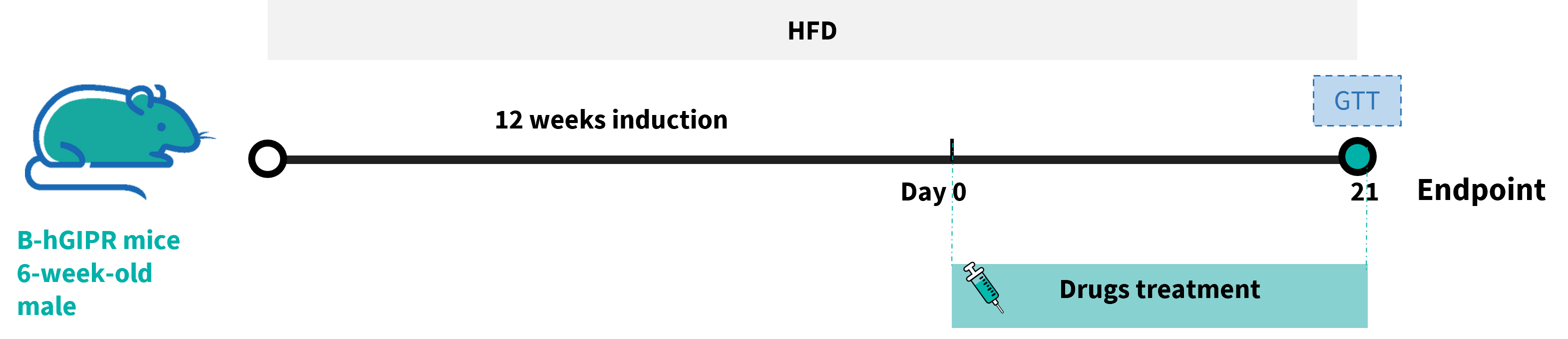
Efficacy study in HFD induced GIPR humanized mice. GIPR humanized mice were fed with a high-fat diet (HFD) for 12 weeks to induce obesity in the mice.
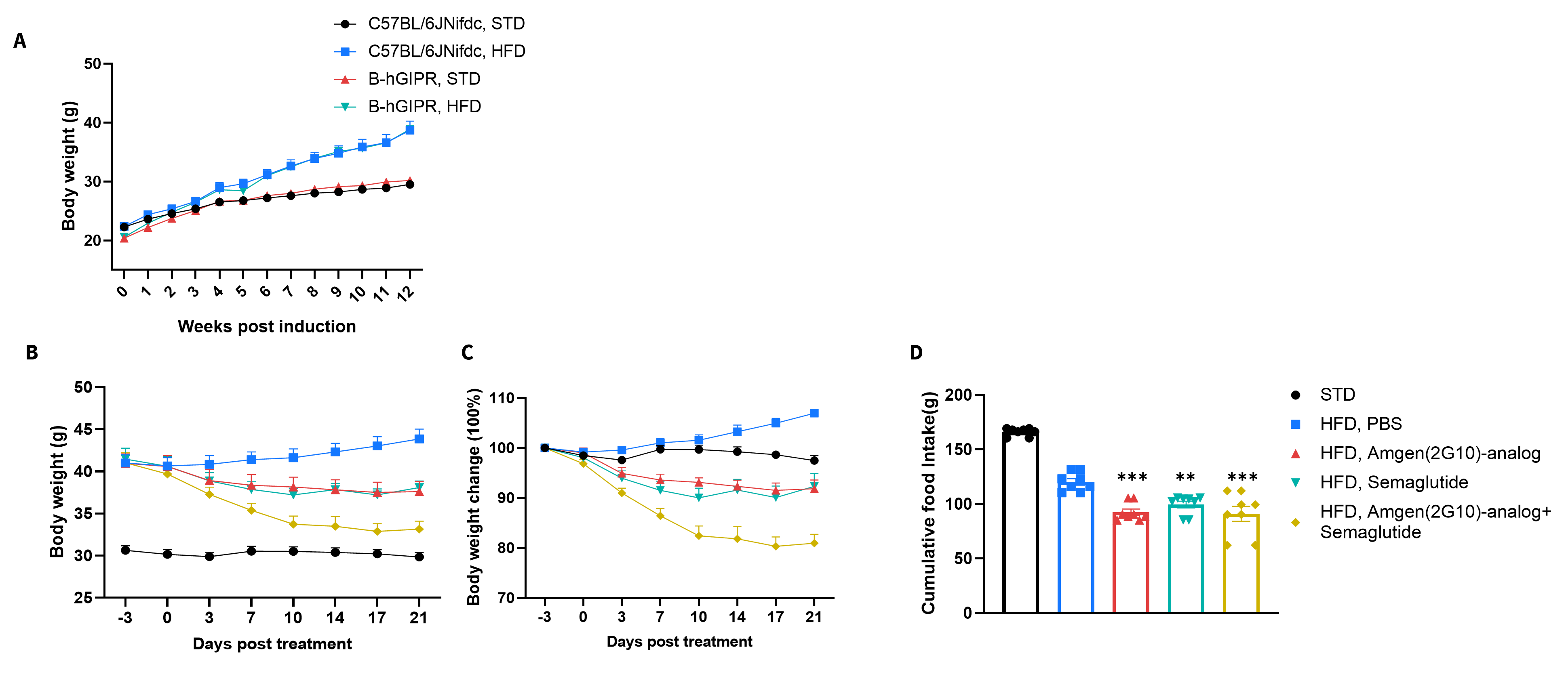
Efficacy study in HFD induced GIPR humanized mice. GIPR humanized mice were fed with a high-fat diet for 12 weeks to induce mice obesity. (A) Body weight change after HFD induction. (B-C) Body weight change after hGIPR-antibody Amgen (2G10) analog (in house), Semaglutide, and combination treatment. (D) Effect of hGIPR-antibody Amgen (2G10), Semaglutide, and combination treatment on food intake on Day 21. Values are expressed as mean ± SEM. Significance was determined by the Ordinary one-way ANOVA compared with HFD PBS group. *p<0.05, **p<0.01,***p<0.001.
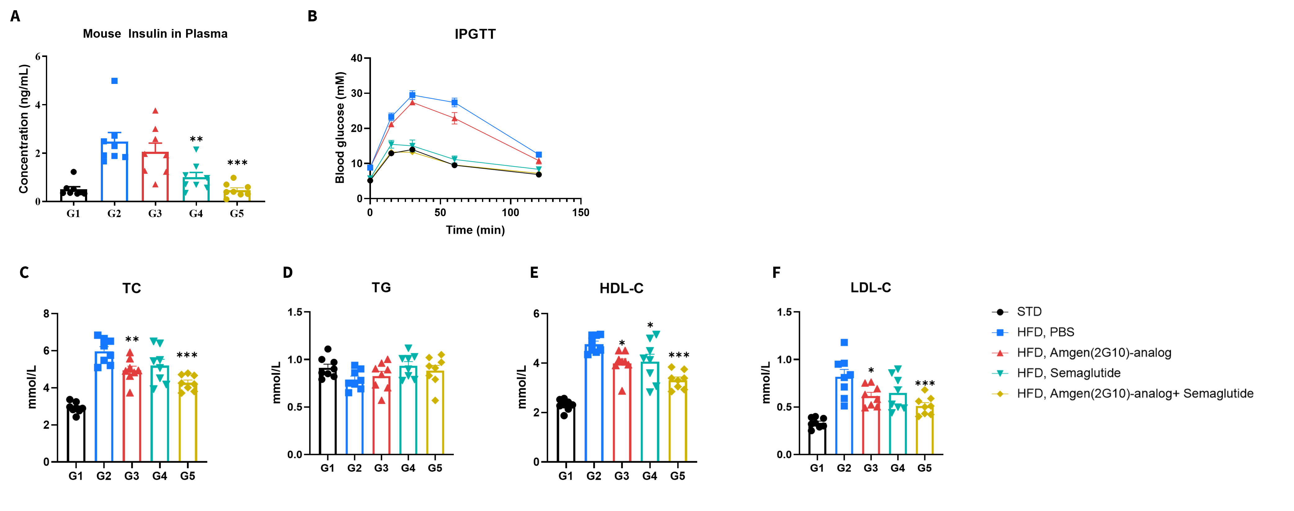
Efficacy study in HFD induced GIPR humanized mice. (A) Fasting insulin after treatment in plasma after 6 hours of fasting. (B) After fasting for 6 hours, mice were intraperitoneally injected with 1.5 g/kg D-glucose (15% solution, 10 μL/g) for Glucose Tolerance Tests after treatment. (C-F) Blood biochemical analysis after treatment. Values are expressed as mean ± SEM. The Ordinary one-way ANOVA determined significance compared with the HFD PBS group. *p<0.05, **p<0.01,***p<0.001.
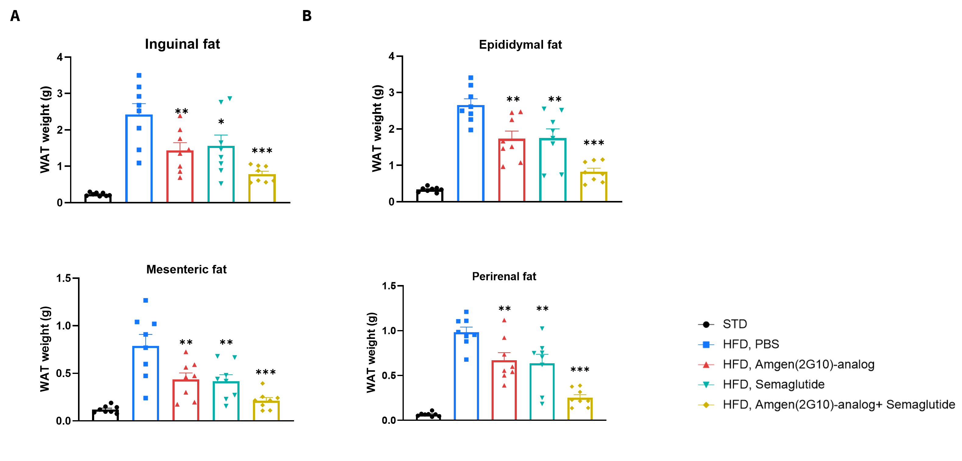
Efficacy study in HFD induced GIPR humanized mice. (A-D) Weight of white adipose tissue at the end of the treatment. Values are expressed as mean ± SEM. Significance was determined by the Ordinary one-way ANOVA compared with HFD PBS group. *p<0.05, **p<0.01,***p<0.001 .
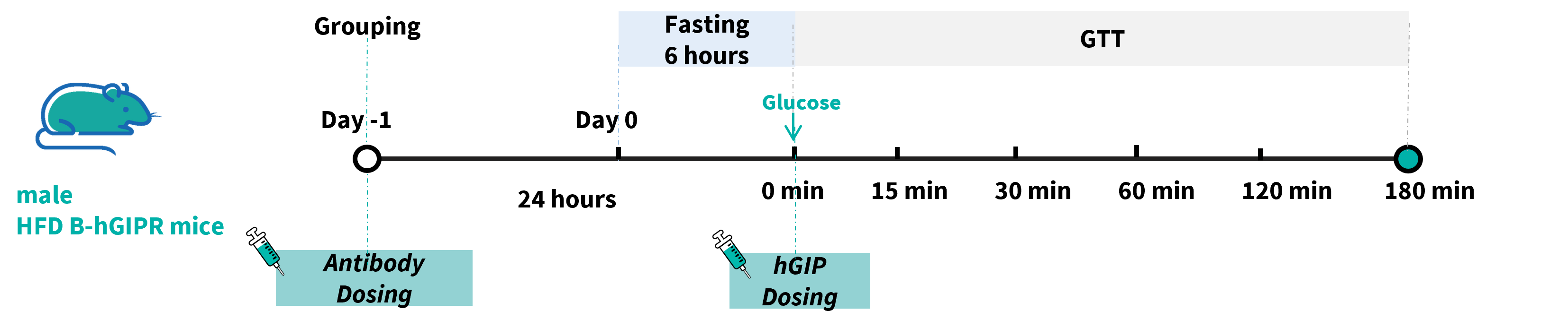
In vivo function of hGIP and GIPR-antibody in HFD induced GIPR humanized mice. GIPR humanized mice were fed with a high-fat diet for 12 weeks to induce obesity in the mice and were randomized into groups and received vehicle or GIPR antibody Amgen (2G10) analog (in house). Approximately 24 hours later, saline or 0.25 mg/kg human GIP (Cat. HY-P0276, MCE) was given via i.p. injection, then immediately followed by an IPGTT at 1.5 g/kg (30% solution, 5 μL/g).
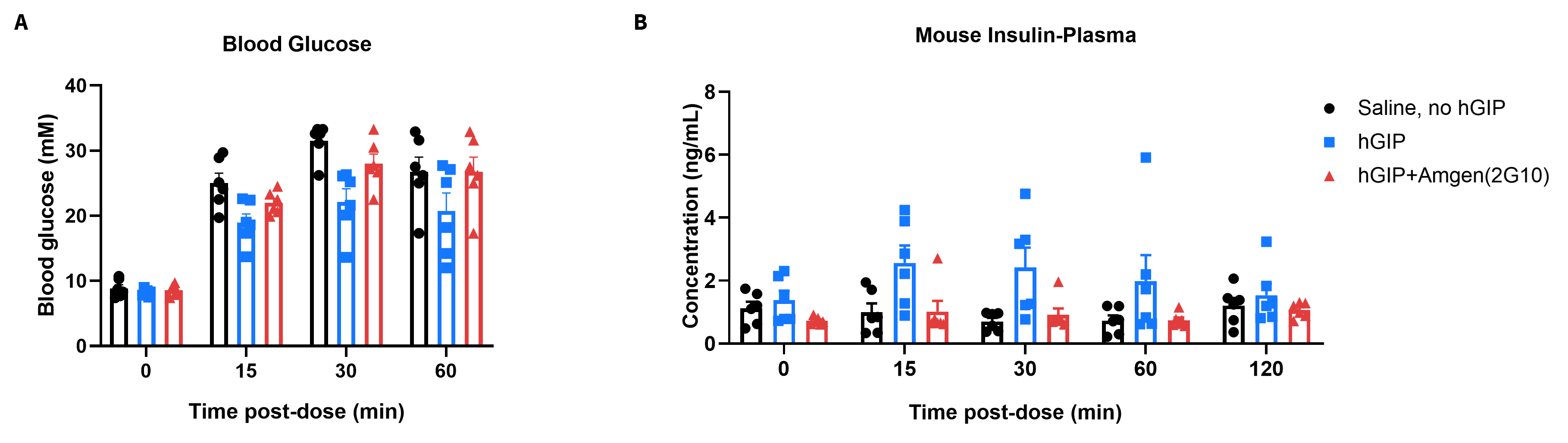
In vivo function of hGIP and GIPR-antibody in HFD induced GIPR humanized mice. GIPR humanized mice were fed with a high-fat diet for 12 weeks to induce obesity in the mice and were randomized into groups and received vehicle or GIPR antibody Amgen (2G10) analog (in house). Approximately 24 hours later, saline or 0.25 mg/kg human GIP (Cat. HY-P0276, MCE) was given via i.p. injection, then immediately followed by an IPGTT at 1.5 g/kg (30% solution, 5 μL/g). (A) Blood glucose and (B) plasma insulin were measured during IPGTT. Data represent means ± SEM.