OSMR: Dual anti-inflammatory and anti-fibrotic therapeutic effects

OSM
OSMR

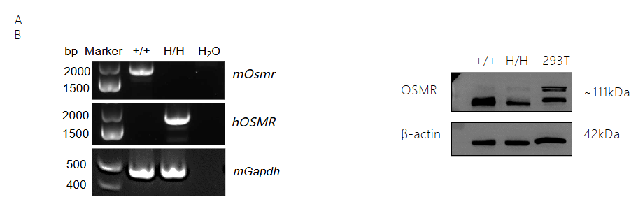
Strain specific analysis of OSMR gene expression in wild-type mice and B-hOSM/hOSMR mice. Kidney was collected from wild-type C57BL/6 mice (+/+) and homozygous B-hOSM/hOSMR mice (H/H), and analyzed by RT-PCR and western blot. (A) mRNA expression. Mouse Osmr mRNA was exclusively detectable in wild-type mice. Human OSMR mRNA was exclusively detectable in homozygous B-hOSM/hOSMR mice. (B) Protein expression. The OSMR protein were detected both in wild-type mice and homozygous B-hOSM/hOSMR mice. 293T cells were used as positive control (PC).
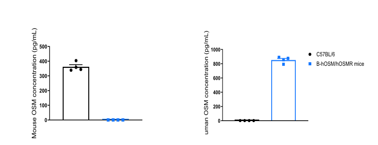
Strain specific OSM expression analysis in homozygous B-hOSM/hOSMR mice by ELISA. Spleen were isolated from wild-type C57BL/6 mice (+/+) and homozygous B-hOSM/hOSMR mice (H/H), and stimulated with anti-mouse CD3 and anti-mouse CD28 antibodies in vitro. Cell culture supernatant were collected and analyzed by species-specific OSM ELISA kit. Mouse OSM was detectable in wild-type mice. Human OSM was exclusively detectable in homozygous B-hOSM/hOSMR mice but not in wild-type mice. ND: Not detected.
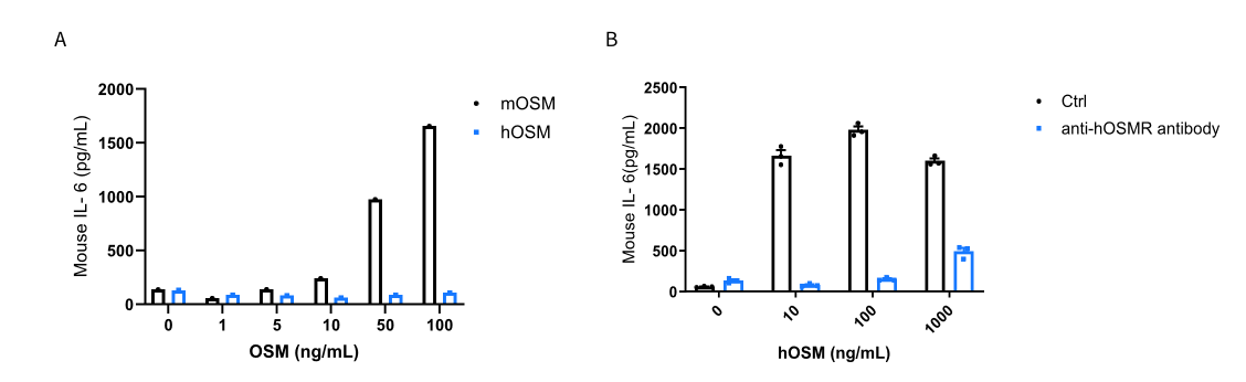
Human OSM induced IL-6 secretion is blockade by anti-hOSMR antibody in MEFs of homozygous B-hOSM/hOSMR mice. Mouse embryonic fibroblasts (MEFs) were derived from either C57BL/6 mice or B-hOSM/hOSMR mice, and stimulated with different concentrations of mOSM (0-100ng/mL) or hOSM (0-1000ng/mL) for 24h. Supernatants from each well were collected and analyzed for IL-6 expression by ELISA. (A) In the MEFs of C57BL/6 mice, IL-6 can be induced by mOSM in a dose-dependent manner, but not by hOSM. It demonstrates that hOSM can’t be recognized by mouse receptor. (B) In the MEFs of B-hOSM/hOSMR mice, hOSM can induce IL-6 secretion, and the function can be blocked by anti-hOSMR antibodies (in house). The results show that the OSMR signaling function was not influenced by gene humanization. Cells were incubated with antibodies (10μg/mL) for 30 minutes, and stimulated with hOSM.
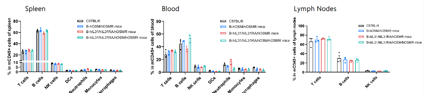
Analysis of leukocyte subpopulations by flow cytometry in immune organs and blood. Splenocytes, peripheral blood, and lymph nodes were isolated from female C57BL/6, B-hOSM/hOSMR mice, B-hIL31/hIL31RA/hOSMR mice and B-hIL31/hIL31RA/hOSM/hOSMR mice (female, 6 or 7-week-old, n = 3). Single live cells were gated on the CD45⁺ population and analyzed by flow cytometry as indicated. Values are expressed as mean ± SEM.
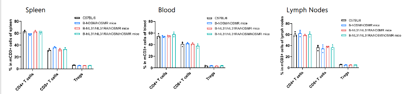
Analysis of T-cell subpopulations by flow cytometry in immune organs and blood. Splenocytes, peripheral blood, and lymph nodes were isolated from female C57BL/6, B-hIL31/hIL31RA/hOSMR mice, B-hOSM/hOSMR mice and B-hIL31/hIL31RA/hOSM/hOSMR mice (female, 6 or 7-week-old, n = 3). Single live cells were gated on the CD3⁺ T-cell population and analyzed by flow cytometry as indicated. Values are expressed as mean ± SEM.