B-hTNFA/hTNFR2/hTNFR1/hIL6/hIL6R mice

C57BL/6-Tnftm1(TNF)Bcgen Tnfrsf1atm1(TNFRSF1A)Bcgen Tnfrsf1btm1(TNFRSF1B)Bcgen /Il6tm1(IL6)Bcgen Il6rtm2(IL6R)Bcgen/Bcgen • 114254

B-hTNFA/hTNFR2/hTNFR1/hIL6/hIL6R mice

Catalog Number: 114254
Strain Name: C57BL/6-Tnftm1(TNF)Bcgen Tnfrsf1atm1(TNFRSF1A)Bcgen Tnfrsf1btm1(TNFRSF1B)Bcgen /Il6tm1(IL6)Bcgen Il6rtm2(IL6R)Bcgen/Bcgen
Strain Background: C57BL/6
NCBI gene ID: 7124,7132,7133,3569,3570 (Human)
Aliases: DIF; TNFA; IMD127; TNFSF2; TNLG1F; TNF-alpha; FPF; p55; p60; TBP1; TNF-R; TNFAR; TNFR1; p55-R; CD120a; TNFR55; TNFR60; TNF-R-I; TNF-R55; p75; TBPII; TNFBR; TNFR2; CD120b; TNFR1B; TNFR80; TNF-R75; p75TNFR; TNF-R-II; CDF; HGF; HSF; BSF2; IL-6; BSF-2; IFNB2; IFN-beta-2; IL6Q; gp80; CD126; HIES5; IL-6R; IL6RA; IL6RQ; IL-1Ra; IL-6RA; IL6QTL; IL-6R-1
---
ライセンスオプション提供可能
B-hTNFA/hTNFR2/hTNFR1/hIL6/hIL6R mice

このページで

  • Description
  • Targeting strategy
  • Phenotypic analysis
  • Efficacy

ポスター

すべて表示

    出版物

      Description

      TNFA/TNFR1/TNFR2: A master pathway of regulating inflammation, immune responses, and cell survival

      • Gene Information: Tumor necrosis factor (TNF) is a protein-coding gene located on chromosome 6p21.33. It encodes a multifunctional proinflammatory cytokine that belongs to the tumor necrosis factor (TNF) superfamily. TNF receptor superfamily member 1A (TNFRSF1A, TNFR1) encodes a member of the TNF receptor superfamily of proteins located on chromosome 12p13.31. TNF receptor superfamily member 1B (TNFRSF1B, TNFR2) encodes a member of the TNF receptor superfamily of proteins located on chromosome 1p36.22.
      • Protein Expression: TNF is primarily expressed by macrophages. TNF is synthesized as a membrane-bound precursor and cleaved into a soluble circulating protein. TNFR1 is expressed on almost all nucleated cells. TNFR2 is expressed on CD4+ T cells, CD8+ T cells, Tregs, endothelial cells, microglia, specific neuronal subpopulations, oligodendrocytes, cardiomyocytes, and human mesenchymal stem cells.
      • Signaling Pathway: TNF exerts its effects primarily through two receptors: TNFR1 and TNFR2.
      • Therapeutic Inhibition: By blocking a central driver of the immune system's inflammatory response, adalimumab inhibits downstream inflammation and improves clinical outcomes, including reducing other inflammatory markers and reducing tissue damage in various autoimmune conditions.

      IL6/IL6R: A key cytokine signaling axis in inflammation and its therapeutic intervention

      • Gene Information: Interleukin 6 (IL6) is a protein-coding gene located on chromosome 7p15.3, encoding a pleiotropic cytokine that serves as a core mediator of the acute phase response. Its cognate receptor alpha subunit, IL6R (CD126), is encoded by a gene situated on chromosome 1q21.3.
      • Protein Expression: IL6 is transiently produced and secreted primarily by activated macrophages, endothelial cells, and fibroblasts in response to stimulation. The receptor, IL6R, exists in two main forms: a transmembrane protein (mIL6R) constitutively expressed on hepatocytes and specific leukocyte subsets, and a functional soluble isoform (sIL6R) generated via alternative splicing or proteolytic shedding.
      • Signaling Pathway: IL6 exerts its biological effects by binding to either mIL6R (classic signaling) or sIL6R (trans-signaling). Both pathways require the subsequent recruitment of the shared transmembrane signal transducer gp130 (IL6ST), forming a hexameric signaling complex that activates the downstream intracellular JAK-STAT3 cascade.
      • Therapeutic Inhibition: By blocking the interaction within this cytokine axis, therapeutic monoclonal antibodies—such as tocilizumab (targeting IL6R) or siltuximab (targeting IL6)—effectively halt hyperactive downstream inflammatory cascades, delivering proven clinical benefits in treating rheumatoid arthritis, systemic juvenile idiopathic arthritis, and induced cytokine release syndrome.
      Targeting strategy

      TNFA

      • The targeted mouse Tnfa whole genomic sequences including 5'UTR, 3'UTR and coding region were replaced by human TNFA whole genomic sequences in B-hTNFA/hTNFR2/hTNFR1/hIL6/hIL6R mice.

      TNFR2

      • The exons 2~6 of mouse Tnfr2 gene that  encode the extracellular domain were replaced by human TNFR2 exons 2~6 in B-hTNFA/hTNFR2/hTNFR1/hIL6/hIL6R mice.
      • Expression of the human TNFR2 protein is driven by the native mouse Tnfr2 promoter, while endogenous mouse Tnfr2 transcription and translation are disrupted.

      TNFR1

      • The exons 2~6 of mouse Tnfr1 gene that  encode the extracellular domain were replaced by human TNFR1 exons 2~6 in B-hTNFA/hTNFR2/hTNFR1/hIL6/hIL6R mice.
      • Expression of the human TNFR1 protein is driven by the native mouse Tnfr1 promoter, while endogenous mouse Tnfr1 transcription and translation are disrupted.

      IL6

      • The exons 1-5 of mouse Il6 gene that encode whole protein is replaced by human counterparts in B-hTNFA/hTNFR2/hTNFR1/hIL6/hIL6R mice.
      • The promoter, 5’UTR and 3’UTR region of the mouse Il6 are also replaced by human counterparts.

      IL6R

      • The CDS of human IL6R gene was inserted after the start codon ATG of mouse Il6ra gene in B-hTNFA/hTNFR2/hTNFR1/hIL6/hIL6R mice.
      • Expression of the human IL6R protein is driven by the native mouse Il6r promoter, while endogenous mouse Il6r transcription and translation are disrupted.
      TNFA Protein Expression Analysis in Serum
      • Mouse TNFA was detected exclusively in wild-type C57BL/6 mice.
      • Human TNFA was detected in homozygous B-hTNFA/hTNFR2/hTNFR1/hIL6/hIL6R mice, but not in wild-type mice.

      Strain specific TNFA expression analysis in wild-type C57BL/6JNifdc mice and homozygous humanized B-hTNFA/hTNFR2/hTNFR1/hIL6/hIL6R mice by ELISA. Serum were collected from wild-type C57BL/6JNifdc mice (+/+) and homozygous B-hTNFA/hTNFR2/hTNFR1/hIL6/hIL6R mice(H/H;H/H;H/H;H/H;H/H), the production of TNFA in serum were assessed after 2 h of stimulation with LPS. Expression levels of mouse and human TNFA were analyzed by ELISA (Mouse TNFA ELISA Kit: Biolegend, 430904; Human TNFA ELISA Kit: Biolegend, 430204). Mouse TNFA was only detectable in wild-type C57BL/6JNifdc mice, and human TNFA was only detectable in homozygous B-hTNFA/hTNFR2/hTNFR1/hIL6/hIL6R mice. Values are expressed as mean ± SEM.
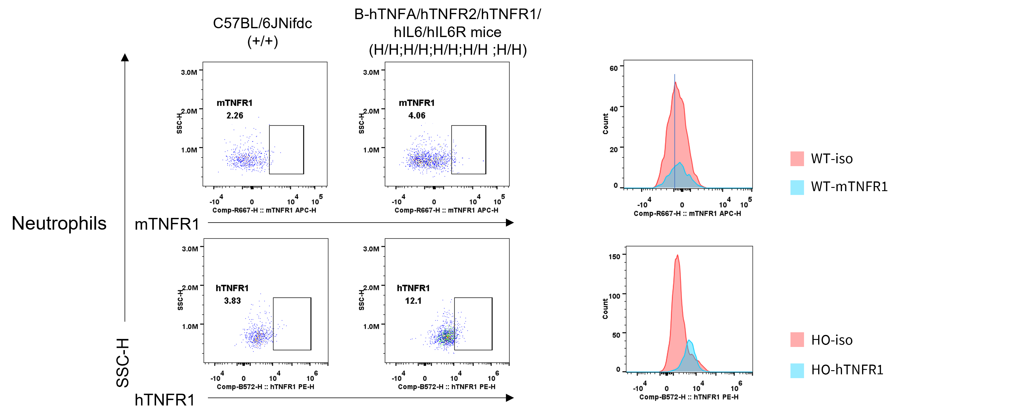
      TNFR1 Protein Expression in Spleen
      • Human TNFR1 was exclusively expressed on neutrophils in B-hTNFA/hTNFR2/hTNFR1/hIL6/hIL6R mice.

      Strain specific TNFR1 expression analysis in wild-type C57BL/6JNifdc mice and homozygous B-hTNFA/hTNFR2/hTNFR1/hIL6/hIL6R mice by flow cytometry. Splenocytes were collected from wild-type C57BL/6JNifdc mice(+/+) and homozygous B-hTNFA/hTNFR2/hTNFR1/hIL6/hIL6R mice(H/H;H/H;H/H;H/H;H/H). The protein expression was analyzed with anti-mouse TNFR1 antibody (Biolegend, 113005) and anti-human TNFR1 antibody(Biolegend, 369903) by flow cytometry.  Mouse TNFR1 was detectable in neutrophils of wild-type mice. Human TNFR1 was detectable in neutrophils of homozygous B-hTNFA/hTNFR2/hTNFR1/hIL6/hIL6R mice.
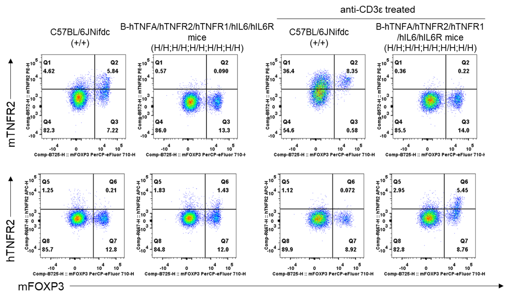
      TNFR2 Protein Expression in Spleen
      • Human TNFR2 was exclusively expressed on regulatory T cells in B-hTNFA/hTNFR2/hTNFR1/hIL6/hIL6R mice.

      Strain specific TNFR2 expression analysis in wild-type C57BL/6JNifdc mice and homozygous B-hTNFA/hTNFR2/hTNFR1/hIL6/hIL6R mice by flow cytometry. Splenocytes were collected from wild-type C57BL/6JNifdc mice and homozygous B-hTNFA/hTNFR2/hTNFR1/hIL6/hIL6R mice, stimulated with anti-CD3ε in vivo (7.5 μg/mice, stimulation for 24 hours, i.p.). The protein expression was analyzed with anti-mouse TNFR2 antibody (Biolegend, 113405) and anti-human TNFR2 antibody (Biolegend, 358406) by flow cytometry. Mouse TNFR2 was only detectable in wild-type mice. Human TNFR2 was only detectable in homozygous B-hTNFA/hTNFR2/hTNFR1/hIL6/hIL6R mice.

      IL6 Protein Expression Analysis in Serum
      • Mouse IL6 was detected exclusively in wild-type C57BL/6 mice
      • Human IL6 was detected in homozygous B-hTNFA/hTNFR2/hTNFR1/hIL6/hIL6R mice, but not in wild-type mice.

      Strain specific IL6 expression analysis in wild-type C57BL/6JNifdc mice and homozygous B-hTNFA/hTNFR2/hTNFR1/hIL6/hIL6R mice by ELISA. Serum was collected from wild-type C57BL/6JNifdc  mice (+/+) (female, n=3, 6-week-old), homozygous B-hTNFA/hTNFR2/hTNFR1/hIL6/hIL6R mice(H/H; H/H ; H/H ; H/H ; H/H) (H/H; H/H) (female, n=3, 6-week-old), the production of IL6 in serum were assessed after 3h of stimulation with LPS (20 μg/200uL, i.p.) in vivo. Expression level of human IL6 were analyzed by ELISA (Human IL-6 ELISA MAX TM Deluxe Set (430508)). Human IL6 was only detectable in homozygous homozygous B-hTNFA/hTNFR2/hTNFR1/hIL6/hIL6R mice (n=3). Values are expressed as mean ± SEM.
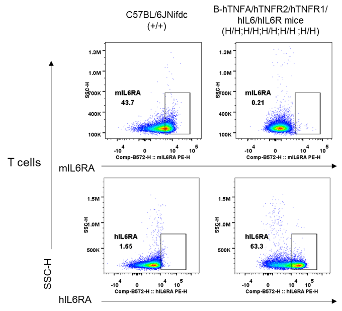
      Membrane IL6R Protein Expression in Spleen
      • Human IL6R was exclusively expressed on T cells in B-hTNFA/hTNFR2/hTNFR1/hIL6/hIL6R mice.

      Strain specific IL6R expression analysis in wild-type C57BL/6JNifdc mice, homozygous B-hTNFA/hTNFR2/hTNFR1/hIL6/hIL6R mice by flow cytometry. Splenocytes were collected wild-type C57BL/6JNifdc mice(+/+) and homozygous B-hTNFA/hTNFR2/hTNFR1/hIL6/hIL6R mice (H/H;H/H;H/H;H/H;H/H). The protein expression was analyzed with anti-mouse CD126 (IL-6Rα chain) Antibody (Biolegend, 115805) and anti-human CD126 (IL-6Rα) Antibody (Biolegend, 352803) by flow cytometry. Mouse IL6R was detectable in T cells of wild-type mice. Human IL6R was detectable in T cells of homozygous B-hTNFA/hTNFR2/hTNFR1/hIL6/hIL6R mice. WT: C57BL/6JNifdc; HO: B-hTNFA/hTNFR2/hTNFR1/hIL6/hIL6R mice.

      Soluble IL6R Protein Expression in Serum
      • Soluble human IL6R was exclusively expressed in B-hTNFA/hTNFR2/hTNFR1/hIL6/hIL6R mice.

      Strain specific IL6R expression analysis in wild-type C57BL/6JNifdc mice and homozygous B-hTNFA/hTNFR2/hTNFR1/hIL6/hIL6R mice by ELISA. Serum was collected from wild-type C57BL/6JNifdc mice (+/+) (female, n=3, 6-week-old), homozygous B-hTNFA/hTNFR2/hTNFR1/hIL6/hIL6R mice (H/H; H/H ; H/H ; H/H ; H/H) (female, n=3, 6-week-old). Expression level of human IL6R were analyzed by ELISA (Human IL6R ELISA Kit (DR600)). Human IL6R was detectable in homozygous B-hTNFA/hTNFR2/hTNFR1/hIL6/hIL6R mice(n=3). Values are expressed as mean ± SEM.

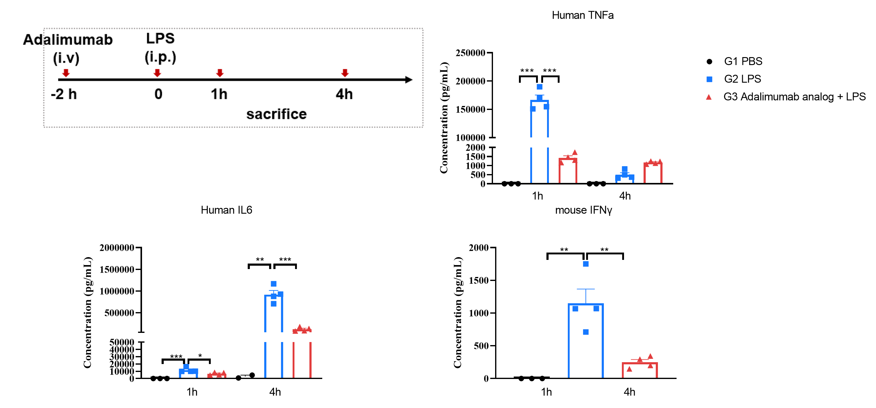
      In Vivo Efficacy of Anti-human TNFA Antibody in LPS-induced Response
      • Anti-human TNFA significantly decreased mouse IL6 and mouse IFNγ expression after LPS injection, indicating a successful inhibition of the downstream activation caused by hTNFA.

      Human TNFA, human IL6, and mouse IFNγ expression analysis in homozygous humanized B-hTNFA/hTNFR2/hTNFR1/hIL6/hIL6R mice by ELISA. Mice received LPS injection (i.p.) two hours following PBS or adalimumab analog (i.v., in house) treatment. Serum was collected from homozygous B-hTNFA/hTNFR2/hTNFR1/hIL6/hIL6R mice one hour and four hours after LPS injection. Expression level of human TNFA, human IL6, and mouse IFNγ, were analyzed by ELISA (ELISA MAX™ Deluxe Set Human TNF-α, Biolegend, 430204; ELISA MAX™ Deluxe Set Human IL-6, Biolegend, 430508; ELISA MAX™ Deluxe Set Mouse IFN-γ, Biolegend, 430804). Human TNFA level was significantly increased one hour and four hours after LPS injection. Human IL6 level was significantly increased one and four hours after LPS injection. Mouse IFNγ level was significantly increased four hours after LPS injection. Pre-treatment of adalimumab analog (in house) significantly decreased human TNFA expression at one hour after LPS injection, human IL6 one and four hours after LPS stimulation, and mouse IFNγ expression at four hours after LPS injection, indicating a successful inhibition of the downstream activation caused by hTNFA. Values are expressed as mean ± SEM. Significance was determined by one-way ANOVA test . * p < 0.5, **p < 0.01, ***p < 0.001.

      * When publishing results obtained using this animal model, please acknowledge the source as follows: The animal model [B-hTNFA/hTNFR2/hTNFR1/hIL6/hIL6R mice] (Cat# 114254) was purchased from Biocytogen.