あなたもお好きかもしれません

Systemic lupus erythematosus (SLE) remains one of the most complex autoimmune diseases to tackle. Affecting over 3.4 million people worldwide, with a staggering 90% being women (Siegel and Sammaritano 2024), SLE is marked by immune dysregulation, autoantibody production, and widespread organ inflammation (Arnaud, Chasset, and Martin 2024). The disease's heterogeneity and elusive triggers—ranging from genetic and hormonal factors to environmental stimuli—have made treatment notoriously difficult (Su et al. 2024). Despite decades of research and some progress with immunosuppressants and biologics, the unmet need for more precise, targeted, and effective therapies persists.
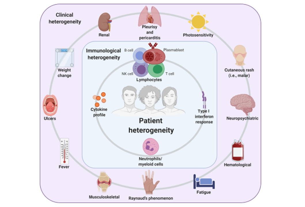
Heterogeneity in SLE (Allen, Rus, and Szeto 2021)
The clinical manifestation of lupus spans an extraordinary spectrum. Some patients may experience primarily cutaneous symptoms, while others grapple with life-threatening nephritis or CNS involvement (Stull, Sprow, and Werth 2023). This variation has historically limited the translatability of findings from traditional mouse models (Richard and Gilkeson 2018). Biocytogen is helping to bridge this gap with a suite of next-generation humanized mice and SLE disease models, designed to accelerate the development and validation of novel SLE therapies.
Our humanized models are genetically engineered to express key human targets involved in SLE pathogenesis, providing a translationally relevant platform for preclinical studies. In particular, our dual- and multi-target humanized models support the evaluation of drug candidates that modulate multiple pathways simultaneously to address disease heterogeneity.
SLE Mouse Models:
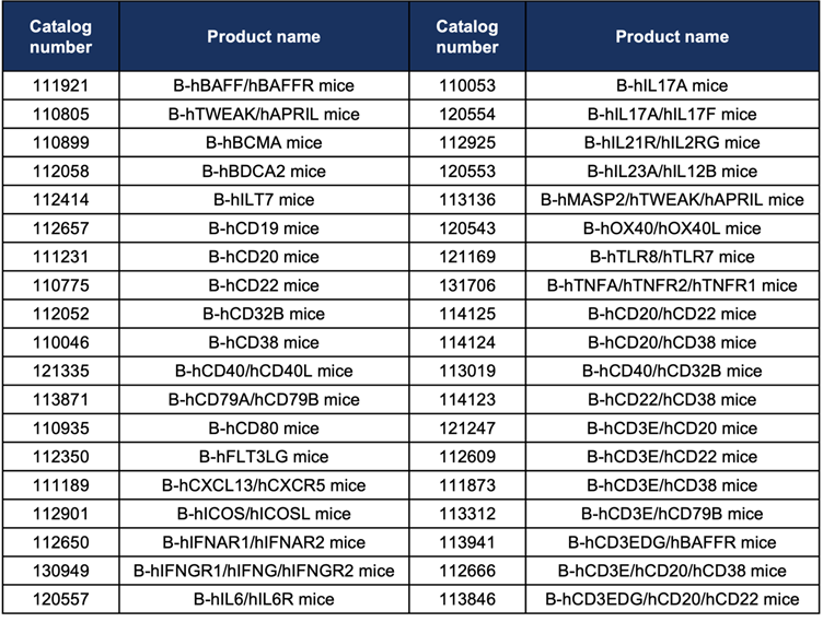
Featured Humanized Mouse Models:
B-hBAFF/hBAFFR mice: Targeting the BAFF/BAFFR axis, crucial for B-cell survival and autoantibody production.
B-hTWEAK/hAPRIL mice: Useful for studying tissue inflammation and fibrosis, with APRIL and TWEAK known to be elevated in SLE patients.
B-hBCMA mice: Supporting research into long-lived plasma cells, a key source of pathogenic antibodies.
B-hBDCA2 and B-hILT7 mice: Targeting plasmacytoid dendritic cells (pDCs), major producers of type I interferons in SLE.
B-hCD19 mice: For B cell-targeted therapies.
B-hCD40 mice: Allowing CD40-based immunomodulatory strategies.
Case Study Highlights
Case Study 1: Pristane-induced SLE Model in B-hCD40 Mice
Pristane is used to induce SLE by initiating chronic immune activation and autoantibody production, effectively modeling key pathological features of human SLE. In a pristane-induced SLE model using B-hCD40 mice, treatment with an anti-CD40 antibody led to a significant reduction in anti-dsDNA IgG levels—a key biomarker associated with lupus flares and renal involvement. This model highlights the potential of anti-CD40 therapies in modulating autoimmune responses. 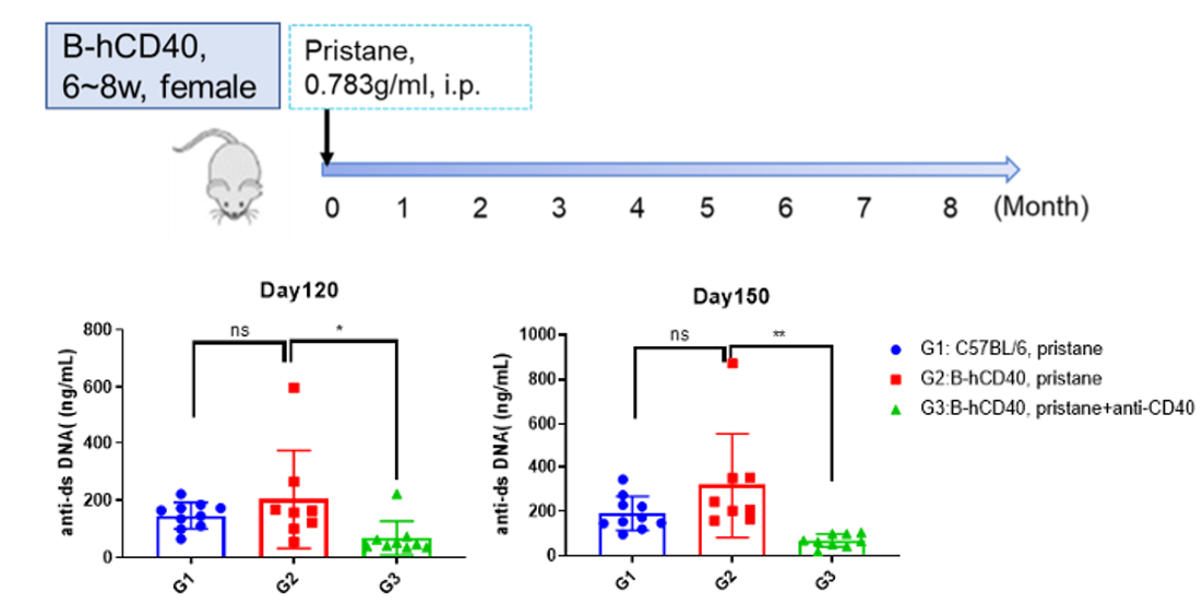
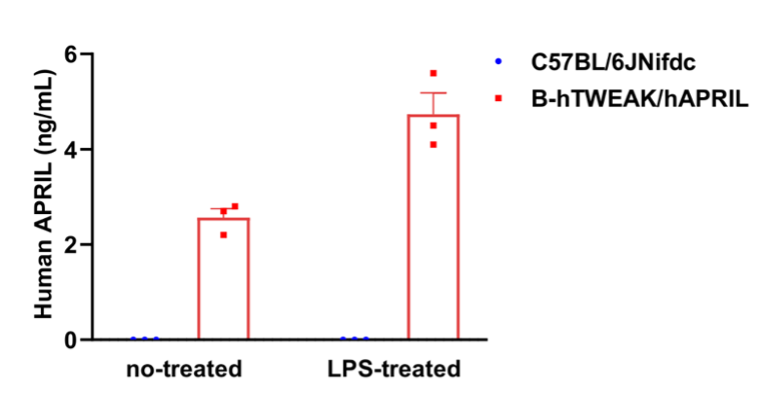
Case Study 2: B-hTWEAK/hAPRIL Mice
APRIL and TWEAK are cytokines elevated in SLE patients, contributing to B cell survival, autoantibody production, and tissue inflammation—making them key players in disease pathogenesis. Biocytogen's B-hTWEAK/hAPRIL mice express human TWEAK and APRIL, enabling in vivo evaluation of therapeutics targeting these cytokines in a human-relevant context.
Case Study 3: MRL/lpr Spontaneous SLE Mouse Model
The MRL/lpr mouse is a widely used spontaneous SLE model that develops hallmark features of lupus, making it a valuable platform for evaluating immunomodulatory therapies in preclinical research.
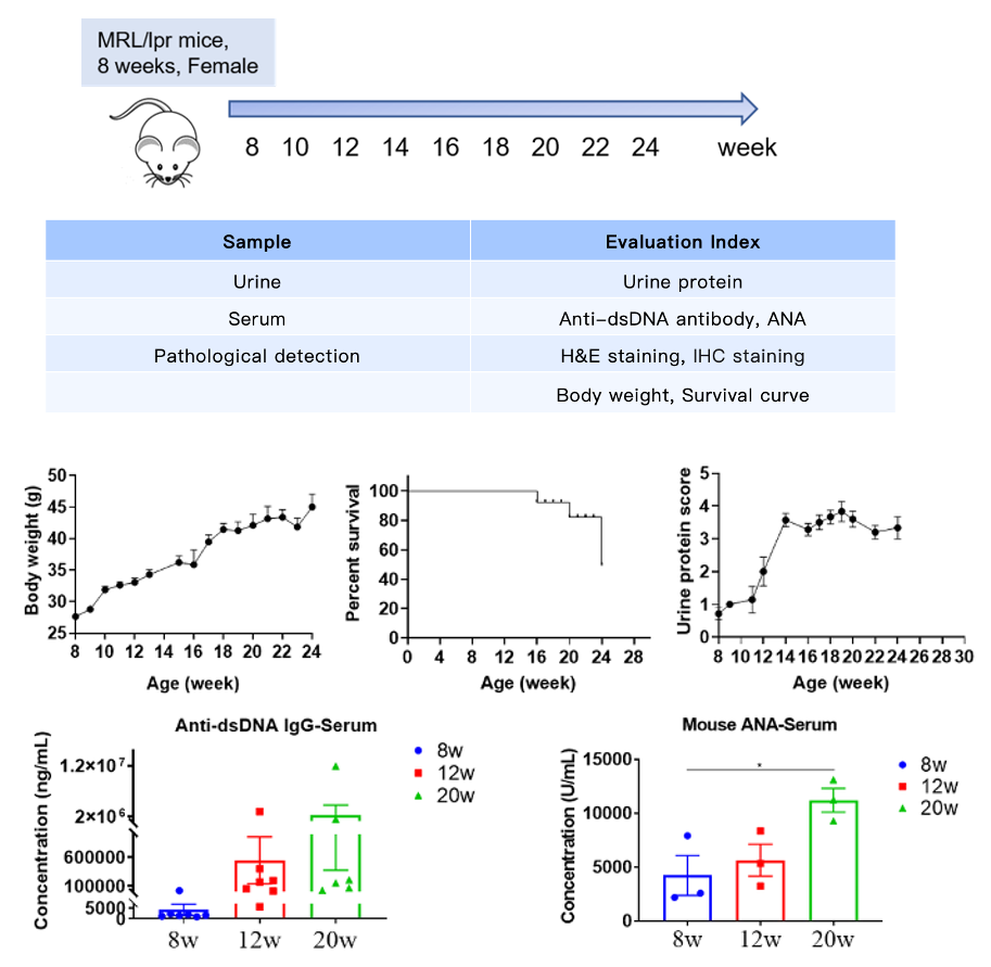
Changes in body weight, urine protein, survival curve, serum anti-dsDNA antibody, and ANA at different time points.
Histologic Assessment of Spontaneous SLE in MRL/lpr Mice
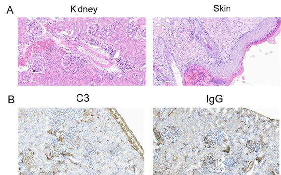
Histopathological analysis of kidneys and skin in 20-week-old MRL/lpr spontaneous SLE mice. (A) Kidney sections exhibit glomerular hypertrophy, cellular proliferation, and mesangial matrix expansion. Skin sections show epidermal hyperplasia, thickening, hyperkeratosis with parakeratosis, and dermal inflammatory infiltrates. (B) Renal tissues display C3 and IgG deposition.
Biocytogen’s Humanized Mice for SLE Targets
Explore How Biocytogen’s Cutting-Edge Humanized Models Can Support Your SLE Therapeutic Pipeline!
Reference:
Siegel, Caroline H., and Lisa R. Sammaritano. "Systemic lupus erythematosus: a review." Jama 331.17 (2024): 1480-1491.
Arnaud, Laurent, François Chasset, and Thierry Martin. "Immunopathogenesis of systemic lupus erythematosus: an update." Autoimmunity Reviews (2024): 103648.
Su, Xu, et al. "Systemic lupus erythematosus: pathogenesis and targeted therapy." Molecular Biomedicine 5.1 (2024): 54.
Allen, Marilyn E., Violeta Rus, and Gregory L. Szeto. "Leveraging heterogeneity in systemic lupus erythematosus for new therapies." Trends in molecular medicine 27.2 (2021): 152-171.
Stull, Courtney, Grant Sprow, and Victoria P. Werth. "Cutaneous involvement in systemic lupus erythematosus: a review for the rheumatologist." The Journal of rheumatology 50.1 (2023): 27-35.
Richard, Mara Lennard, and Gary Gilkeson. "Mouse models of lupus: what they tell us and what they don’t." Lupus science & medicine 5.1 (2018): e000199.